ITL-3582ee74b124
Watch the video
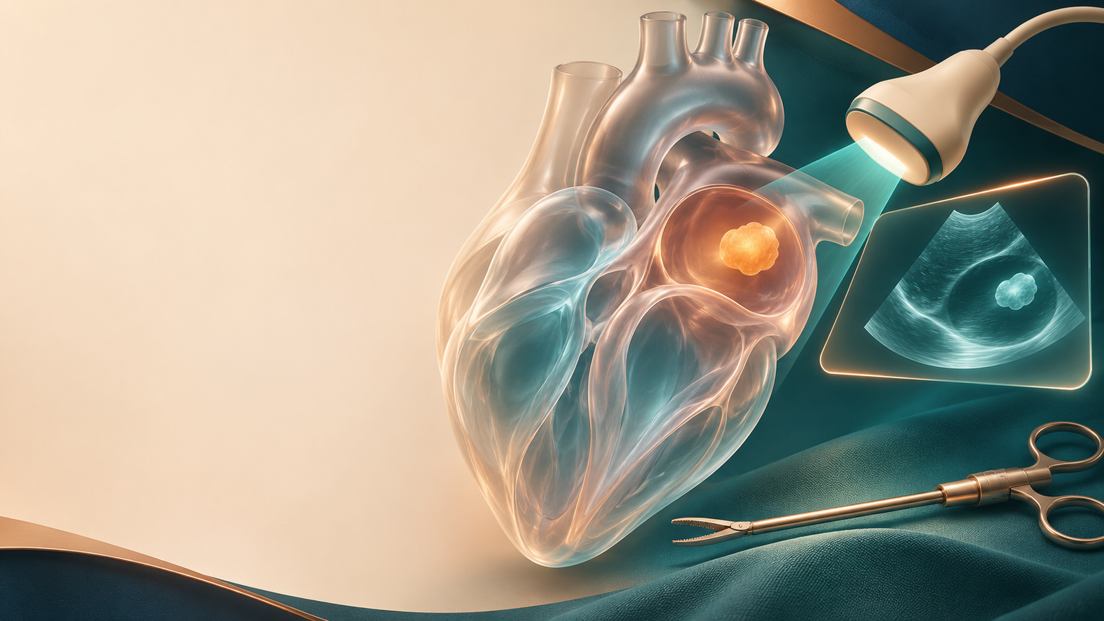
Figure 1. The approach should follow the anatomy and physiology of the mass. Conceptual artwork, including the simulated imaging panel; not a patient scan.
Key points
- “Left atrial mass” is an imaging finding, not a histologic diagnosis; thrombus, tumor and other mimics require different decisions.[1]
- Before choosing access, define the attachment, mobility, valve interaction and relationship to surrounding structures—not size alone.
- TEE clarifies dynamic anatomy; CMR and CT can address tissue characterization and extension when those questions remain unresolved.[1]
- A 2026 series supports feasibility in selected patients, but its 26 minimally invasive cases had no sternotomy control group.[2]
- One-lung ventilation, cannulation and conversion readiness belong in the same planning discussion as complete excision.
Replace the diagnostic label with an operative map
The referral says “left atrial myxoma, minimally invasive excision.” Before accepting that plan, the anesthesiologist and surgeon need a shared description of what must be removed and what may need repair.
A pedunculated mass attached to the left side of the interatrial septum is compatible with a myxoma. That familiar appearance is useful, but it does not make every left atrial mass a myxoma. The differential also includes thrombus and tumorlike structures. An infiltrative lesion or broad attachment can create a very different operation from a localized stalk-based lesion.[1]
The preoperative imaging summary should describe the attachment site and extent, mobility through the cardiac cycle, dimensions in more than one plane, proximity to the mitral apparatus and evidence of inflow obstruction. It should also identify other lesions or structural disease that may require treatment. This turns “a mass is present” into information that can change the surgical route.
Choose additional imaging to answer a remaining question
Echocardiography is usually the first test. TEE is particularly useful when localization is uncertain or a mobile lesion needs better definition. Three-dimensional imaging can clarify attachment and relationships to nearby structures.[1]
CMR adds tissue characterization and assessment of invasion; CT can define spatial relationships, calcification and coronary anatomy. These tests are complementary rather than a mandatory sequence for every patient. A stable patient with uncertain tissue origin may benefit from further characterization; an obstructive lesion requiring urgent treatment should not be delayed merely to complete an imaging checklist. The imaging review specifically recognizes hemodynamic instability as a limitation to CMR.[1]
For the operative team, the next test is useful when its answer could change the diagnosis, urgency, extent of resection or reconstruction. More images without a decision attached do not necessarily improve the plan.
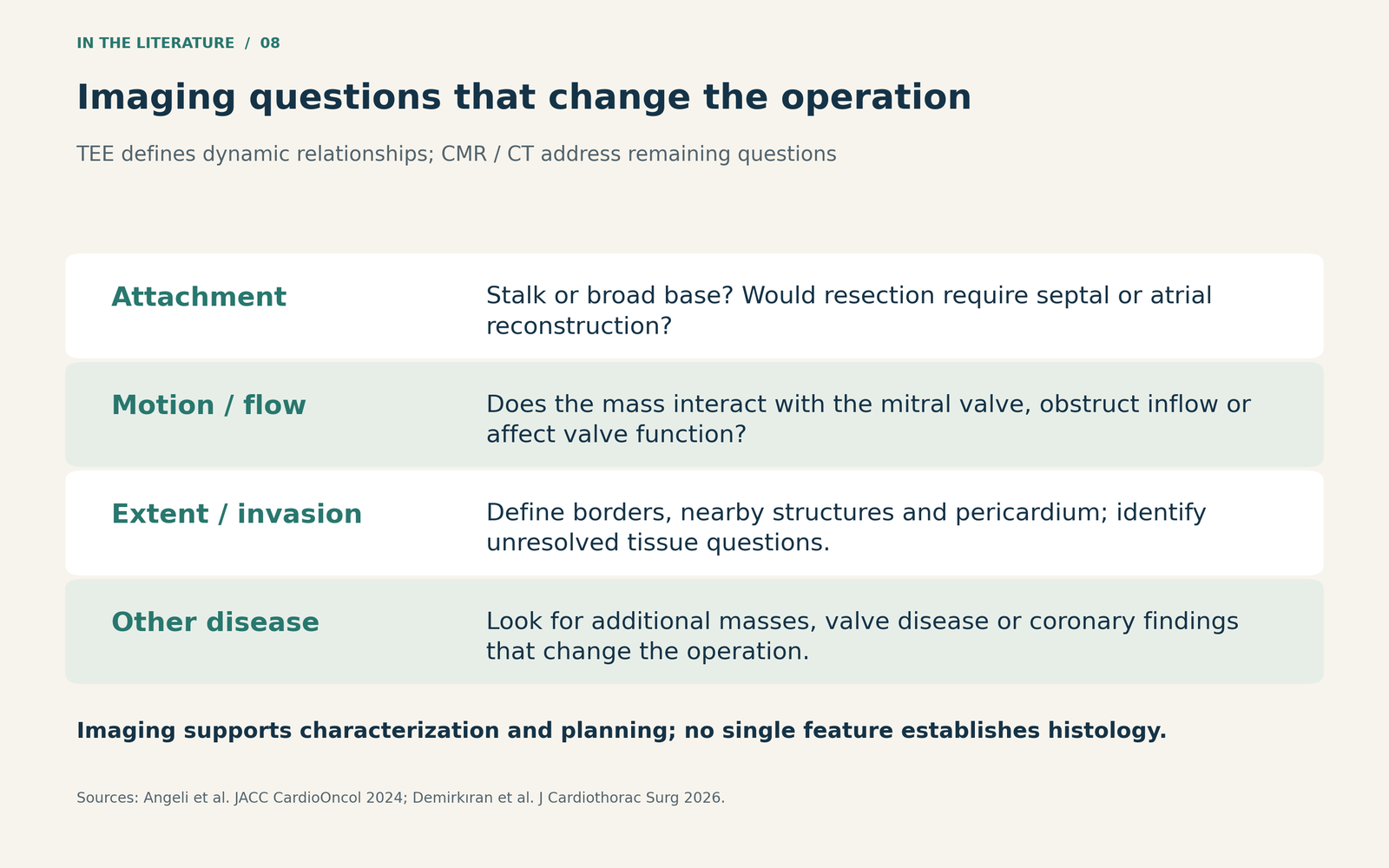
Figure 2. An operative map assembled from complementary imaging. The findings inform planning; none of these features alone establishes histology.[1, 2]

What does the newer minimally invasive series establish?
Demirkıran and colleagues reported 26 selected patients treated between 2015 and 2023: 17 underwent robotic-assisted surgery and nine right minithoracotomy. Twenty-one masses were in the left atrium. Final pathology included 23 myxomas, two papillary fibroelastomas and one teratoma with somatic malignant transformation.[2]
The center selected access using tumor size, location, neighboring structures and suspected invasiveness. Patients treated by conventional sternotomy were excluded. Consequently, this study describes what the team accomplished in selected minimally invasive cases; it cannot determine whether that approach reduces complications, hospital stay or recurrence compared with sternotomy.[2]
One minithoracotomy patient required conversion to sternotomy to repair bleeding associated with clamp injury to the left atrial appendage and adjacent posterior wall. There was one in-hospital death from COVID-19 pneumonia. The reported median follow-up was 32 months, with a range of 9–104 months; no tumor recurrence was reported. The longest follow-up is not the follow-up of every patient.[2]
The largest resected mass measured 85 × 35 mm, but it was a right atrial lesion. That observation is neither a validated upper size limit nor evidence that every similarly sized left atrial mass can be removed through the same access. Size matters through exposure, extraction and reconstruction requirements; it is not a stand-alone eligibility rule.[2]
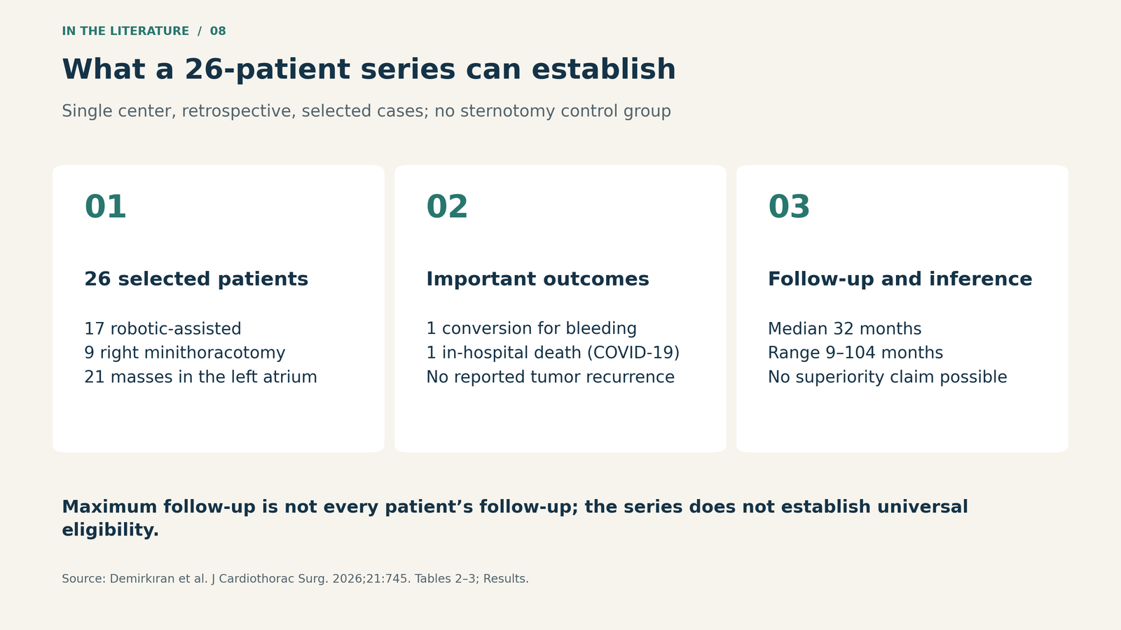
Figure 3. A feasibility series with 17 robotic and nine minithoracotomy procedures. The data do not compare minimally invasive surgery with sternotomy.[2]

Decide whether the patient can tolerate the route
Access planning extends beyond the tumor. The 2026 series used one-lung ventilation and routine TEE. Four patients had central rather than peripheral arterial cannulation; peripheral arterial disease influenced that choice in one patient. The center generally favored sternotomy when coronary bypass or complex reconstruction for an invasive mass was anticipated.[2] These are descriptions of that team's practice, not universal contraindications.
A useful preoperative discussion therefore connects four elements: the planned resection and repair, respiratory reserve for the intended exposure, a feasible cannulation route, and a workable conversion plan. Prior right-sided chest surgery or adhesions may alter access. Additional valve disease may alter the procedure. Suspected infiltration may change the required margins and reconstruction.
For anesthesia, the value of TEE is to keep the actual anatomy and hemodynamic effect visible as the plan is implemented. A mass interacting with the mitral valve deserves a specific discussion of obstruction and the response to instability. After excision, reassessment should address residual mass, valve function, any repaired septal or atrial defect and ventricular function. This is a clinical synthesis of the imaging and surgical evidence, rather than a tested checklist.[1, 2]
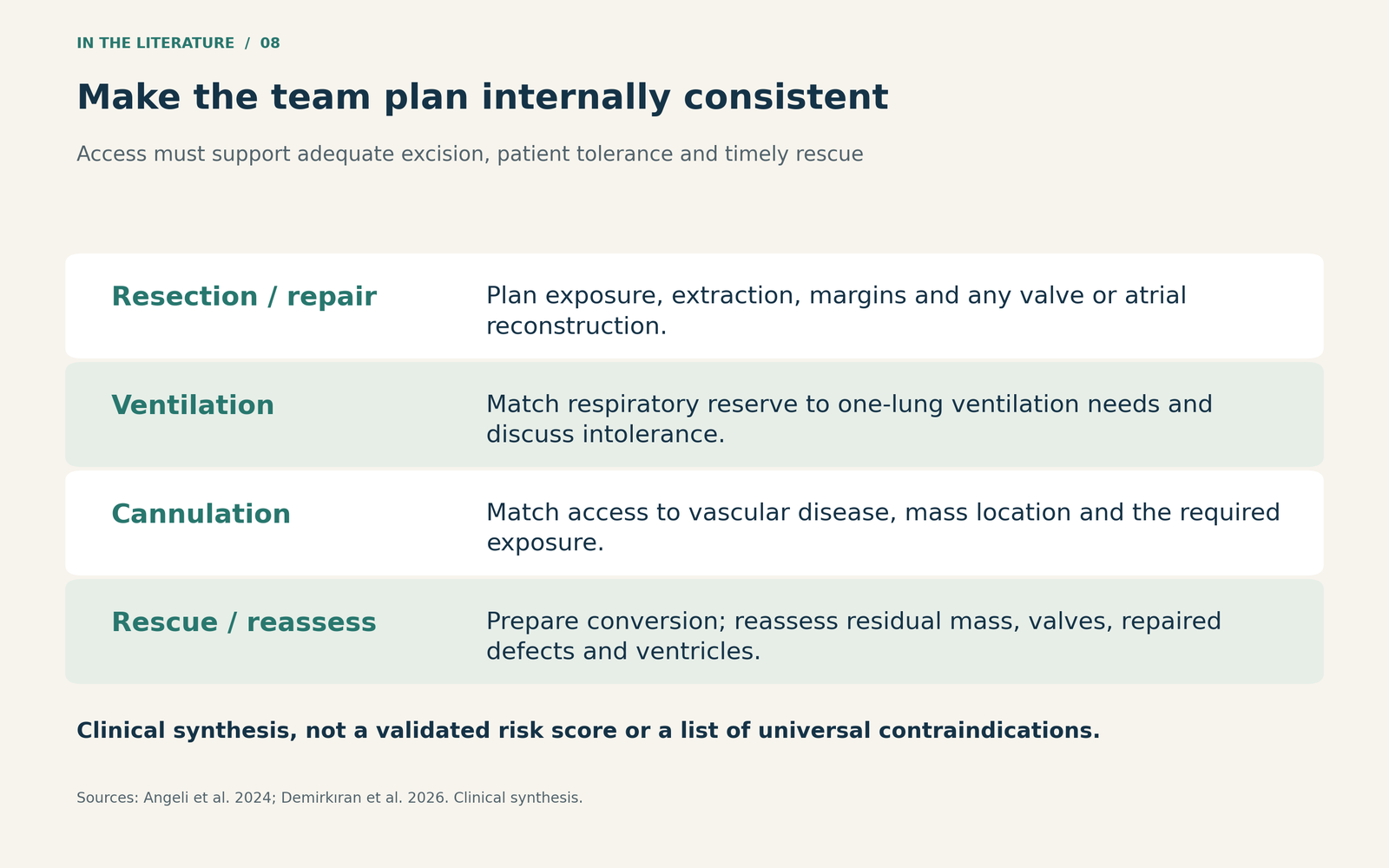
Figure 4. The incision is one part of the plan. Clinical synthesis; the goal is an adequate operation with a prepared rescue pathway.[1, 2]

Three takeaways
- Describe an operation, not just a mass. Attachment, valve interaction and invasion determine what the team must accomplish.
- Keep feasibility separate from comparative benefit. A successful selected series cannot establish that one access route is safer or faster for all patients.
- Choose access with the whole team. Complete excision, ventilation, cannulation and conversion must remain compatible with the patient's anatomy and physiology.
References
- Angeli F, Bodega F, Bergamaschi L, et al. Multimodality imaging in the diagnostic work-up of patients with cardiac masses: JACC: CardioOncology state-of-the-art review. JACC CardioOncol. 2024;6(6):847–862. doi:10.1016/j.jaccao.2024.09.006.
- Demirkıran T, Özdaş IT, Kadan E, Akyol FB, Varol ME, Karabacak K. Minimally invasive and robotic-assisted excision of intracardiac masses: a single-center retrospective study. J Cardiothorac Surg. 2026;21:745. doi:10.1186/s13019-026-04527-6.