
ITL-461eb7294038
Juan Li, MD · Updated November 18, 2026
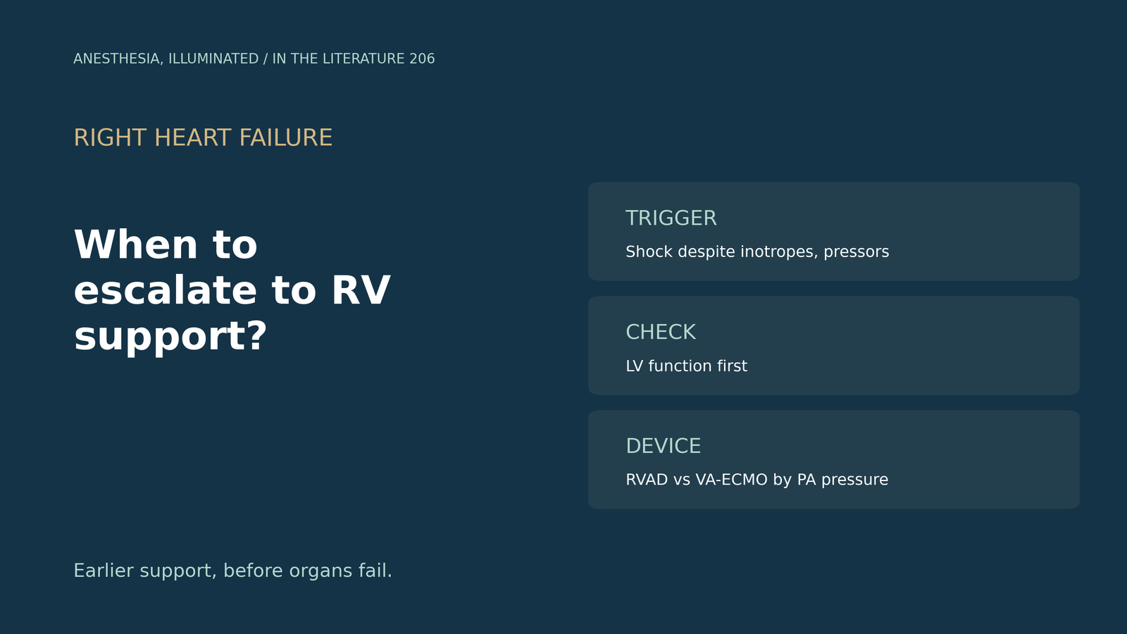
Key points
- Escalate when drugs are failing. Hypotension needing vasoconstrictors with poor hemodynamics should prompt consideration of MCS, and worsening RHF should lead to rapid RV support.[1]
- Do not wait. Support should be prompt when cardiac index stays below 2.2 despite inotropes, and the faster it is instituted, the better.[2]
- Numbers help, but none is decisive. There is no hemodynamic gold standard; indicators include RA/PCWP above 0.86 or PAPi of 0.9 or less.[3]
- Look at the left ventricle first. LV function must be assessed before RV support, because isolated RV support can overload the LV.[4]
- Earlier appears better. In observational data, 90-day survival was 79% with immediate versus 46% with delayed RVAD.[5]
After LVAD implantation, the right ventricle dilates, the CVP rises to 18 mmHg and blood pressure falls despite inotropes, vasopressors and an inhaled pulmonary vasodilator. When is it time for temporary RV mechanical support, and which device fits?[1], [3]
When to escalate
The AHA statement on right-sided heart failure advises that hypotension needing vasoconstrictors with persistent poor hemodynamics should prompt consideration of MCS, and that worsening RHF should lead to rapid deployment of RV support. RA/PCWP above 0.63 is associated with RHF after LVAD, and 42% to 75% of acute RHF patients may recover sufficient function.[1] Welker and colleagues advise prompt support when cardiac index stays below 2.2 despite inotropes and state that the faster support is instituted, the better.[2] Geller and colleagues note that there is no hemodynamic gold standard for RV failure, list RA/PCWP above 0.86 or PAPi of 0.9 or less among indicators, and advise starting temporary MCS as soon as possible in selected candidates.[3]
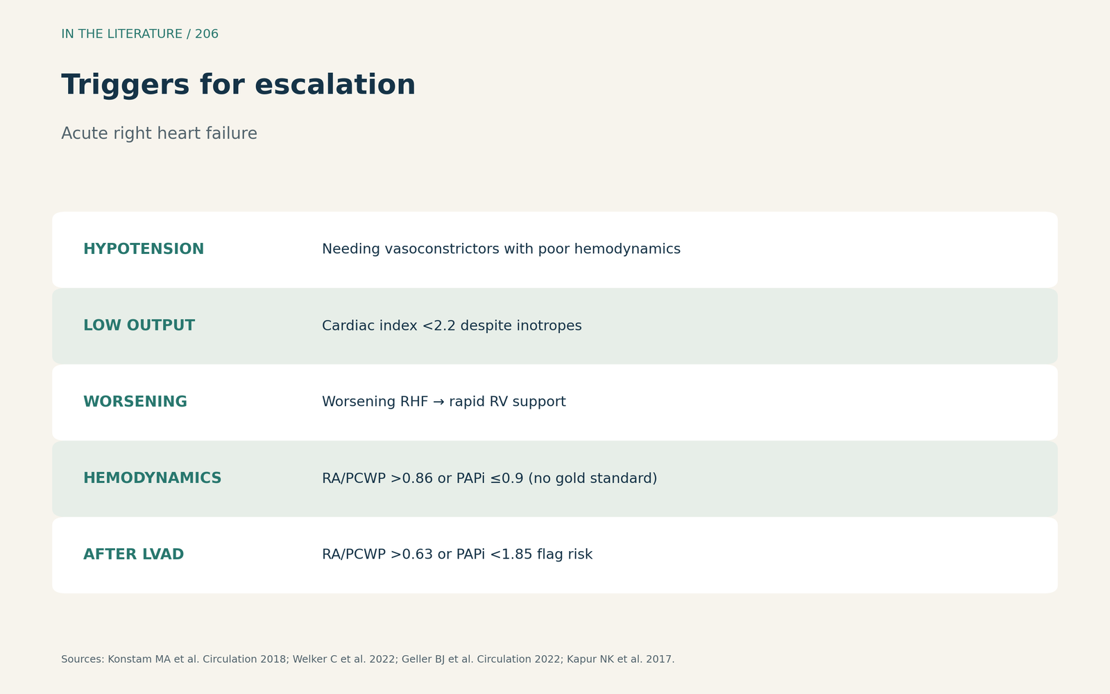
Figure 1. Persistent shock despite drugs. Synthesized from Konstam et al., Welker et al. and Geller et al.[1], [2], [3] View larger.

Which device
Kapur and colleagues define PAPi as PA pulse pressure divided by RA pressure; a PAPi below 1.85 predicted RV failure after LVAD, and RA/PCWP above 0.86 was associated with RV infarction. LV function must be assessed before RV support, because isolated RV support can overload the LV.[4] The AHA statement warns that RVAD flow will further raise pulmonary pressure when afterload is high.[1] Manca and colleagues suggest RVADs when RAP is high with low PA pressures and VA-ECMO with severe pulmonary hypertension and biventricular involvement.[6] Geller and colleagues add that VA-ECMO may be considered with RV dysfunction plus hypoxemia or more severe shock, and Welker and colleagues favour VA-ECMO with LV venting for biventricular failure.[3], [2]
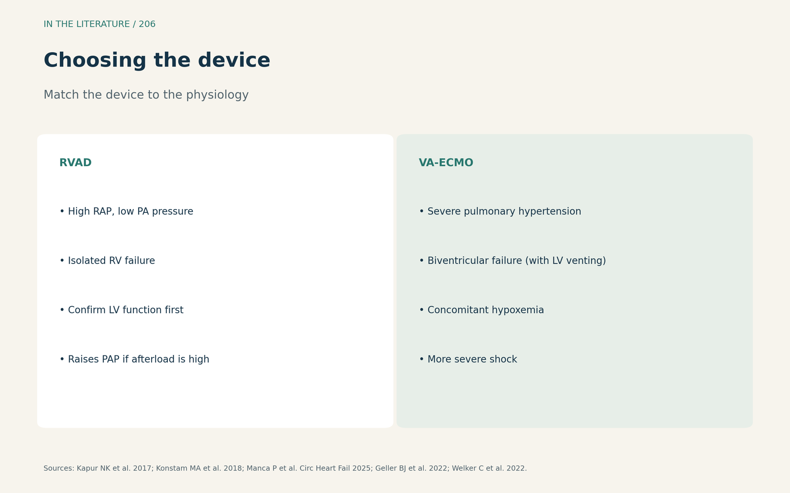
Figure 2. Low PA pressure: RVAD; high PA pressure or both ventricles: VA-ECMO. Synthesized from Kapur et al., Konstam et al., Manca et al., Geller et al. and Welker et al.[4], [1], [6], [3], [2] View larger.

What timing and outcomes look like
Abdelshafy and colleagues reviewed 31 studies with 1598 patients: temporary RVAD weaning succeeded in 23% to 100% and 30-day survival ranged from 46% to 100%. Ninety-day survival was 79% with immediate versus 46% with delayed RVAD, and they advise insertion before secondary organ damage develops.[5] Manca and colleagues note that in-hospital mortality of acute RV failure approaches 70% in some settings.[6]
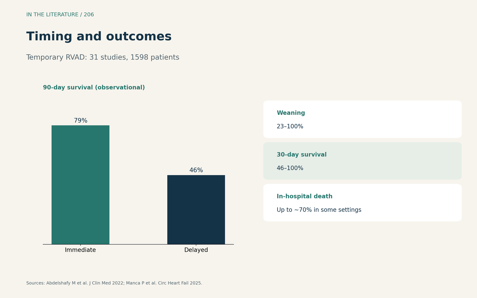
Figure 3. Wide ranges, a timing signal. Redrawn from Abdelshafy et al. and Manca et al.[5], [6] View larger.

Three takeaways
- Escalate when hypotension and low output persist despite inotropes and vasopressors.[1], [2]
- Use PAPi and RA/PCWP as support, not rules; check LV function before RV support.[3], [4]
- RVAD for isolated RV failure with low PA pressure; VA-ECMO for pulmonary hypertension, both ventricles or hypoxemia.[6], [3]
References
- Konstam MA, Kiernan MS, Bernstein D, et al. Evaluation and management of right-sided heart failure: a scientific statement from the American Heart Association. Circulation. 2018;137(20):e578–e622. doi:10.1161/CIR.0000000000000560
- Welker C, Huang J, Nunez-Gil IJ, Villavicencio MA, Ramakrishna H. Percutaneous right ventricular mechanical circulatory support: analysis of recent data. J Cardiothorac Vasc Anesth. 2022;36(8):2783–2788. doi:10.1053/j.jvca.2022.01.022
- Geller BJ, Sinha SS, Kapur NK, et al. Escalating and de-escalating temporary mechanical circulatory support in cardiogenic shock: a scientific statement from the American Heart Association. Circulation. 2022;146(6):e50–e68. doi:10.1161/CIR.0000000000001076
- Kapur NK, Esposito ML, Bader Y, et al. Mechanical circulatory support devices for acute right ventricular failure. Circulation. 2017;136(3):314–326. doi:10.1161/CIRCULATIONAHA.116.025290
- Abdelshafy M, Caliskan K, Guven G, et al. Temporary right ventricular assist devices: a systematic review. J Clin Med. 2022;11(3):613. doi:10.3390/jcm11030613
- Manca P, Nuzzi V, Mulè M, et al. Gaps and knowledge in the contemporary management of acute right ventricular failure. Circ Heart Fail. 2025;18(9):e012030. doi:10.1161/CIRCHEARTFAILURE.124.012030