ITL-d1f284a5ed92
Watch the video
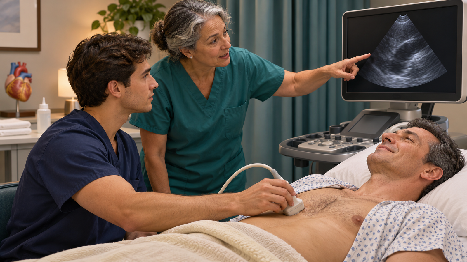
Figure 1. Supervised scanning links image acquisition to feedback. Course attendance is the start of an assessment process. Conceptual illustration.
Key points
- Define the task first: basic cardiac POCUS/FoCUS and a comprehensive echocardiogram have different scopes.
- Acquiring a recognizable view, interpreting pathology, and making a sound clinical decision are distinct abilities.
- A US residency survey found that 60% of responding programs reported no formal POCUS knowledge or skills assessment; it was a 2022 survey, published in 2025, with a 31% response rate.
- A 2026 mastery-learning study supports assessment, feedback, and retesting for image acquisition; it did not establish independent bedside diagnostic competence in residents.
- A useful local assessment should include image quality, interpretation, clinical integration, and recognition of when expert help is needed.
A resident who has completed a weekend FoCUS course assesses hypotension in recovery. A parasternal view looks reassuring, but the right ventricle is poorly seen. The resident reports, “The heart is normal.”
The teaching opportunity is immediate: did the examination answer the clinical question, and what remains unassessed? A limited examination should not imply exclusion of disease that was never adequately evaluated. The 2024 multisociety nomenclature statement makes that distinction central to clear reporting.[1]
Define what the resident is being asked to do
Basic cardiac POCUS/FoCUS uses focused views, principally grayscale imaging, to answer a defined set of questions. Advanced cardiac POCUS extends the modalities and complexity of assessment. Consultative echocardiography describes a different workflow involving a consulting team; it may itself be limited or comprehensive.[1]
These distinctions matter when designing a curriculum. “Learn TTE” is too vague an objective. A better task is: acquire adequate focused views, recognize a specified abnormality or an inadequate examination, explain its relevance, and communicate the next step. That task does not automatically include comprehensive valve quantification or advanced hemodynamic measurements.
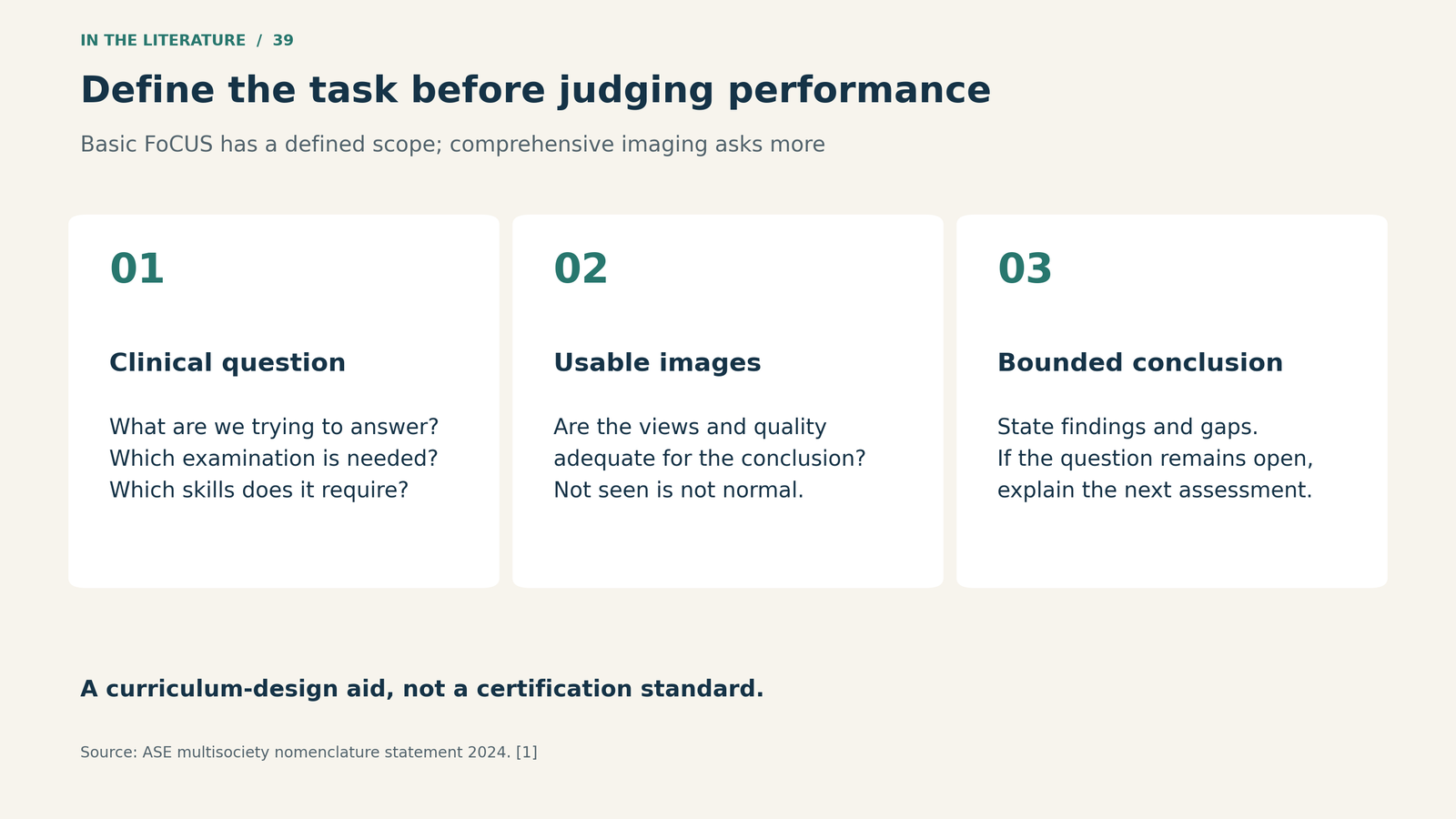
Figure 2. Define the intended question, image requirement, and reporting scope before assessing performance. This is a curriculum-design aid, not a certification standard.[1]

The missing link may be assessment
Edwards and colleagues surveyed 157 US anesthesiology residency programs in 2022 and received 48 completed responses. Most respondents used several teaching methods, but 60% reported no formal assessment of POCUS knowledge or skills. Faculty time, trained faculty, and resident time were common barriers.[2]
Because this was a small, self-selected, self-reported sample, it does not establish that 60% of all programs lacked assessment in 2025 or today. It does expose a practical problem: a program can deliver lectures and scanning opportunities without demonstrating what each trainee can reliably do.
A case log documents experience. A certificate documents completion of a specified process. Neither alone shows that a resident can obtain diagnostic-quality images in a difficult patient, distinguish uncertainty from normality, or integrate a finding into care. Those abilities need to be observed for the intended task.
A recent study makes the feedback loop concrete
Evans and colleagues evaluated a mastery-learning curriculum in 18 participants: 12 anesthesiology fellows and six attending anesthesiologists, at one pediatric institution. The scheduled curriculum used two three-hour sessions combining simulation and standardized-patient scanning. Image-acquisition performance was assessed with a 20-item checklist, with a study-specific passing standard of 85% (17/20).[3]
None passed at baseline. Twelve of 18 passed the first posttest; among the 15 who attended both scheduled sessions, that was 12/15. Six participants needed additional practice and retesting before all 18 reached the standard. This is useful evidence for adapting practice to demonstrated performance rather than assuming everyone learns at the same rate.[3]
The boundary matters. Testing used the same standardized patient at baseline and follow-up, with posttests one to two months after training. The skills assessment concentrated on acquisition and anatomical identification, not real-time interpretation of pathology in sick patients. This uncontrolled study did not establish long-term clinical transfer or patient benefit, and its participants were not a residency cohort.[3]
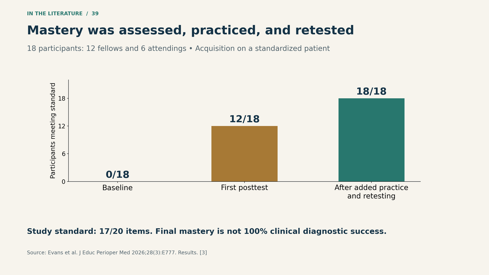
Figure 3. Achievement of the study's image-acquisition standard. The final result includes additional deliberate practice and retesting; it is not a 100% clinical diagnostic success rate.[3]

What should the next supervised assessment include?
A practical local assessment can use four linked tasks. This is a proposed teaching structure, informed by the scope and assessment evidence, rather than a validated national pass rule.[1, 3]
Acquire: Observe the resident selecting views, orienting the probe, optimizing the image, and saving interpretable clips. Include more than an easy normal volunteer; recognize when the available windows cannot answer the question.
Interpret: Use normal and abnormal clips, as well as technically limited examples. Ask what the image supports, what it does not establish, and which finding would require confirmation. An uninterpretable RV view should not be scored as a normal RV.
Integrate: Present the hemodynamic context and ask how the finding changes the working diagnosis or next investigation. A visible effusion, for example, is an imaging finding; its hemodynamic significance requires clinical and echocardiographic assessment rather than a label alone.
Communicate and escalate: Require a concise report of indication, views and quality, findings, uncertainty, and the plan for additional expertise or imaging. ASE recommends documentation, image archiving according to local capabilities, and periodic review; these make feedback possible after the bedside encounter.[1]
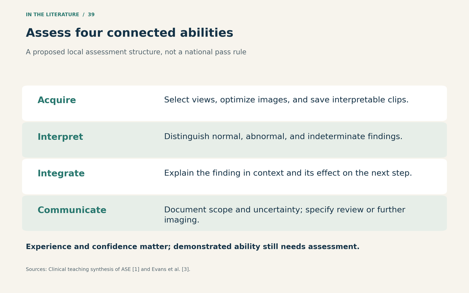
Figure 4. A proposed assessment structure. Record performance in each domain instead of treating a scan count or confidence score as the entire assessment.[1, 3]

Return to the resident in recovery
A more useful report would describe the views obtained, the observed LV appearance, and the fact that RV assessment was inadequate, followed by a plan for senior review or further imaging if the unanswered question matters to management. This is not a failed educational encounter: recognizing the boundary is part of competent practice.
Before reducing supervision, the team should know which tasks have been demonstrated and how uncertain or unexpected findings will be reviewed. Build ongoing review of saved examinations into training, with refresher practice when performance warrants it. The practical goal is reliable care within a defined scope, supported by a system that can detect and correct errors.
Three takeaways
- Specify the task before setting the threshold. Basic FoCUS acquisition and comprehensive diagnostic echocardiography are different training goals.
- Assess performance, then tailor practice. The 2026 study supports a feedback-and-retest process for acquisition, not automatic clinical independence after a course.
- Teach the limits explicitly. A useful clinician knows when an image answers the question, when it does not, and how to obtain help.
References
- Kirkpatrick JN, Panebianco N, Díaz-Gómez JL, Adhikari S, Bremer ML, Bronshteyn YS, et al. Recommendations for cardiac point-of-care ultrasound nomenclature. J Am Soc Echocardiogr. 2024;37(9):809–819. doi:10.1016/j.echo.2024.05.001.
- Edwards J, Ahn D, Alcaraz D, Chiles C, Khuu T, Soni NJ, et al. Point-of-care ultrasound training among anesthesiology residency programs in the United States. BMC Anesthesiol. 2025;25:105. doi:10.1186/s12871-025-02929-y.
- Evans MA, Rosenfeld B, Ballard HA, Rowland MJ, Kilner KJ, Patel S, et al. A simulation-based mastery learning curriculum to address cardiac point-of-care ultrasound requirements of anesthesiology board certification. J Educ Perioper Med. 2026;28(3):E777. doi:10.46374/VolXXVIII_Issue3_Toaz.