ITL-74955fc0312d
Watch the video
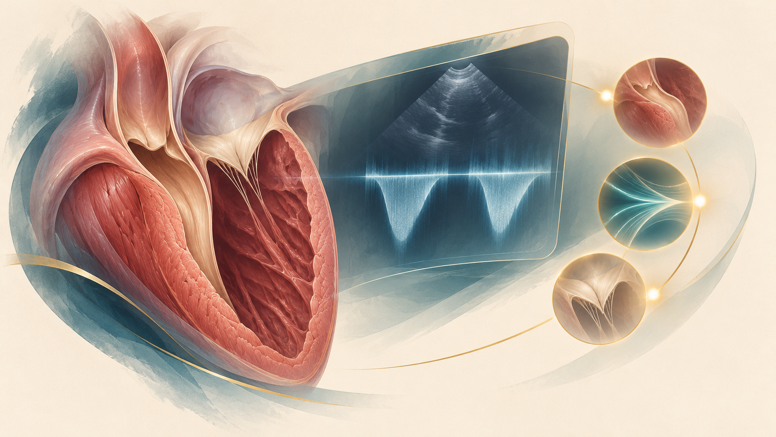
Figure 1. A low number is one part of the assessment. Conceptual artwork, not a diagnostic echocardiogram.
Key points
- General anesthesia changes loading conditions and contractility; intraoperative resting gradients can underrepresent preoperative obstruction.
- Assess the Doppler signal, systolic anterior motion (SAM), and mitral regurgitation together before interpreting the gradient.
- Protocol-based intraoperative provocation can reveal residual obstruction, but pharmacologic stress can also exaggerate gradients.
- A high provoked gradient requires anatomical explanation; it is not an automatic instruction to remove more septum.
- Complete the examination for new aortic regurgitation, ventricular septal defect, and coronary-cameral flow before concluding the assessment.
After separation from bypass, the left ventricular outflow tract (LVOT) gradient looks reassuring. The practical question is whether the obstruction has been adequately relieved under conditions that meaningfully test the result—and whether the intervention has introduced another problem. The 2022 ASE imaging recommendations place intraoperative TEE in both roles: evaluating myectomy adequacy and identifying complications.[1]
First make sure the gradient means what you think it means
Before bypass, define the distribution and longitudinal extent of septal thickening, the SAM–septal contact site, and abnormalities of the mitral leaflets, chordae, or papillary muscles. This anatomical map gives the post-bypass examination a specific target. A number without that map cannot distinguish incomplete relief of obstruction from another source of accelerated flow.[1]
For the post-bypass assessment, document the hemodynamic setting and obtain a well-aligned LVOT continuous-wave Doppler signal, often from the deep transgastric view. Use color and, where needed, pulsed-wave Doppler to localize acceleration. Separate the LVOT signal from the mitral regurgitant jet; also consider an intracavitary gradient in a small, hyperdynamic ventricle. Review SAM and mitral regurgitation at the same time.[1, 2]
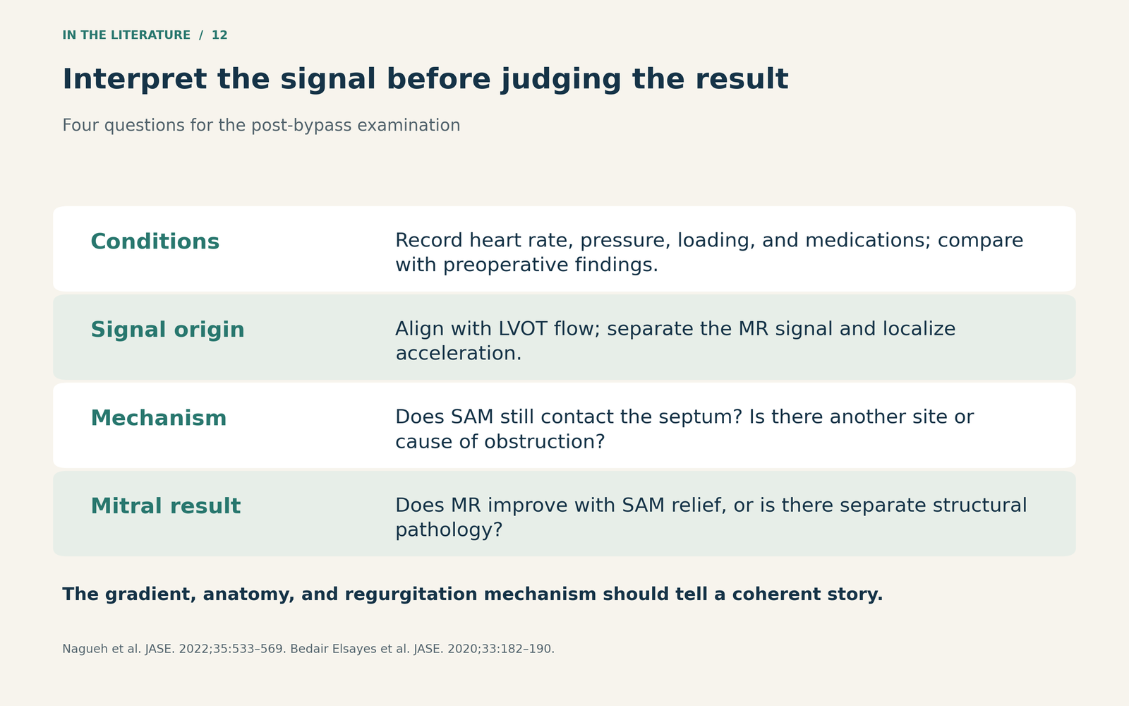
Figure 2. Establish the measurement context before judging the surgical result. These questions synthesize ASE imaging guidance and the provocative-testing study.[1, 2]

SAM-related regurgitation often improves when obstruction is relieved. Persistent regurgitation from intrinsic mitral pathology requires a separate explanation and may require a different intervention. Its mechanism matters more than simply reporting that “MR remains.”[1]
What resting measurements can miss
In a single-center retrospective series of 293 myectomy patients, 82 of the 197 patients with significant preoperative resting obstruction showed mild or no obstruction under general anesthesia. After bypass, 7 patients had a resting gradient above 30 mmHg, while 63 had a residual gradient above 30 mmHg only during pharmacologic provocation.[2]
Provoked gradients, persistent SAM, or significant mitral regurgitation contributed to a second bypass run in 41 patients (14%). Thus, resting measurements alone could miss findings that changed intraoperative management in this program.[2]
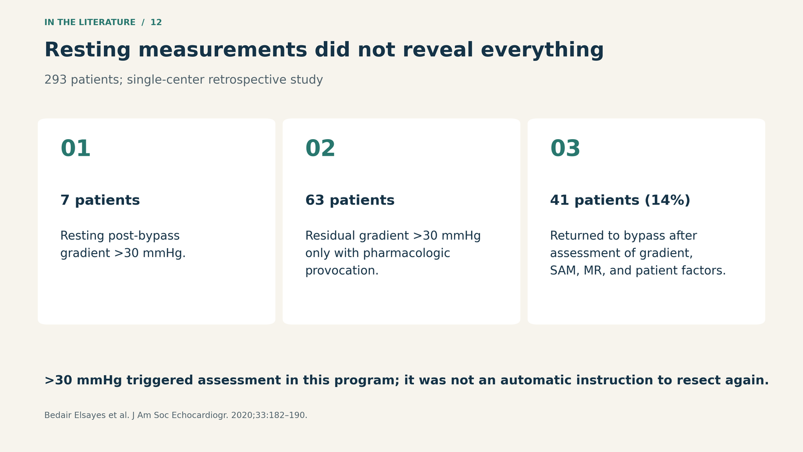
Figure 3. Findings from the Bedair Elsayes series. The 7 and 63 patients represent different post-bypass findings; the 41 reinterventions were selected using combined anatomical and hemodynamic assessment, not the gradient alone.[2]

This was an institutional protocol, not a randomized comparison proving that provocation or reintervention improves long-term outcomes. The study's threshold of greater than 30 mmHg was a trigger for assessment within that protocol, not a universal mandate for another resection. The authors also found that pharmacologic provocation could produce higher gradients than preoperative physiological testing.[2]
If provocation raises the gradient, look for the mechanism
A reproducible gradient accompanied by persistent SAM–septal contact and relevant mitral regurgitation is different from accelerated flow without demonstrable LVOT narrowing. Reassess loading conditions, cavity size, Doppler alignment, and the actual site of acceleration. A further bypass run must weigh the residual mechanism against the benefit already achieved and the risks of additional resection, including septal injury.[1, 2]
A 2026 retrospective study of 36 patients, available here as an abstract only, adds a useful caution: 22% showed elevated postoperative peak gradients without demonstrable LVOT obstruction on the study's three-dimensional assessment. This supports integrating geometry with Doppler rather than using pressure gradients alone; the abstract does not establish a universal three-dimensional area cutoff or prove improved clinical outcomes.[3]
Finish with the structures, not just the outflow number
The ASE recommendations call for exclusion of new aortic regurgitation, ventricular septal defect, and coronary-cameral fistula after myectomy. Immediate septal thickness measurements can also be affected by tissue edema, so they should not become an isolated reason for additional resection.[1]
A published 2024 case illustrates the importance of timing: coronary-cameral flow was seen during terminal warm cardioplegia, then was no longer demonstrated after bypass or on follow-up TTE one week later. This is a detection example, not evidence that all postoperative fistulas are harmless or can be left untreated.[4]
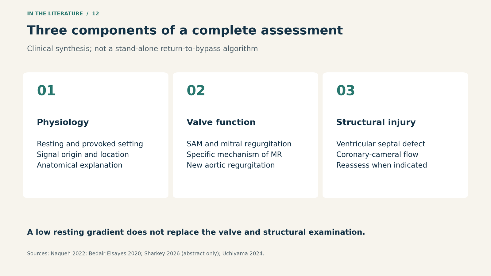
Figure 4. The final assessment should explain residual physiology, valve function, and possible new structural injury. It is a clinical synthesis, not a stand-alone return-to-bypass algorithm.[1, 2, 3, 4]

A useful final report therefore connects the gradient and testing conditions, the residual anatomical mechanism, the mitral result, and the complication survey. “Low resting gradient” becomes one finding within that conclusion.
Three takeaways
- Interpret a low gradient in context. General anesthesia and Doppler alignment can mask obstruction; correlate with the preoperative study and current anatomy.
- Use provocation to ask a better anatomical question. A higher gradient warrants explanation, not automatic additional resection.
- Close the assessment with a structural survey. Document mitral and aortic valve function and look specifically for septal defects and abnormal coronary-cameral flow.
References
- Nagueh SF, Phelan D, Abraham T, Armour A, Desai MY, Dragulescu A, et al. Recommendations for multimodality cardiovascular imaging of patients with hypertrophic cardiomyopathy: an update from the American Society of Echocardiography, in collaboration with the American Society of Nuclear Cardiology, the Society for Cardiovascular Magnetic Resonance, and the Society of Cardiovascular Computed Tomography. J Am Soc Echocardiogr. 2022;35(6):533–569. doi:10.1016/j.echo.2022.03.012.
- Bedair Elsayes A, Basura A, Zahedi F, Moreno-Duarte I, Rowin EJ, Maron M, et al. Intraoperative provocative testing in patients with obstructive hypertrophic cardiomyopathy undergoing septal myectomy. J Am Soc Echocardiogr. 2020;33(2):182–190. doi:10.1016/j.echo.2019.08.021.
- Sharkey A, Ahmed U, Manji AA, Mahmood F, Saeed S, Valencia J, et al. Peak gradients and left ventricular outflow tract area in hypertrophic obstructive cardiomyopathy: a 3-dimensional echocardiography study. J Cardiothorac Vasc Anesth. 2026;40(4):1112–1121. doi:10.1053/j.jvca.2026.01.014. Abstract only.
- Uchiyama K, Tsubokawa T. Iatrogenic coronary artery fistula that developed during septal myectomy was detected intraoperatively through transesophageal echocardiography. JA Clin Rep. 2024;10:27. doi:10.1186/s40981-024-00711-6.