
ITL-1157d94c3c96
Juan Li · In the Literature · Updated November 16, 2026
Watch the video

Key points
- A normal ejection fraction is reassuring, but does not establish complete myocardial recovery.
- Global longitudinal strain complements EF; neither should be interpreted without symptoms, valve function and loading conditions.
- Pre-TAVR GLS predicts risk in some cohorts. That evidence is different from showing recovery after the procedure.
- Before interpreting a serial GLS change, check image quality, tracking software, blood pressure and rhythm.
- Persistent symptoms deserve reassessment even when EF is normal or improving.
What has actually recovered?
After TAVR, the prosthesis has a satisfactory gradient and the report describes a normal left ventricular ejection fraction (LVEF). Can the team conclude that the myocardium has recovered? EF alone cannot answer that question. EF describes the fraction of ventricular volume ejected; global longitudinal strain (GLS) describes longitudinal deformation. The 2025 ASE/EACVI consensus recommends GLS as a complementary measure of global LV function. It does not replace EF. [1]
In the usual negative convention, GLS becoming more negative means greater longitudinal shortening. Both EF and strain are affected by loading conditions. Removing severe aortic obstruction changes afterload, so a better measurement after TAVR does not, by itself, prove that fibrosis has reversed or that all myocardial disease has resolved. [1]
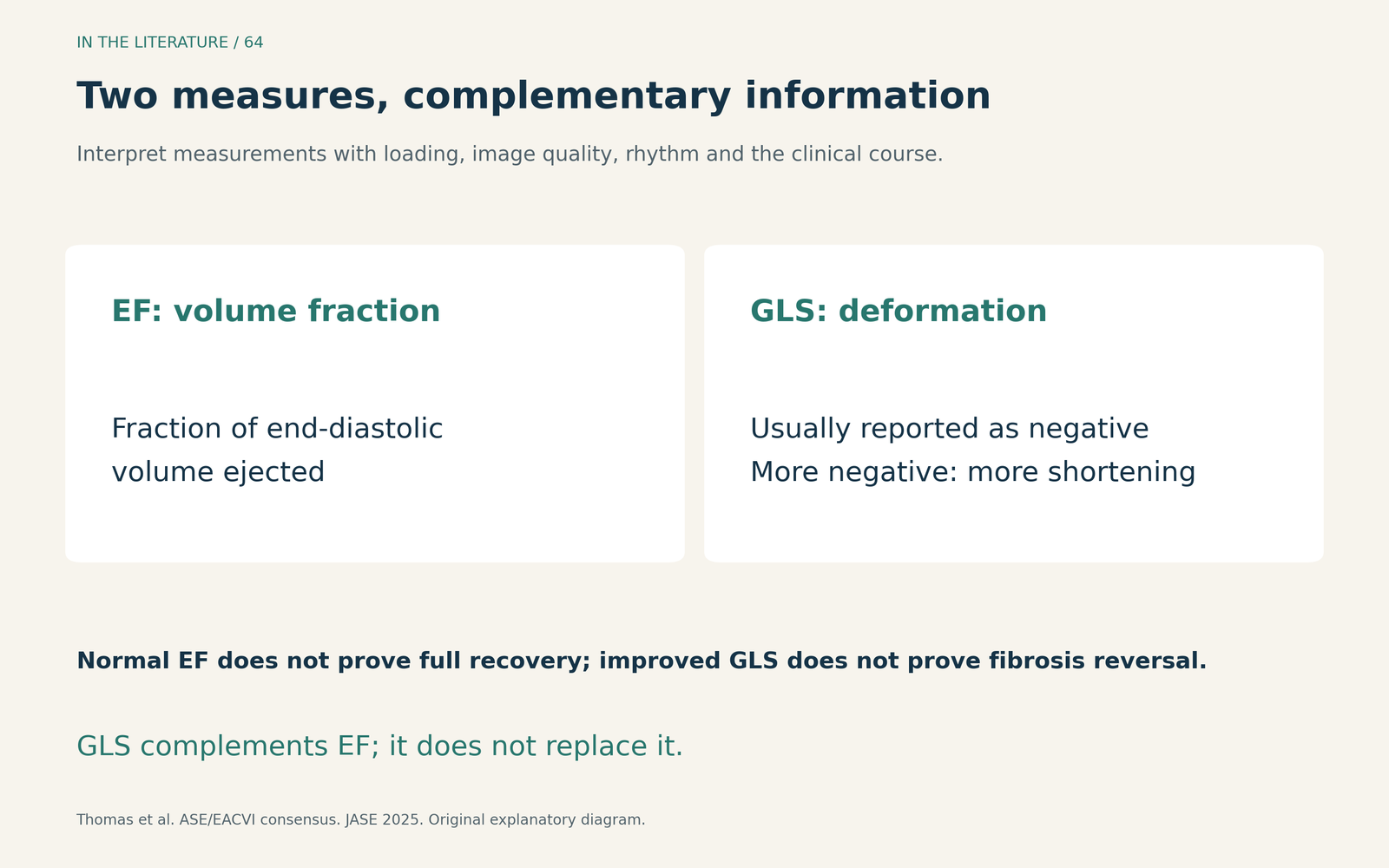
Figure 1. Original explanatory diagram. EF and GLS provide complementary information; image quality, loading and rhythm affect interpretation. [1] View figure

Preserved EF can coexist with an adverse myocardial signal
Lee and colleagues studied 344 TAVI patients with baseline LVEF ≥50%. The 86 patients in the worst baseline GLS quartile, defined as GLS >−16%, had a higher adjusted hazard of all-cause death: HR 2.26, 95% CI 1.11–4.60. Median follow-up was 18 months. This was an observational association despite preserved EF, not proof that treating the GLS value improves survival. [2]
Two distinctions matter at the bedside. First, the −16% boundary was derived from that cohort; it is not a universal post-TAVR recovery target. Second, post-TAVI GLS was not measured in this study. Its prognostic finding cannot establish how quickly strain improves after valve replacement. [2]
Improvement is possible, but it is not uniform
Kassem and colleagues' 2025 retrospective study provides serial measurements. Of 417 consecutive TAVR procedures, 80 selected patients had suitable baseline and follow-up images and met the study criteria. Follow-up echocardiography occurred at 8–16 months, averaging approximately one year. Mean EF increased from 59.2% to 62.1%, while mean GLS became more negative, from −16.8% to −19.2%. [3]
This was a mixed-EF cohort, with important exclusions including new left bundle branch block or a cardiac device and severe ischemic cardiomyopathy with EF <20%. The means describe those selected patients. They do not establish complete recovery, a survival benefit, or the benefit of a GLS-guided treatment strategy. [3]
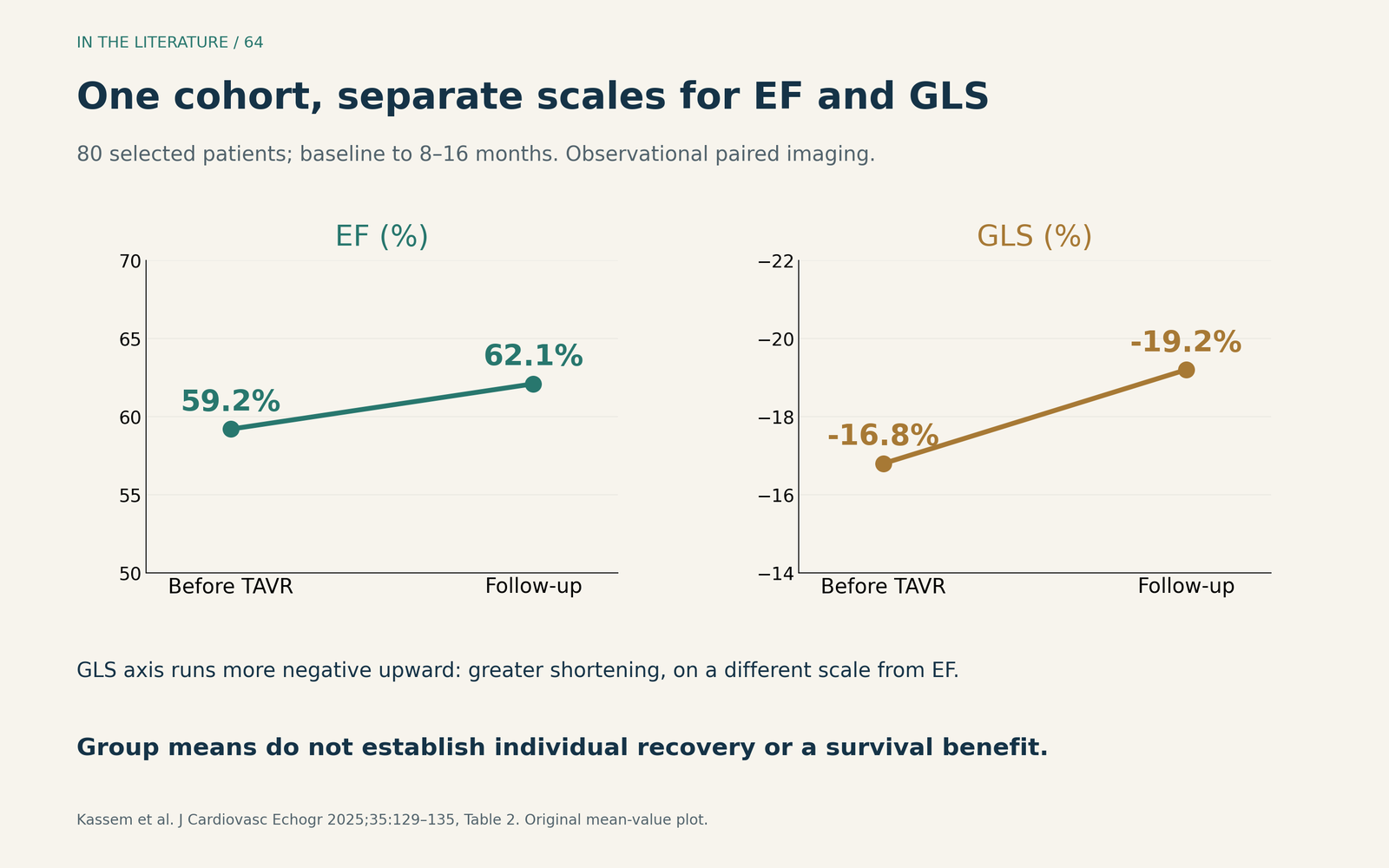
Figure 2. Original display of the reported group means, using separate scales for EF and GLS. Follow-up was 8–16 months. Greater negative GLS indicates greater longitudinal shortening; these observations do not promise the same response in an individual patient. [3] View figure

The newer literature also argues against expecting one trajectory. In Kislitsina and colleagues' 2026 observational analysis, the study included 195 TAVR patients, of whom 176 had paired GLS changes available. The TAVR group did not show a significant improvement in GLS. The strain dataset excluded patients who died before follow-up or lacked usable follow-up images. This is another selected imaging cohort, not a randomized comparison of myocardial recovery. [4]
For patients starting with reduced EF, a 2026 study by Tanaka and colleagues offers a further update, available here as a formal abstract only. It analyzed 121 patients with baseline EF <50%, with serial assessment through one year. The model-estimated EF increase was 12.3 percentage points; ischemic heart disease and a larger baseline LV end-diastolic diameter were associated with more limited recovery. The full methods and uncertainty estimates were unavailable, so this estimate should not become an individual prediction. Its study visit schedule is not a guideline follow-up schedule. [5]
Make serial measurements comparable
Before calling a change “recovery” or “deterioration,” examine how the measurements were obtained. The ASE/EACVI consensus advises ideally using the same tracking-software company and release for serial GLS. Review apical views, foreshortening, tracking and region-of-interest placement; consider blood pressure, loading conditions, rhythm and acquisition timing. Cross-vendor comparison requires care rather than an automatic assumption of equivalence. [1]
Record EF and GLS alongside valve hemodynamics and the patient's functional course. A single abnormal GLS result is a reason to understand the measurement and clinical context, not a stand-alone diagnosis of irreversible injury. Conversely, an improved value should not end the evaluation of ongoing breathlessness. GLS provides continuous prognostic information; the evidence reviewed here does not establish a universal post-TAVR “cured” cutoff. [1, 6]
When the numbers and the patient disagree
The 2025 ESC/EACTS guideline supports an early postprocedural echocardiographic baseline and lifelong follow-up. For bioprosthetic valves, TTE should be performed within three months, at one year and annually thereafter, with earlier reassessment when new cardiovascular symptoms arise. This schedule evaluates the prosthesis and the heart; it should not be reduced to tracking EF alone. [6]
If symptoms persist despite apparently satisfactory valve function, reassess ventricular function, other valve disease and possible myocardial pathology. CMR can help characterize remodeling, scar or diffuse fibrosis when that information addresses a clinical question. It is not required routinely for every asymptomatic TAVR recipient. Persistent heart failure or reduced EF also calls for appropriate guideline-directed heart-failure treatment and adjustment, rather than waiting for a strain number to normalize. [6]
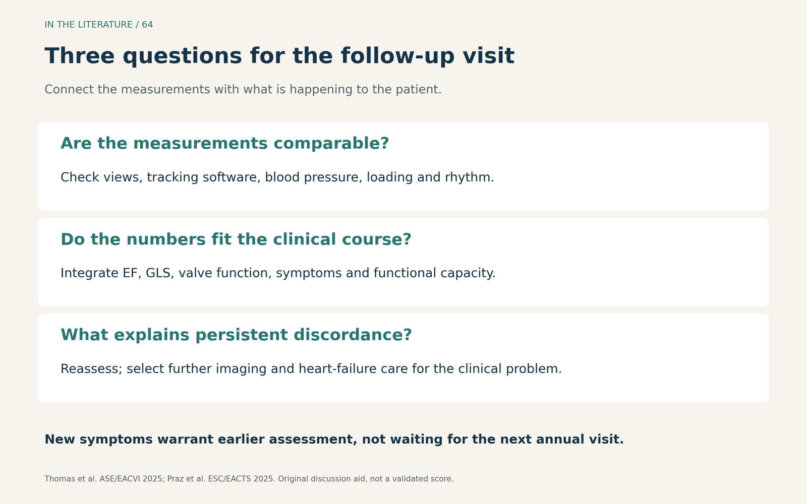
Figure 3. Original clinical discussion aid: establish measurement comparability, integrate symptoms and valve findings, and investigate persistent discordance. This is not a validated score or a GLS-based treatment algorithm. [1, 6] View figure

Three takeaways
- Use EF and GLS together. A normal EF does not establish complete myocardial recovery, and GLS does not replace EF.
- Compare like with like. Check technique, loading and rhythm before interpreting serial strain; cohort averages are not individual recovery targets.
- Follow the patient as well as the images. Persistent symptoms warrant reassessment even when EF improves; no single GLS cutoff closes the case.
References
- Thomas JD, Edvardsen T, Abraham T, et al. Clinical Applications of Strain Echocardiography: A Clinical Consensus Statement From the American Society of Echocardiography Developed in Collaboration With the European Association of Cardiovascular Imaging of the European Society of Cardiology. J Am Soc Echocardiogr. 2025;38(11):985–1020. doi:10.1016/j.echo.2025.07.007. PubMed.
- Lee SH, Oh JK, Lee SA, et al. Incremental Prognostic Value of Left Ventricular Global Longitudinal Strain in Patients with Preserved Ejection Fraction Undergoing Transcatheter Aortic Valve Implantation. J Am Soc Echocardiogr. 2022;35(9):947–955.e7. doi:10.1016/j.echo.2022.04.013. PubMed.
- Kassem J, Ghazal R, Fakhreddin O, et al. Assessment of Left Ventricular Global Longitudinal Strain Improvement Following Transcatheter Aortic Valve Replacement. J Cardiovasc Echogr. 2025;35(2):129–135. doi:10.4103/jcecho.jcecho_99_24. PubMed.
- Kislitsina ON, Chris Malaisrie S, Thomas JD, et al. Left and Right Heart Remodelling at 1 Year After Transcatheter Versus Surgical Aortic Valve Replacement: A Speckle-Tracking Echocardiography Study. Interdiscip Cardiovasc Thorac Surg. 2026;41(6):ivag134. doi:10.1093/icvts/ivag134. PubMed.
- Tanaka K, Kageyama S, Sugiyama H, et al. Serial Changes in Left Ventricular Ejection Fraction After Transcatheter Aortic Valve Implantation: Associations With Ischemic Heart Disease and Left Ventricular End-Diastolic Diameter. Echocardiography. 2026;43(9):e70626. doi:10.1111/echo.70626. PubMed. Formal abstract only.
- Praz F, Borger MA, Lanz J, et al. 2025 ESC/EACTS Guidelines for the management of valvular heart disease. Eur J Cardiothorac Surg. 2025;67(8):ezaf276. doi:10.1093/ejcts/ezaf276. PubMed.