
ITL-c3b850f23e6b
Juan Li · In the Literature · Updated November 16, 2026
Watch the video

Key points
- Prosthesis–patient mismatch (PPM) means a normally functioning valve has an effective orifice too small for the patient's body size; a high gradient alone does not establish it.
- Interpret indexed effective orifice area (EOAi) with BMI, the valve's model and size, leaflet motion and flow status.
- Measured and predicted PPM classify patients differently. Record the method rather than treating the labels as interchangeable.
- A newly rising gradient or abnormal leaflet motion requires evaluation for another mechanism, even when PPM is also present.
- Recent outcome studies do not justify either dismissing PPM or treating its label as an automatic indication for reintervention.
Begin with the finding that needs explaining
A follow-up echocardiogram reports an unexpectedly high mean gradient after TAVR. The useful clinical question is whether a normally functioning prosthesis is too small for this patient, or whether measurement, flow or valve dysfunction explains the finding.
The 2024 ASE imaging guideline defines PPM by the relationship between effective orifice area (EOA) and body size. The valve's intrinsic structure and motion should be normal. Conversely, low-flow PPM may produce a normal gradient: neither a high nor a normal gradient settles the diagnosis alone. [1]
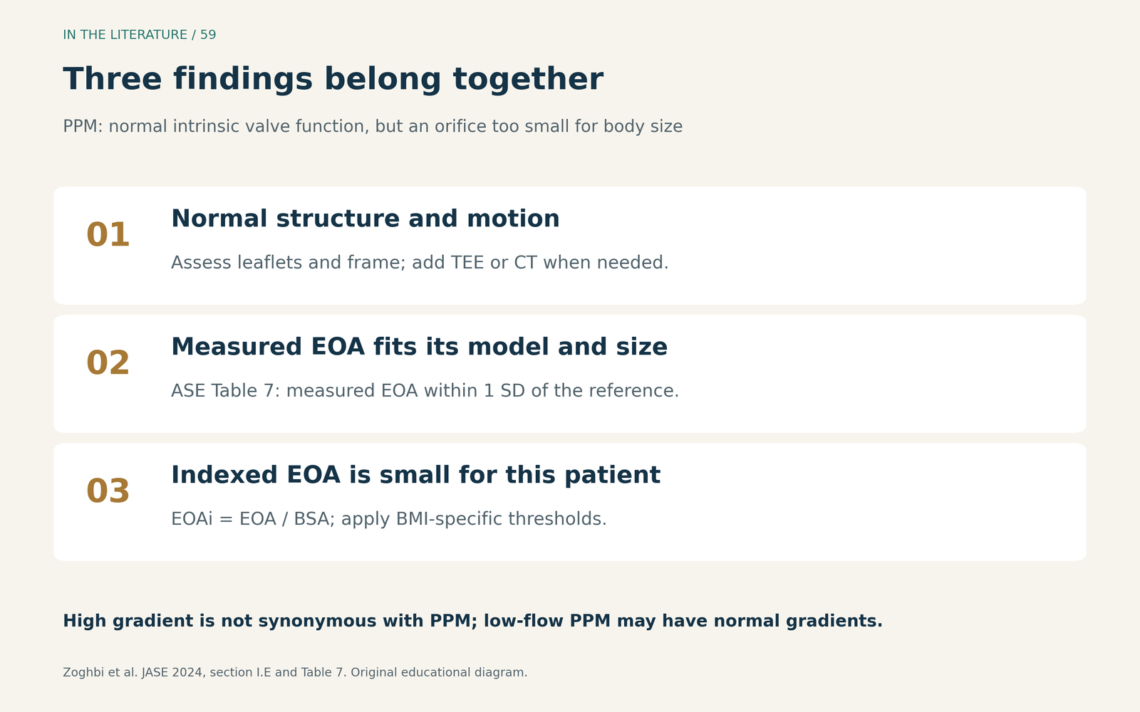
Figure 1. Original explanation of the ASE framework. EOA describes the functional orifice; EOAi is EOA divided by body surface area (BSA). Normal leaflet function can coexist with an orifice inadequate for body size. [1] View figure

Apply the threshold only after checking its premises
The ASE aortic PPM table uses severe PPM at EOAi ≤0.65 cm²/m² when BMI is below 30 kg/m², and ≤0.55 cm²/m² when BMI is 30 or greater. The corresponding moderate ranges, reported to two decimal places, are 0.66–0.85 and 0.56–0.70 cm²/m². Applying the nonobese threshold to everyone can overstate PPM in obesity. [1]
These numbers come with an essential condition: normal valve structure and motion, with measured EOA within one standard deviation of the reference EOA for that valve model and size. An area substantially below its expected value should prompt investigation of measurement or impaired valve performance before it is attributed entirely to patient–prosthesis size mismatch. [1]
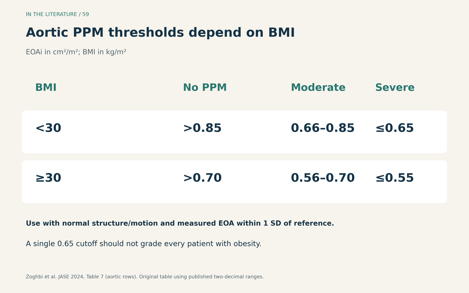
Figure 2. Original table of the published aortic thresholds. All EOAi values are in cm²/m². The normal-structure, normal-motion and reference-EOA conditions apply to both BMI groups. [1] View figure

Why measured and predicted PPM can disagree
Measured EOA comes from the patient's Doppler examination and continuity equation. Predicted EOA uses reference performance for a valve model and size. The former reflects individual flow and implantation but is vulnerable to measurement error; the latter cannot fully represent the particular valve's expansion or the patient's physiology. Low flow can also reduce measured valve opening and create an appearance of severe mismatch. [2]
The IMPPACT TAVR registry, reported by Guthoff and colleagues in 2024, included 38,808 native-AS TAVR patients and excluded valve-in-valve procedures. Among the 18,025 patients with both measurements available, severe PPM occurred in 4.1% by measured EOA versus 0.5% by predicted EOA. This is a change in classification within a shared cohort, not proof that one method is a diagnostic gold standard. [2]
In mortality analyses restricted to patients with follow-up beyond 30 days, severe versus nonsevere measured PPM had an adjusted two-year mortality HR of 1.14 (95% CI 0.94–1.38); the corresponding predicted-PPM HR was 0.92 (0.52–1.62). Neither showed a significant association. The cohort's mean age was 81 years, and the analysis does not establish that PPM is harmless in younger patients over longer follow-up. [2]
Tomii and colleagues' 2025 Bern registry analysis examined flow-dependent reference values in 1,510 balloon-expandable TAVR recipients. It classified discharge flow using stroke-volume index <35 mL/m² versus ≥35 mL/m², then applied flow-specific reference EOA values. Incorporating flow changed PPM classification, particularly in low-flow patients. The analysis reported no consistent adverse-outcome signal through five years, but severe predicted PPM was uncommon and the results cannot be generalized to all valve platforms. It is a method under evaluation, not a replacement for an integrated clinical assessment. [3]
Choose the next assessment from the pattern
Start by checking the LVOT diameter and pulsed-wave Doppler sampling location, the continuous-wave Doppler tracing and the comparison with the valve's expected performance. Interpret EOA alongside the Doppler velocity index (DVI), jet contour, acceleration time and flow state. No single DVI cutoff should replace the complete TAVR assessment. [1]
A stable small EOAi with normal structure is different from a new gradient increase accompanied by falling EOA/DVI or abnormal leaflet motion. The latter should prompt assessment for obstruction, thrombosis, degeneration or another mechanism. ASE’s hemodynamic criteria for structural valve deterioration require changes from a stable postimplantation baseline with corroborating morphology; an isolated high gradient is insufficient. TEE or CT can clarify morphology when transthoracic findings do not explain the physiology. [1]
The practical implication is to resolve the mechanism before planning another intervention. When results remain discordant and would change treatment, additional imaging or invasive hemodynamic assessment may help. Predicted EOA should inform that discussion, rather than override the patient's actual findings. [1, 3]
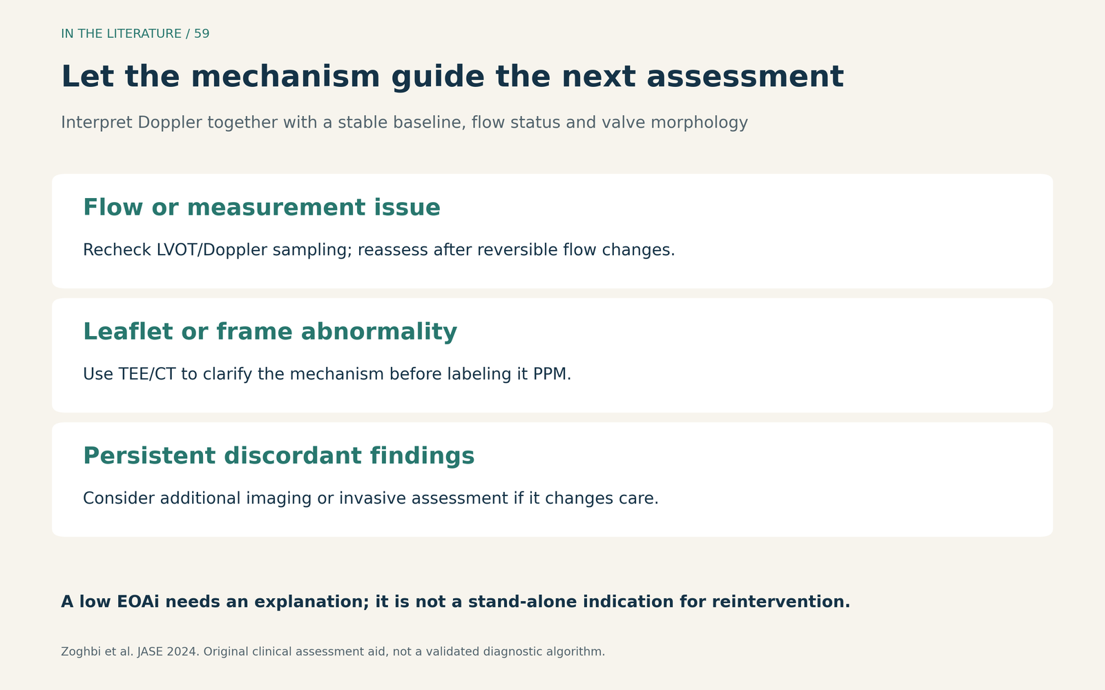
Figure 3. Original clinical assessment aid based on the imaging framework. These are prompts for interpretation, not a validated diagnostic algorithm. A low EOAi alone does not establish the mechanism or the benefit of reintervention. [1, 3] View figure

What do the newest outcome studies add?
Two recent reports help explain why the PPM label should neither be ignored nor made the sole explanation for symptoms. Only their complete published abstracts were available for this article.
Suruga and colleagues, JACC Cardiovascular Interventions 2025, studied 512 patients younger than 75 years with STS scores below 4%, receiving newer-generation balloon-expandable valves for native AS at one center. PPM occurred in 200 patients: 162 moderate and 38 severe. Over a median 1,034 days, PPM was associated with higher all-cause mortality (HR 2.55, 95% CI 1.3–5.0) and heart-failure hospitalization (HR 4.43, 2.0–9.9). The abstract does not identify these HRs as adjusted. These associations warrant attention but do not prove that correcting PPM improves outcomes. [4]
Cozzi and colleagues' November 2026 SCOPE I analysis included 662 patients. Although the parent valve trial was randomized, the comparison by PPM category was post hoc and observational. Good health status meant a KCCQ overall summary score ≥75 without a decline of >10 points from baseline. In the multivariable model, severe PPM was associated with lower odds of being alive with good health status at three years (OR 0.51, 95% CI 0.27–0.96). After additional adjustment for 30-day mitral regurgitation, the estimate was 0.59 (0.30–1.15), no longer statistically significant. This model sensitivity supports evaluating accompanying disease; it does not prove that mitral regurgitation fully explains the association or that PPM has no effect. [5]
Three takeaways
- Confirm the diagnosis before assigning its severity. Normal prosthetic structure, expected EOA and a small BMI-adjusted EOAi belong together.
- Document how and when PPM was assessed. Measured versus predicted area, flow status and the stable baseline can change the interpretation.
- Treat the clinical problem behind the label. Persistent symptoms or worsening hemodynamics need a mechanism-based assessment; observational associations alone do not establish a benefit from reintervention.
References
- Zoghbi WA, Jone PN, Chamsi-Pasha MA, et al. Guidelines for the Evaluation of Prosthetic Valve Function With Cardiovascular Imaging: A Report From the American Society of Echocardiography Developed in Collaboration With the Society for Cardiovascular Magnetic Resonance and the Society of Cardiovascular Computed Tomography. J Am Soc Echocardiogr. 2024;37(1):2–63. doi:10.1016/j.echo.2023.10.004
- Guthoff H, Abdel-Wahab M, Kim WK, et al. Impact of Measured and Predicted Prosthesis-Patient Mismatch After Transcatheter Aortic Valve Replacement. JACC Cardiovasc Interv. 2024;17(22):2626–2635. doi:10.1016/j.jcin.2024.08.041
- Tomii D, Heg D, Nakase M, et al. Flow Status-Based Predicted Prosthesis-Patient Mismatch in Patients Undergoing Transcatheter Aortic Valve Replacement With a Balloon-Expandable Valve. Struct Heart. 2025;9(4):100379. doi:10.1016/j.shj.2024.100379
- Suruga K, Patel V, Nagasaka T, et al. Prosthesis-Patient Mismatch in Young and Low-Risk Patients After Newer Generation Balloon-Expandable Transcatheter Aortic Valve Replacement. JACC Cardiovasc Interv. 2025;18(12):1512–1523. doi:10.1016/j.jcin.2025.05.003. Abstract only.
- Cozzi O, Tomii D, Lanz J, et al. Prosthesis-Patient Mismatch and Patient-Centered Outcomes After TAVR. JACC Cardiovasc Interv. 2026;19(21):2857–2871. doi:10.1016/j.jcin.2026.07.049. Abstract only.