ITL-0cada010f600
Juan Li · In the Literature · Updated November 16, 2026
Watch the video
Key points
- A higher Doppler than invasive gradient after TAVR does not, by itself, establish prosthetic obstruction.
- Pressure recovery explains part of the difference; acquisition technique, valve geometry and flow also matter.
- Compare mean with mean, and record timing: an immediate catheter measurement and a discharge echocardiogram are not interchangeable snapshots.
- An invasive gradient associated with mortality in a registry is not automatically a target that should be lowered by postdilation.
- Persistent unexplained discordance should lead to an integrated assessment of hemodynamics, symptoms and valve morphology before another intervention.
Start with what each measurement represents
The implanted valve appears well seated, but Doppler reports a higher gradient than the catheter tracing. Before deciding whether the valve needs another balloon inflation, establish what was measured, where it was measured and under which flow conditions.
Doppler estimates the pressure drop associated with the highest transvalvular jet velocity. Downstream, some kinetic energy becomes pressure again: pressure recovery. A catheter positioned beyond that recovery measures a smaller net pressure difference. Thus, the two methods can legitimately produce different values. The 2024 ASE guideline also emphasizes flow, prosthesis size and type, and the limitations of the simplified Bernoulli equation. When proximal LVOT velocity exceeds 1.5 m/s, its contribution should not be ignored in the Doppler gradient calculation. Pressure recovery is one explanation, not a diagnosis that should be assigned to every discrepancy. [1]
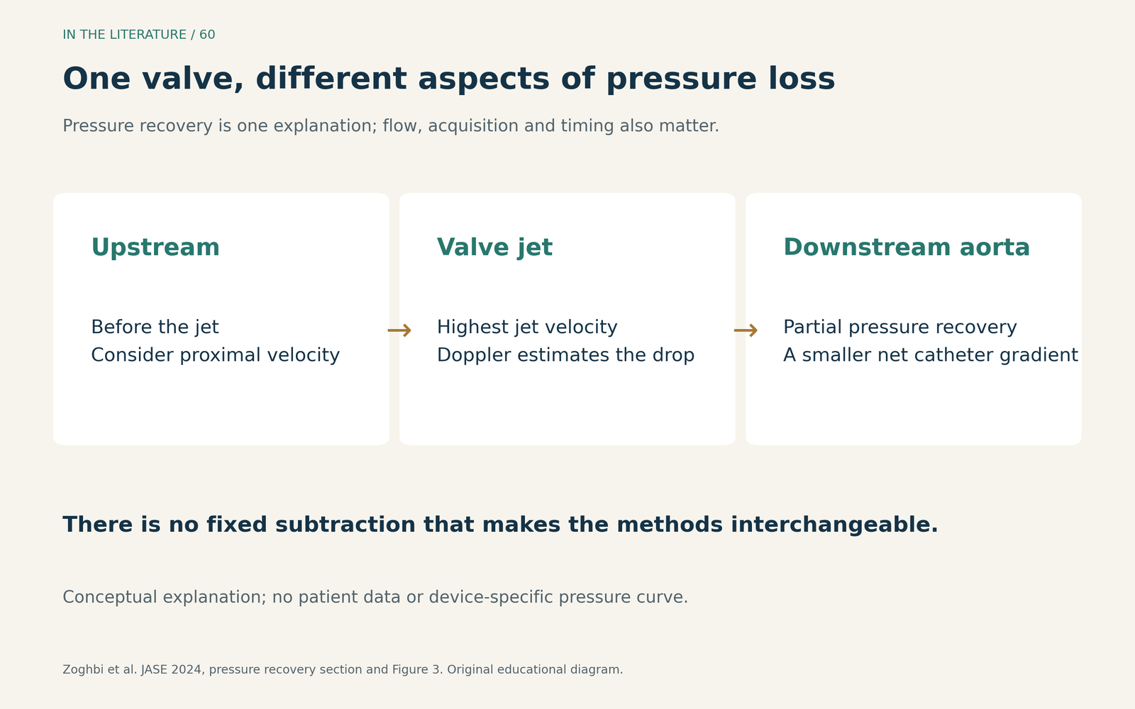
Figure 1. Original conceptual explanation, without patient data or a simulated pressure curve. Doppler and a downstream catheter interrogate different aspects of pressure loss. Catheter location and the extent of recovery matter. [1] View figure

The measurements cannot be substituted for one another
In Abbas and colleagues' 2023 multicenter study, paired intraprocedural measurements were available after 823 native-valve TAVR procedures and 77 valve-in-valve procedures. The postimplantation Doppler and invasive examinations were performed within 5–10 minutes of each other, rather than as strictly simultaneous beat-matched acquisitions. [2]
After native-valve TAVR, median Doppler mean gradient was 4 mmHg (IQR 3–6) versus 0 mmHg (0–3) invasively. After valve-in-valve TAVR, the corresponding values were 10 mmHg (7–15) and 4 mmHg (0–10). Individual agreement was wide; a population-average difference cannot serve as a correction factor for the next patient. These are observed cohort distributions, not proposed normal ranges. [2]
The investigators did not directly correct their measurements for proximal LVOT velocity or pressure recovery. Their findings therefore support noninterchangeability, but do not prove that pressure recovery has been excluded as a mechanism. A high residual Doppler gradient that would prompt an additional procedure warrants confirmation of the actual hemodynamic problem. [2]
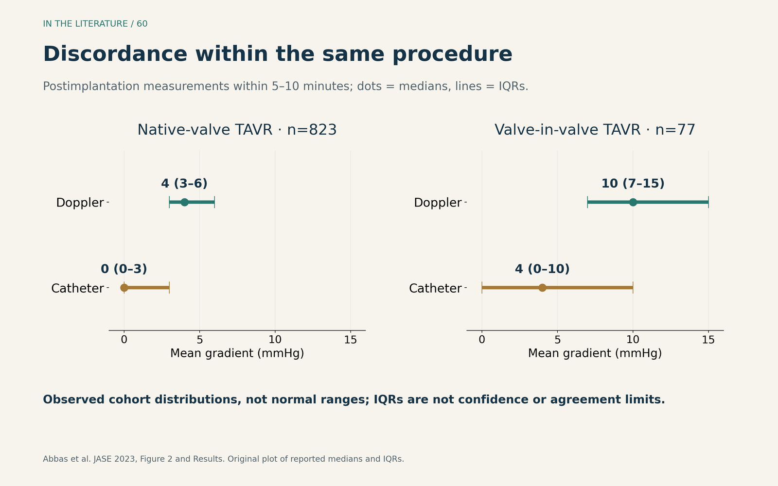
Figure 2. Original plot of published medians and interquartile ranges from Abbas et al.; the two postimplantation groups are shown separately. Horizontal intervals are IQRs, not confidence intervals or limits of agreement. Measurements were obtained within 5–10 minutes. [2] View figure

Match the metric—and the physiologic moment
First confirm that both numbers are mean gradients. A catheter peak-to-peak gradient is a different measurement. Then check when each study occurred and document blood pressure, heart rate and flow state. The invasive mean gradient is calculated from the ventricular–aortic pressure difference during systole; its interpretation still depends on sound acquisition. [1, 3]
Pfenniger and colleagues' 2025 analysis of 1,353 registry patients compared immediate peri-TAVI invasive measurements with postprocedural echocardiography. Median post-TAVI mean gradients were 9 mmHg (IQR 6–12) by echo and 4 mmHg (2–7) invasively; the postprocedural correlation was weak (r=0.23). The discharge echo and immediate catheter assessment should not be treated as measurements under identical conditions. [3]
A rise from an immediate procedural measurement to a later echo can reflect changing flow as well as a different method. Conversely, an unexplained rise on serial echocardiograms obtained under stable conditions deserves investigation. ASE recommends integrating the gradient with effective orifice area, Doppler velocity index (DVI), jet contour, acceleration time and valve morphology, using a stable postimplantation examination for comparison. [1]
A prognostic association is not a balloon-inflation target
In van den Dorpel and colleagues' 2025 single-center registry, 1,227 patients underwent TAVI for native aortic stenosis. Invasive gradients were obtained immediately after implantation; echo was performed within 24 hours. An invasive mean gradient >10 mmHg was associated with higher one-year all-cause mortality in the reported multivariable analysis: HR 1.91 (95% CI 1.11–3.65). The cutoff came from a spline fitted within that same cohort. [4]
This finding argues against ignoring a convincingly elevated invasive gradient. It does not establish that reducing every value above 10 mmHg improves survival, nor that postdilation is the appropriate response. The study was observational, and its different measurement times also matter. Its lack of a significant echo-gradient mortality association does not make a persistently high Doppler gradient harmless. The clinical task remains to explain the finding. [4]
Resolve the discrepancy before choosing a treatment
The 2025 Heart Valve Collaboratory imaging framework recommends invasive assessment when echo findings conflict substantially with symptoms or the severity of suspected valve dysfunction remains uncertain. It also identifies sources of invasive error: zeroing, calibration, catheter position and multiple side holes. A catheter value should be checked, not accepted as infallible. [5]
A useful sequence is:
- Confirm the acquisition. Review multiple Doppler windows, tracing quality and proximal LVOT sampling; check the pressure recordings, catheter positions and whether measurements were made under comparable conditions. A poorly placed pulsed-wave sample can distort both EOA and DVI. If invasive clarification is needed, ASE favors simultaneous upstream and downstream pressure recordings with two catheters. [1, 5]
- Interpret flow and the baseline. Look for a reversible high-flow state, significant regurgitation or subvalvular obstruction. For serial assessment, compare with a stable baseline rather than assuming the immediate procedural gradient defines lifelong valve performance. [1, 5]
- Identify the mechanism if concern remains. Abnormal leaflet motion, a new fall in EOA/DVI or unexplained symptoms should prompt targeted imaging. TEE and cardiac CT can help distinguish leaflet pathology, frame underexpansion and other causes; invasive confirmation may clarify discordant hemodynamics when it would change treatment. [1, 5]
The next intervention should follow the demonstrated mechanism and its clinical consequences. A stable, explained difference between modalities is a different problem from new obstruction or an inadequately expanded valve.
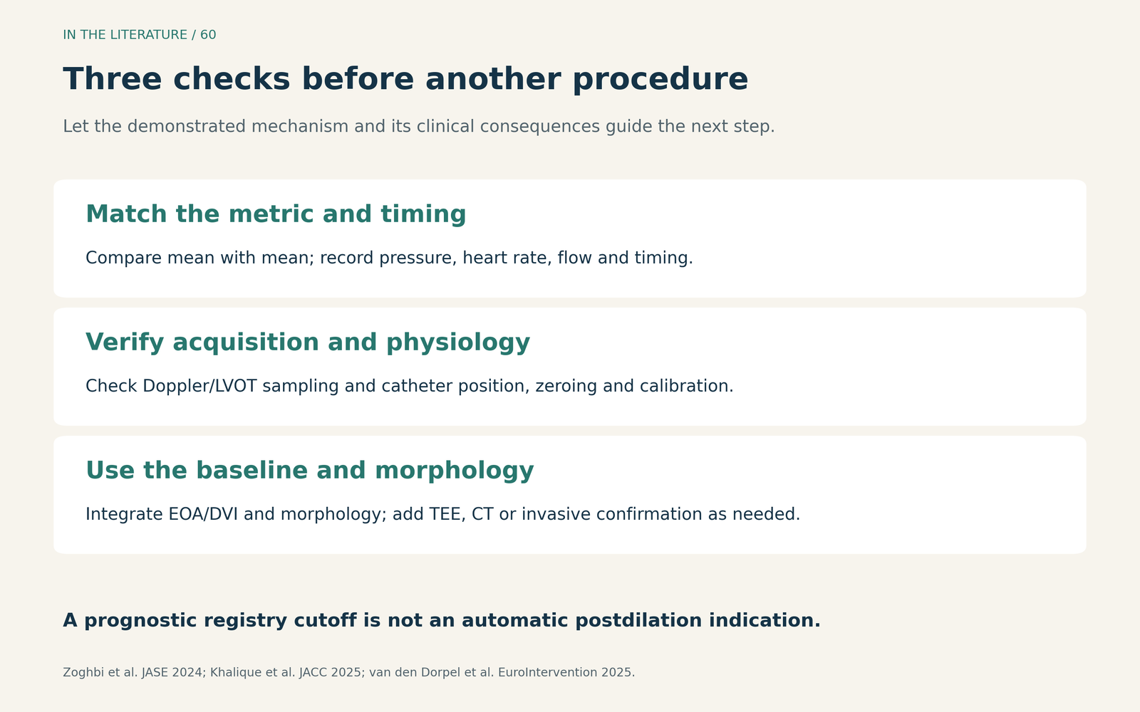
Figure 3. Original clinical assessment aid based on ASE and Heart Valve Collaboratory guidance. This is an interpretation sequence, not a validated score or an automatic treatment algorithm. [1, 5] View figure

Three takeaways
- Name the measurement before interpreting the number. Mean versus peak-to-peak, sampling location and timing can change what a gradient means.
- Explain discordance with the whole examination. Flow, EOA/DVI, waveform shape, morphology and a stable baseline are more useful than a fixed subtraction rule.
- Confirm the mechanism before an additional procedure. Prognostic registry cutoffs identify associations; they do not establish that chasing a lower number benefits the patient.
References
- Zoghbi WA, Jone PN, Chamsi-Pasha MA, et al. Guidelines for the Evaluation of Prosthetic Valve Function With Cardiovascular Imaging: A Report From the American Society of Echocardiography Developed in Collaboration With the Society for Cardiovascular Magnetic Resonance and the Society of Cardiovascular Computed Tomography. J Am Soc Echocardiogr. 2024;37(1):2–63. doi:10.1016/j.echo.2023.10.004
- Abbas AE, Khalili H, Madanat L, et al. Echocardiographic Versus Invasive Aortic Valve Gradients in Different Clinical Scenarios. J Am Soc Echocardiogr. 2023;36(12):1302–1314. doi:10.1016/j.echo.2023.06.016
- Pfenniger A, Stolte T, Reichl JJ, et al. Comparison of invasive and non-invasive gradients before and after TAVI and their implications on clinical outcomes. Cardiovasc Interv Ther. 2025;40:362–377. doi:10.1007/s12928-024-01074-6
- van den Dorpel MMP, Chatterjee S, Adrichem R, et al. Prognostic value of invasive versus echocardiography-derived aortic gradient in patients undergoing TAVI. EuroIntervention. 2025;21(8):e411–e425. doi:10.4244/EIJ-D-24-00341
- Khalique OK, Zaid S, Tang GHL, et al. Best Practices for Imaging of Transcatheter Valve Failure: An Update From the Heart Valve Collaboratory. J Am Coll Cardiol. 2025;85(10):1042–1055. doi:10.1016/j.jacc.2024.12.017