
ITL-571081e347d8
Juan Li, MD · Updated November 18, 2026
Key points
- A stuck bioprosthesis is rare but dangerous. A suture loop entangled on a stent post can restrict leaflet motion, and 3D TEE detects it right after bypass.[1]
- Not every early failure is technical. In two cases, neither tissue nor suture entrapment explained the obstruction, and valve function should be checked by TEE before stopping bypass.[2]
- Structural failure can be subtle. A small disc fracture caused severe intraprosthetic MR years after implantation.[3]
- Later, think thrombus. Bioprosthetic mitral valve thrombosis occurred at 5.05 per 100 patient-years, mostly subclinical.[4]
- Thrombus and pannus look different. Pannus is a fixed, highly echogenic mass, and 3D TEE may find thrombi that 2D TEE misses.[5]
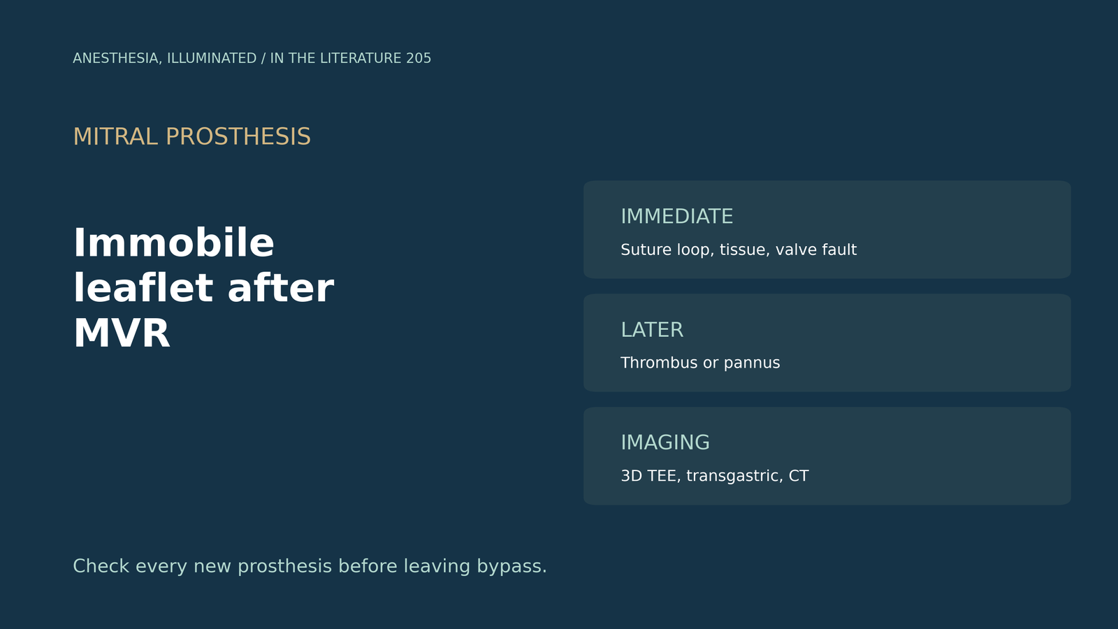
After mitral valve replacement with a bioprosthesis, the post-bypass TEE shows one leaflet that barely opens and a high transmitral gradient. What should be checked, and when should the team go back on bypass?[1], [2]
In the operating room: sutures, tissue and the valve itself
Takeshita and colleagues describe a stuck bioprosthetic valve as rare but potentially fatal. A suture loop entangled on a stent post interferes with leaflet motion; intraoperative 3D TEE detected it right after bypass, the restricted motion led to a second bypass run, and afterwards the valve functioned normally. Residual subvalvular tissue is another reported cause.[1] Takahashi and colleagues report two cases in which neither perivalvular tissue nor suture entrapment explained the obstruction, and the manufacturer found no structural issues in the explanted valves. They note that papillary muscles and chords appear as hyperechoic structures interfering with the valve, that a normal bioprosthesis shows only trace to mild central regurgitation, and that valve function should be checked by TEE before stopping bypass.[2]
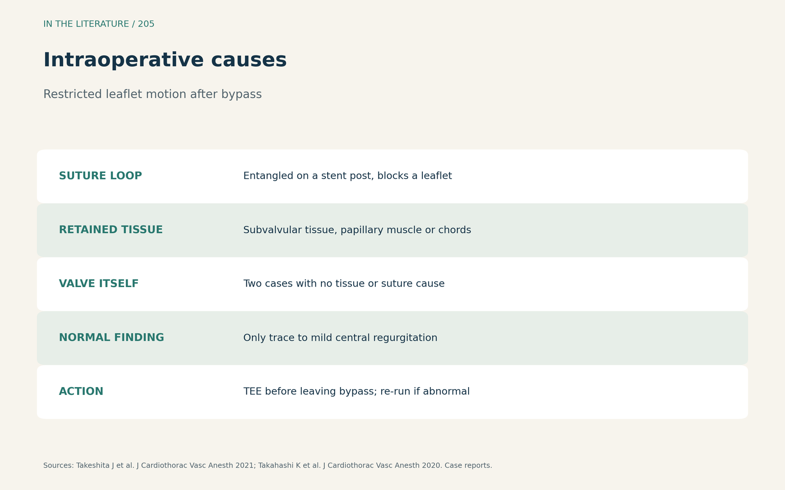
Figure 1. Suture, tissue or the valve itself. Synthesized from Takeshita et al. and Takahashi et al.[1], [2] View larger.

Structural failure
Eiras and colleagues describe severe intraprosthetic MR 13 years after mechanical valve replacement; macroscopic examination of the explant revealed a small disc fracture.[3]
Later: thrombus or pannus?
Zorman and colleagues found bioprosthetic mitral valve thrombosis at 5.05 per 100 patient-years. Subclinical thrombosis was more common than clinical thrombosis, oral anticoagulation was linked to 3.5-fold lower thrombosis, risk persisted beyond 3 months and peaked at 12 to 24 months, and reduced leaflet motion may be seen as early as implantation; the authors call their findings hypothesis-generating.[4] Serban and colleagues describe pannus as a fixed, highly echogenic mass, unlike thrombus, and note that pannus can be treated only with surgery. The transgastric view shows the ventricular side of the prosthesis, 3D TEE may identify thrombi that 2D TEE misses, fluoroscopy best evaluates mechanical leaflet motion, and MDCT had 88% sensitivity for subprosthetic masses.[5]
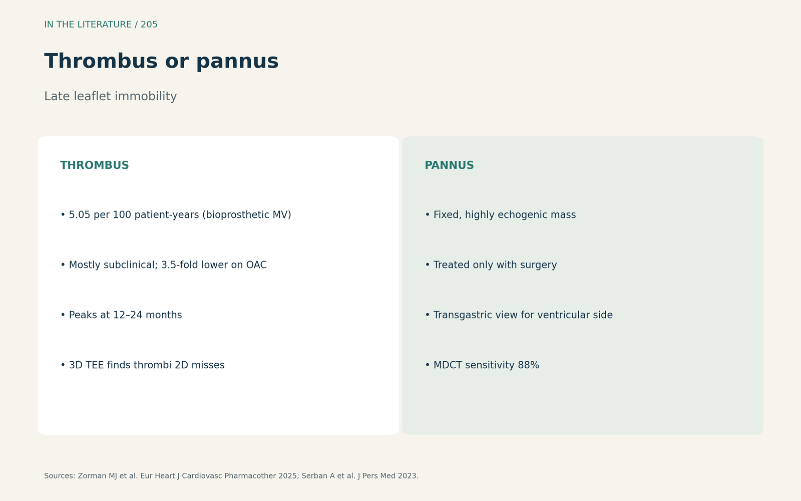
Figure 2. Mobile and soft, or fixed and bright. Synthesized from Zorman et al. and Serban et al.[4], [5] View larger.

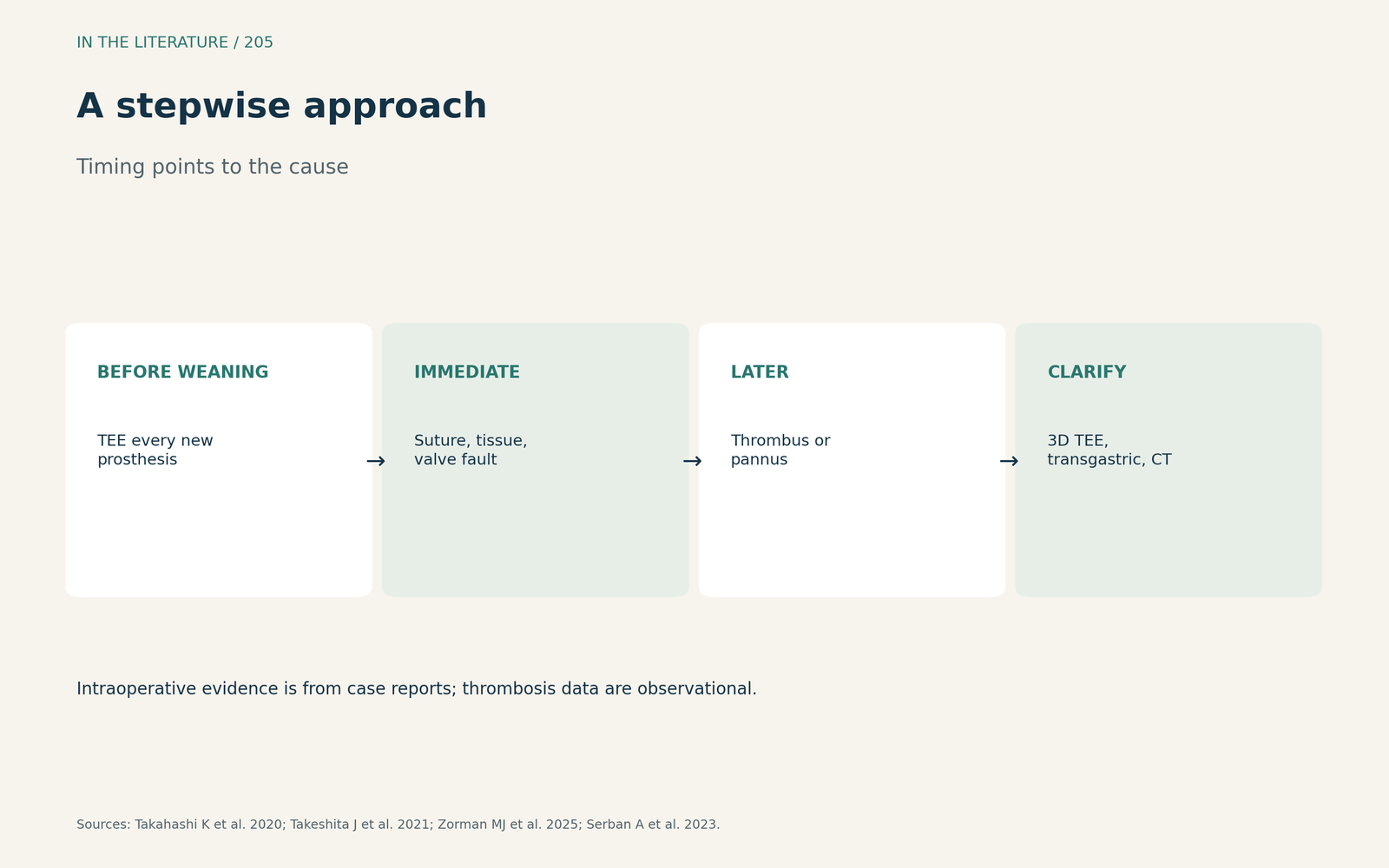
Figure 3. Timing points to the cause. Synthesized from Takahashi et al., Takeshita et al., Zorman et al. and Serban et al.[2], [1], [4], [5] View larger.

Three takeaways
- Check leaflet motion of every new mitral prosthesis with TEE, including 3D, before stopping bypass.[1], [2]
- Immediate immobility suggests a suture loop, retained tissue or a valve fault; later immobility suggests thrombus or pannus.[1], [4]
- Use transgastric views, 3D TEE and CT to separate thrombus from pannus.[5]
References
- Takeshita J, Akiyama K, Anada N, Nakajima Y. Intraoperative diagnosis of a stuck bioprosthetic valve leaflet due to a loop of suture after mitral valve replacement. J Cardiothorac Vasc Anesth. 2021;35(6):1830–1832. doi:10.1053/j.jvca.2020.07.007
- Takahashi K, Sajima T, Ishiguro Y. Intraoperative transvalvular regurgitation immediately after mitral bioprosthetic valve implantation. J Cardiothorac Vasc Anesth. 2020;34(6):1704–1707. doi:10.1053/j.jvca.2019.12.040
- Eiras M, Martínez A, Taboada M, Durán D. Severe mitral regurgitation due to a small fracture of a prosthetic valve. J Cardiothorac Vasc Anesth. 2021;35(2):679–680. doi:10.1053/j.jvca.2020.06.032
- Zorman MJ, Vibhishanan J, Dangas K, et al. Valve thrombosis and antithrombotic therapy after bioprosthetic mitral valve replacement: a systematic review and meta-analysis. Eur Heart J Cardiovasc Pharmacother. 2025;11(3):251–263. doi:10.1093/ehjcvp/pvaf005
- Serban A, Dadarlat-Pop A, Achim A, et al. Diagnosis of left-sided mechanical prosthetic valve thrombosis: a pictorial review. J Pers Med. 2023;13(6):967. doi:10.3390/jpm13060967