ITL-3d900742a96d
Juan Li, MD · Updated November 17, 2026
Watch the video
Key points
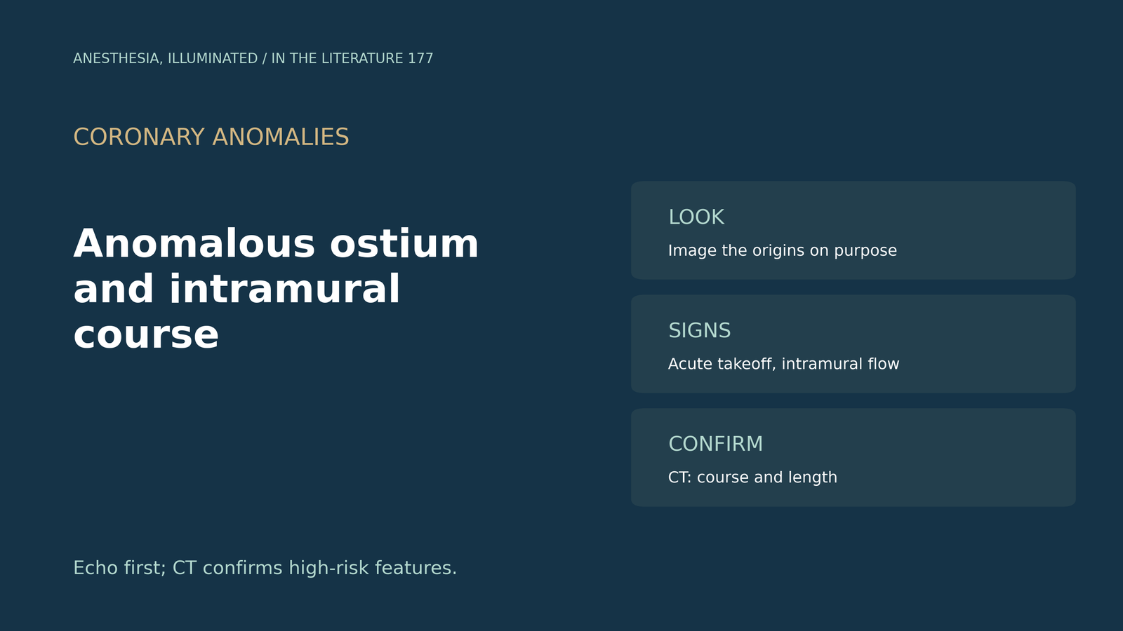
- Echo can see the ostia. In ideal settings TTE identifies the left coronary origin in 98% to 100% and the right in 80% to 96%, without radiation.[1]
- Look for the intramural signs. An acute takeoff angle with diastolic color flow inside the aortic wall correctly defined an intramural course in 94%.[1]
- A protocol finds more. Adding focused interarterial-space imaging found anomalous origins in 1.28% of TTEs, 3.6 times the earlier rate, and six patients had slit-like ostia.[2]
- Know the retroaortic clue. A small circle beneath the noncoronary cusp and a vessel behind the aortic root suggested an anomalous circumflex.[3]
- Confirm with CT. CT defines anatomy and high-risk features, and an intramural length of 3.9 mm or more predicted high-risk lesions.[4], [1]
A young athlete with exertional chest pain has a TTE in which the right coronary origin is hard to see. How can an anomalous origin and an intramural course be detected, and when is CT needed?[1], [2]
What echo can show
Capponi and colleagues note that TTE visualizes the ostia without radiation; in ideal settings it identifies the left coronary origin in 98% to 100% and the right in 80% to 96%. An acute takeoff angle with diastolic color flow in the proximal course within the aortic wall correctly defined an intramural course in 94% in a cited study, and TEE may be more appropriate in some patients.[1] Ahluwalia and colleagues report that prevalence is 0.2% to 0.35% by TTE versus 0.3% to 1.5% by CT; after adding focused interarterial-space imaging, anomalous origin was found in 143 of 11,115 TTEs (1.28%), 3.6 times the earlier rate, and six patients had slit-like ostia.[2]
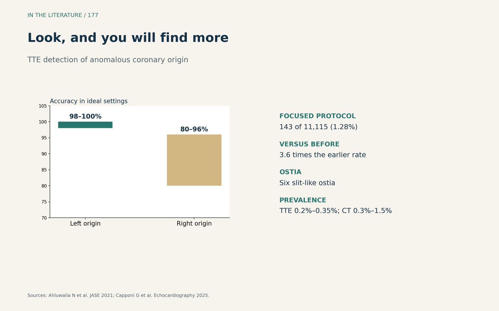
Figure 1. Look, and you will find more. Redrawn from Ahluwalia et al. and Capponi et al.[2], [1] View larger.

Signs of a dangerous course
Mancinelli and colleagues saw a small circle beneath the noncoronary cusp in parasternal long axis and a vessel behind the aortic root in short axis, identifying an anomalous circumflex; reversible inferolateral akinesia appeared on stress, and malignant features include an acute takeoff angle and a dilated aortic root.[3] Narvaez-Guerra and colleagues advise off-axis imaging and anterior tilting from the parasternal short axis to find an anomalous right coronary origin, and they list an intramural course among the high-risk features.[4]
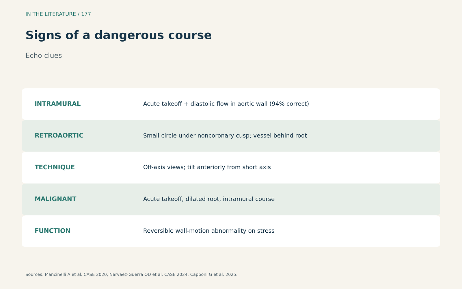
Figure 2. Signs of a dangerous course. Synthesized from Mancinelli et al., Narvaez-Guerra et al. and Capponi et al.[3], [4], [1] View larger.

Confirming with CT
Cardiac CT is fundamental for coronary anatomy and high-risk features.[4] CT detects an intramural course with strong correspondence to surgical findings, and an intramural length of 3.9 mm or more predicted high-risk lesions with sensitivity 77% and specificity 75%; echo remains first line, but CT and MRI complete the assessment.[1]
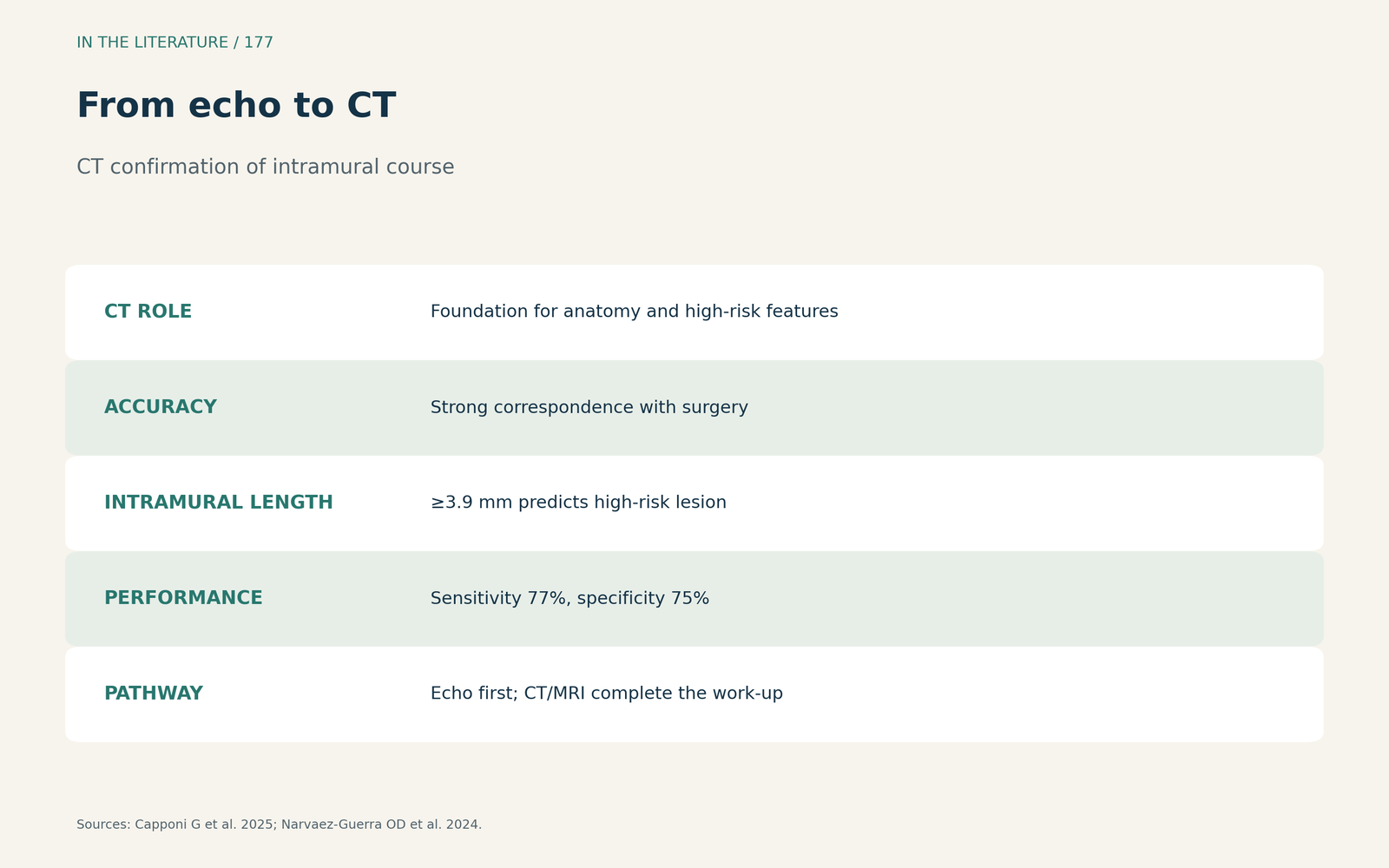
Figure 3. From echo to CT. Synthesized from Capponi et al. and Narvaez-Guerra et al.[1], [4] View larger.

Three takeaways
- Image the coronary origins on purpose. A focused protocol finds more anomalies.[2]
- Acute takeoff plus intramural diastolic flow means think intramural. Look behind and between the great arteries.[1], [3]
- Confirm with CT. It defines the course, ostium and intramural length.[4], [1]
References
- Capponi G, Castaldi B, Sirico D, et al. Multi-modality imaging in coronary anomalies: focus on anomalous aortic origin of coronary arteries. Echocardiography. 2025;42(6):e70213. doi:10.1111/echo.70213
- Ahluwalia N, Stern KW, Parness IA, Lytrivi ID, Srivastava S. Trends in incidental diagnosis of anomalous aortic origin of coronary artery in patients less than 30 years of age: a single-center experience. J Am Soc Echocardiogr. 2021;34(10):1115–1116. doi:10.1016/j.echo.2021.06.016
- Mancinelli A, Golino M, Miglierina E, et al. Three echocardiographic signs to identify anomalous origin of the circumflex coronary artery from the right sinus of Valsalva: a case report. CASE (Phila). 2020;4(5):324–327. doi:10.1016/j.case.2020.07.008
- Narvaez-Guerra OD, Sorour N, Aurigemma GP, et al. A multimodality imaging and multidisciplinary approach to manage anomalous right coronary artery from the pulmonary artery in pregnancy. CASE (Phila). 2024;8(3):122–126. doi:10.1016/j.case.2023.12.013