ITL-7b98da86e5ed
Juan Li, MD · Updated November 17, 2026
Watch the video
Key points
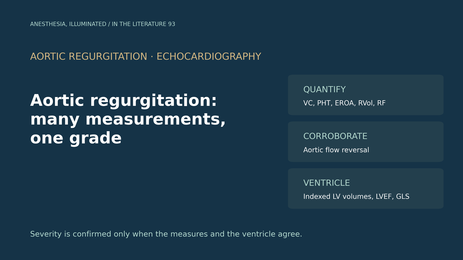
- No single echo parameter grades aortic regurgitation (AR). The British Society of Echocardiography (BSE) confirms severity only when a quantitative or semi-quantitative measure, a corroborative sign and indexed left ventricular (LV) volumes agree; colour jet length or area should not be used.[1]
- Severe thresholds include vena contracta above 0.6 cm, jet width above 65% of the outflow tract, pressure half-time below 200 ms, effective regurgitant orifice area (EROA) above 0.3 cm², regurgitant volume above 60 mL and regurgitant fraction above 50%. Chronic severe AR also requires LV dilation.[1], [2]
- Diastolic flow reversal in the aorta is a useful corroborative sign. A descending aortic end-diastolic velocity cut-off was highly specific but only moderately sensitive, and distal abdominal reversal was feasible and sensitive.[1], [3], [4]
- The ventricle decides timing: LV ejection fraction (LVEF) of 55% or less and severe LV enlargement are guideline triggers, and indexed end-systolic volume of 45 mL/m² or more and abnormal strain mark higher risk.[2], [5], [6]
- Loading, acute AR and eccentric jets distort every index. Small jets seen just after bypass with a new prosthesis are often transient.[1], [7]
A preoperative echocardiogram reports "moderate to severe" aortic regurgitation. The pressure half-time looks severe, the vena contracta looks moderate, and the ventricle is only mildly dilated. In the operating room, transesophageal echocardiography (TEE) under anesthesia suggests something different again. Which measurements should you trust, how do you combine them, and when is the regurgitation severe enough to matter?[1], [2]
Why one number is not enough
Every AR parameter has a technical or physiological weakness. The 2025 BSE practical guideline therefore recommends that, beyond obviously mild AR, quantitative or semi-quantitative methods should be used together with corroborative findings. It states that jet length or jet area from any window should not be used to assess severity.[1]
Its integration rule is explicit: severity is confirmed when at least one quantitative or semi-quantitative characteristic, one corroborative characteristic and indexed LV volumes all agree. When they disagree, or AR cannot be quantified, TEE is the next step, and cardiac magnetic resonance (CMR) can clarify discordant data when transthoracic images are poor.[1] The 2020 American College of Cardiology/American Heart Association (ACC/AHA) valve guideline similarly notes that, when AR is significant, quantitative regurgitant volume and EROA are better predictors of clinical outcome than qualitative measures.[2]
The parameters and their thresholds
The BSE guideline grades each parameter separately:[1]
- Vena contracta: below 0.3 cm mild; above 0.6 cm severe.
- Jet width to outflow tract ratio: below 25% mild; above 65% severe.
- Pressure half-time: above 500 ms mild; below 200 ms severe.
- EROA: below 0.10 cm² mild; above 0.3 cm² severe.
- Regurgitant volume: below 30 mL mild; above 60 mL severe.
- Regurgitant fraction: below 30% mild; above 50% severe.
- Aortic flow reversal: holodiastolic reversal in the descending thoracic aorta with end-diastolic velocity of 20 cm/s or more, or any reversal in the abdominal aorta, supports severe AR.
Values between the mild and severe cut-offs are moderate. The ACC/AHA guideline adds that a diagnosis of chronic severe AR requires evidence of LV dilation.[2] A normal indexed LV volume almost always rules out chronic severe AR.[1]
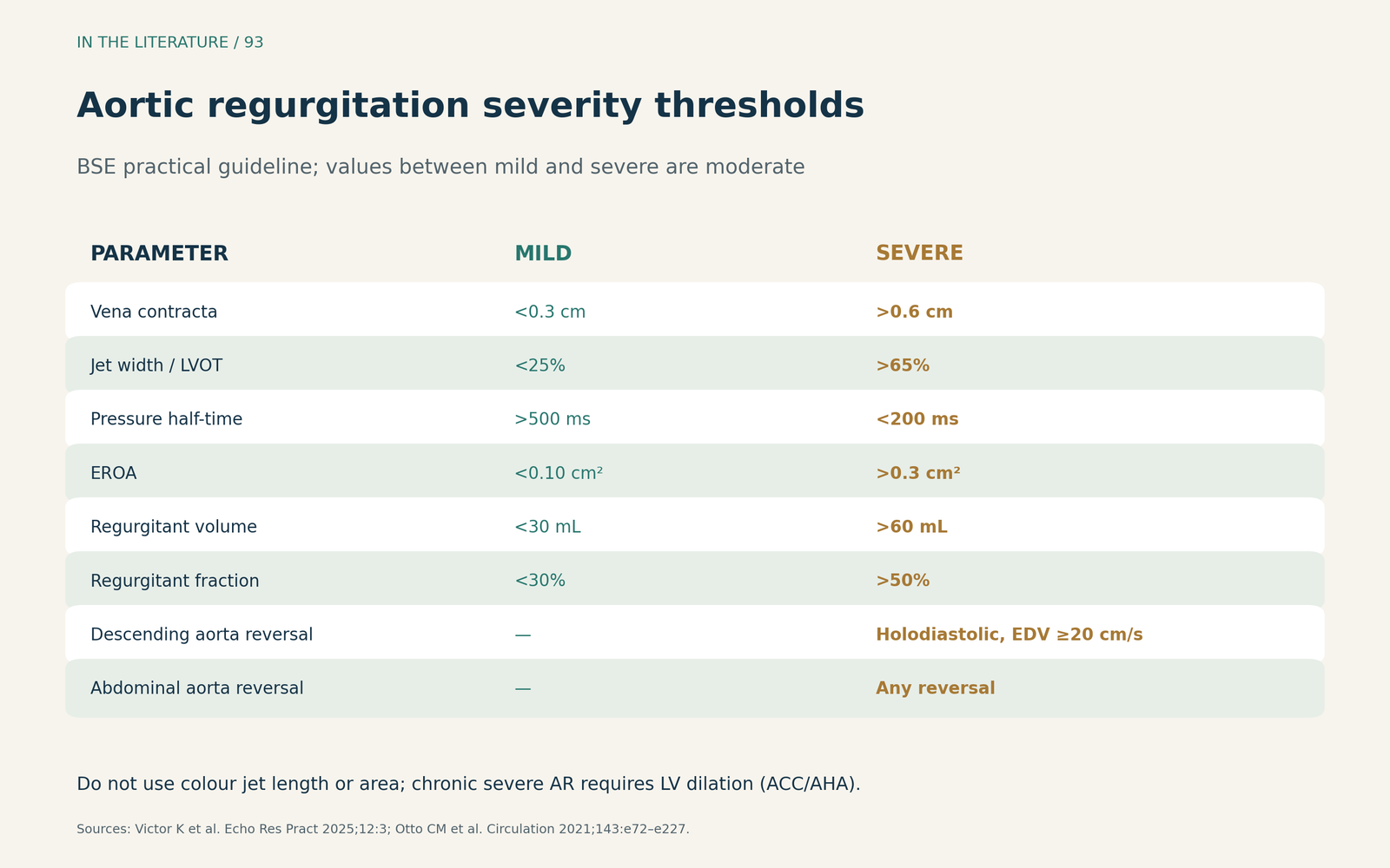
Figure 1. One parameter at a time, then integrate. Thresholds redrawn from the BSE practical guideline; chronic severe AR also requires LV dilation per the ACC/AHA guideline.[1], [2] View larger.

Aortic flow reversal: useful, but not alone
Diastolic flow reversal reflects regurgitant volume leaving the aorta. Knoll and colleagues compared end-diastolic flow reversal in the upper descending aorta with CMR in 66 patients with chronic AR. A cut-off above 18 cm/s had a sensitivity of 57.9% and specificity of 91.5% for severe AR. In multivariable analysis, only end-diastolic flow reversal and vena contracta remained associated with severe AR on CMR.[3]
Suzuki and colleagues assessed holodiastolic reversal in the abdominal aorta in 103 patients with moderate or severe AR. The distal segment was assessable far more often than the proximal segment (94% versus 54%), and distal reversal had a sensitivity of 95%, specificity of 84% and accuracy of 87% against integrative echo grading. The authors concluded it should be a complementary tool rather than a standalone criterion.[4] The BSE guideline also warns that rapid elastic recoil can produce flow reversal without severe AR.[1]
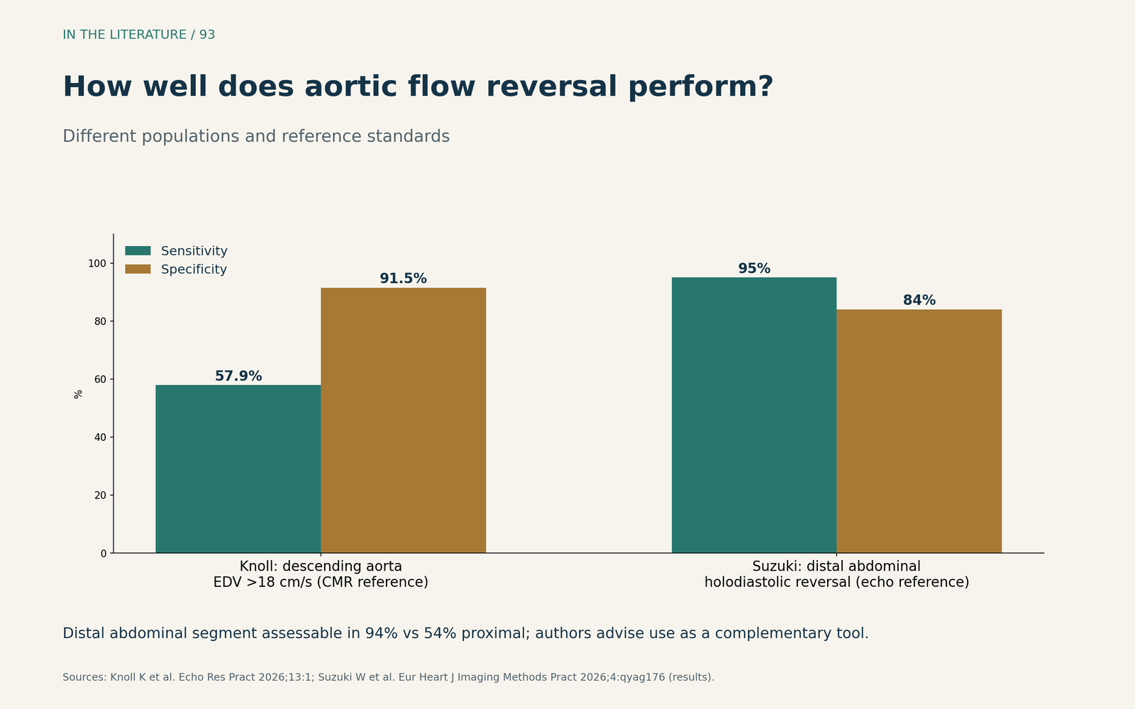
Figure 2. Flow reversal adds evidence but misses or overcalls some cases. Redrawn from the results of Knoll et al. (CMR reference) and Suzuki et al. (integrative echo reference); different populations and reference standards.[3], [4] View larger.

Is the ventricle telling you it is severe?
For chronic AR, the LV response determines when to operate. The ACC/AHA guideline recommends surgery in asymptomatic severe AR with LVEF of 55% or less, and considers it reasonable when LVEF is normal but the LV is severely enlarged (end-systolic dimension above 50 mm or above 25 mm/m²).[2]
Volumes and strain may detect risk earlier. In 1,100 patients with moderate-to-severe or severe AR, Anand and colleagues found that indexed LV end-systolic volume was more strongly associated with outcomes than indexed end-systolic dimension, with mortality risk starting to rise between 44 and 45 mL/m².[5] Yang and colleagues found that, in asymptomatic chronic AR, risk of death rose when automated global longitudinal strain fell below 15%, with a 2.6-fold risk below that value.[6] Both studies were retrospective and single-center, and these thresholds are not yet guideline triggers.
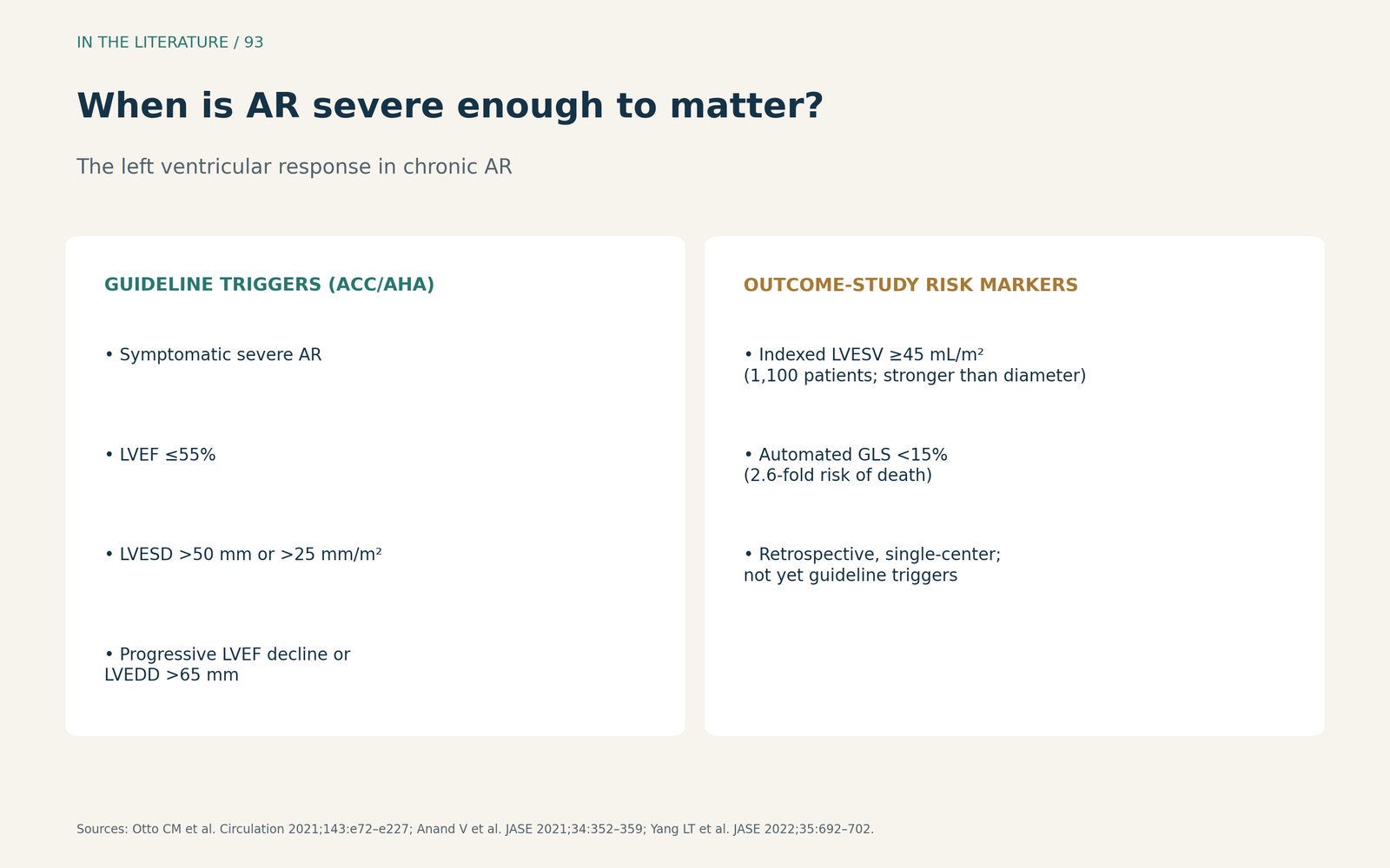
Figure 3. What makes AR "severe enough to matter". Synthesized from the ACC/AHA guideline surgical triggers and the outcome studies of Anand et al. and Yang et al.[2], [5], [6] View larger.

Pitfalls in acute AR and in the operating room
Loading conditions change the numbers. The BSE guideline notes that raised LV diastolic filling pressure can shorten pressure half-time even without severe AR, and that vasodilators have a similar effect. In acute AR, the LV is often not dilated, and rapid pressure equalization can abolish diastolic flow reversal in the descending aorta, so a "normal" ventricle and absent reversal do not exclude severe acute AR. With eccentric jets, small errors in PISA radius are multiplied, and jet-based measurements become less reliable.[1]
After valve replacement, new jets are common. Chemtob and colleagues documented aortic insufficiency in 18 of 21 patients after weaning from cardiopulmonary bypass with one bioprosthesis; jets were trivial or mild, eccentric and mostly resolved after protamine or by chest closure. Because they originated near the sewing ring, they could mimic paravalvular leak.[7] This was a small series of a single valve type.
Three takeaways
- Integrate, do not eyeball. Combine at least one quantitative or semi-quantitative measure, a corroborative sign and indexed LV volumes; ignore colour jet length and area.[1]
- Use flow reversal as support. Descending and abdominal aortic reversal add evidence, but have false negatives and false positives.[1], [3], [4]
- Let the ventricle and the context decide. LV volumes, function and strain determine whether AR matters; loading, acute onset and post-bypass jets demand caution.[2], [5]–[7]
References
- Victor K, Ring L, Tsampasian V, et al. Echocardiographic assessment of aortic regurgitation: a practical guideline from the British Society of Echocardiography. Echo Res Pract. 2025;12(1):3. doi:10.1186/s44156-024-00067-8
- Otto CM, Nishimura RA, Bonow RO, et al. 2020 ACC/AHA guideline for the management of patients with valvular heart disease: a report of the American College of Cardiology/American Heart Association Joint Committee on Clinical Practice Guidelines. Circulation. 2021;143(5):e72–e227. doi:10.1161/CIR.0000000000000923
- Knoll K, Rosner S, Gross S, et al. Assessment of chronic aortic regurgitation using end-diastolic flow reversal in the upper descending aorta: diagnostic accuracy and prediction of aortic valve surgery in a prospective echocardiography and cardiac magnetic resonance imaging study. Echo Res Pract. 2026;13(1):1. doi:10.1186/s44156-025-00101-3
- Suzuki W, Kojima S, Takeda R, et al. Feasibility and diagnostic utility of echocardiographic distal abdominal aortic diastolic flow reversal for assessing aortic regurgitation. Eur Heart J Imaging Methods Pract. 2026;4(4):qyag176. doi:10.1093/ehjimp/qyag176
- Anand V, Yang L, Luis SA, et al. Association of left ventricular volume in predicting clinical outcomes in patients with aortic regurgitation. J Am Soc Echocardiogr. 2021;34(4):352–359. doi:10.1016/j.echo.2020.11.014
- Yang LT, Takeuchi M, Scott CG, et al. Automated global longitudinal strain exhibits a robust association with death in asymptomatic chronic aortic regurgitation. J Am Soc Echocardiogr. 2022;35(7):692–702. doi:10.1016/j.echo.2021.10.018
- Chemtob RA, Sale S, Wyler D, et al. Postpump aortic insufficiency is transient after valve replacement with a novel prosthesis. J Am Soc Echocardiogr. 2021;34(9):1017–1019. doi:10.1016/j.echo.2021.04.020