ITL-4bd88ec956da
Juan Li, MD · Updated November 17, 2026
Watch the video
Key points
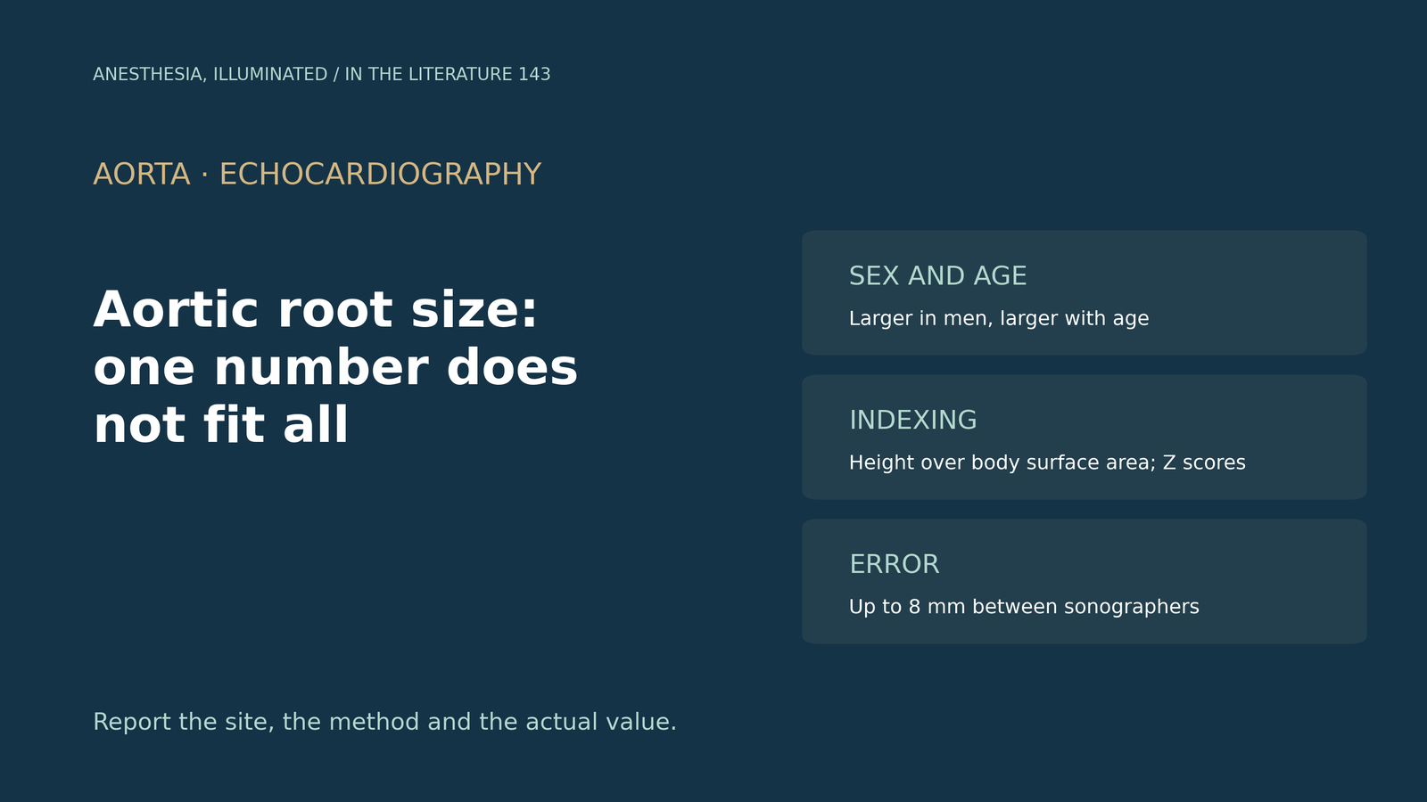
- One number does not fit all. In 1,585 healthy adults from 15 countries, all aortic root dimensions were larger in men than in women, root size increased with age, and upper limits of normal were lower than older guideline values.[1]
- How you index changes the answer. After indexing to body surface area women appeared larger, whereas men remained larger after indexing to height, supporting height over body surface area.[1], [2]
- Z scores express distance from the predicted mean in standard deviations, avoiding jumps between nomograms; for an average adult, the upper limit at the sinuses of Valsalva is about 3.8 cm.[3]
- Measurement noise is real. Different sonographers produced individual differences up to 8 mm, and intraoperative TEE slightly underestimated magnetic resonance measurements.[4], [5]
- Site matters as well as size. Mortality appears to rise from proximal to distal measurement sites, but there is no widely accepted standard yet, so report the site and the actual measurement.[6], [3]
Before aortic valve replacement, intraoperative TEE shows a sinus of Valsalva diameter of 4.1 cm in a short woman and the same value in a tall man. Is either root dilated, and how should the number be interpreted?[1], [3]
Normal values depend on sex and age
The World Alliance of Societies of Echocardiography (WASE) study measured the annulus, sinuses of Valsalva and sinotubular junction in 1,585 healthy adults. All root dimensions were larger in men than in women, the sinus and junction diameters increased with age, and the upper limits of normal, defined as the 97.5th percentile for sex and age, were lower across all age groups than in previous guidance. Asians had the smallest nonindexed dimensions; the differences between races were relatively small but could cause under- or overdiagnosis of dilatation.[1]
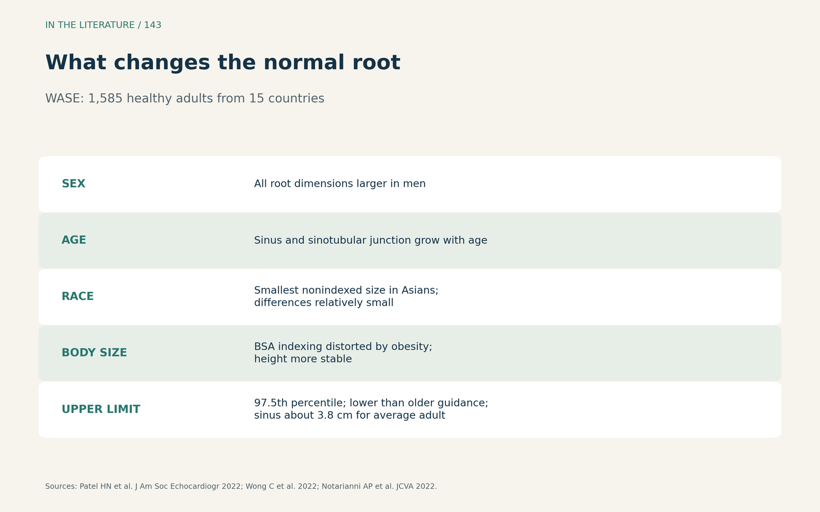
Figure 1. What changes the normal root. Synthesized from the WASE study, Wong et al. and Notarianni et al.[1], [3], [2] View larger.

Indexing and Z scores
Indexing to body surface area makes women's roots appear larger, whereas indexing to height keeps men's roots larger; WASE and accompanying editorials favor height, because body surface area is inflated by obesity.[1], [2] Wong and colleagues argue for a single Z score: the number of standard deviations above or below the mean predicted for age, sex and body size. They show that for the average WASE adult the upper limit at the sinuses of Valsalva is 3.8 cm, so a definition of aneurysm as 50% above normal would require at least 5.7 cm, larger than common surgical thresholds; reports should therefore state the actual measurement.[3] Notarianni and colleagues note that older reference ranges came from small, mostly White samples, and that updated, patient-specific reference ranges were needed.[2]
Measurement error and modality
Bons and colleagues had different sonographers measure the same patients: larger diameters had larger test-retest variability, individual differences reached 8 mm, and they suggest verifying with CT or magnetic resonance when a patient approaches the threshold for surgery.[4] Rong and colleagues compared intraoperative TEE with magnetic resonance in 30 patients having ascending aortic repair and found narrow limits of agreement, although TEE underestimated the magnetic resonance measurements.[5]
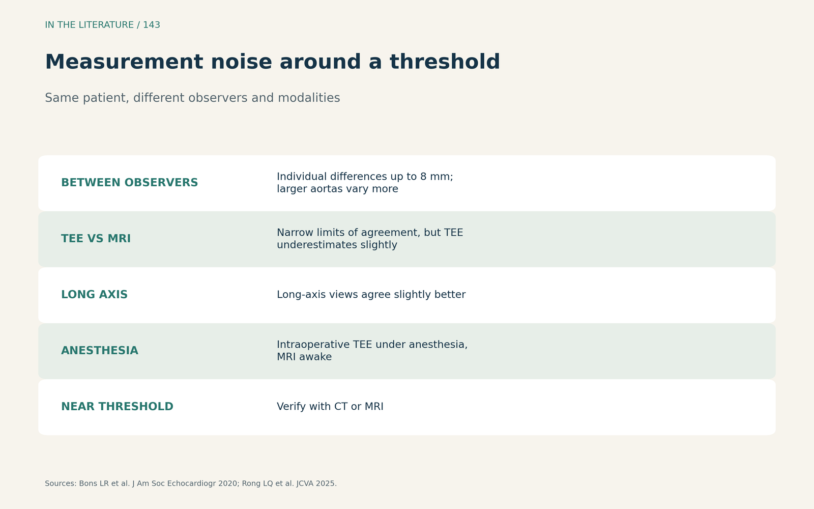
Figure 2. Measurement noise around a threshold. Synthesized from Bons et al. and Rong et al.[4], [5] View larger.

Site of measurement
Alktaish and colleagues, discussing a large registry, note that mortality appears to increase from proximal to distal aortic measurement sites, that the sinuses are measured nearly ubiquitously while other segments are measured much less often, and that there is no widely accepted standard; their advice is to be aware but change nothing yet.[6]
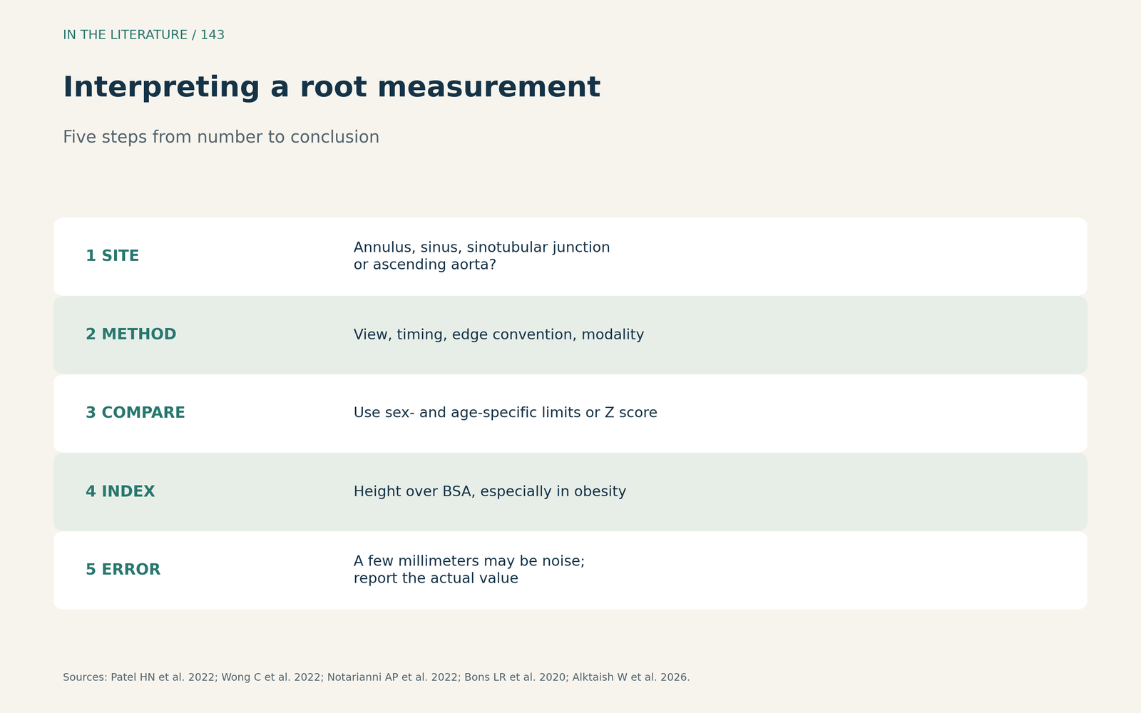
Figure 3. Interpreting a root measurement. Synthesized from the WASE study, Wong et al., Notarianni et al., Bons et al. and Alktaish et al.[1], [3], [2], [4], [6] View larger.

Three takeaways
- Compare like with like. Use normal values for sex and age; contemporary limits are lower than older ones.[1]
- Prefer height and Z scores. Body surface area distorts indexing in obesity; Z scores avoid nomogram jumps.[1], [3], [2]
- Report site, method and error. A few millimeters of difference can be noise; confirm near thresholds.[4], [5], [6]
References
- Patel HN, Miyoshi T, Addetia K, et al. Normal values of aortic root size according to age, sex, and race: results of the World Alliance of Societies of Echocardiography study. J Am Soc Echocardiogr. 2022;35(3):267–274. doi:10.1016/j.echo.2021.09.011
- Notarianni AP, Patel B, Patel PA. Normal aortic root: when one size does not fit all. J Cardiothorac Vasc Anesth. 2022;36(9):3444–3446. doi:10.1053/j.jvca.2022.04.052
- Wong C, Aurigemma GP, Parker MW. Normal aortic dimensions: from A-to-Z score. J Am Soc Echocardiogr. 2022;35(3):275–277. doi:10.1016/j.echo.2021.12.001
- Bons LR, van den Hoven AT, McGhie JS, et al. Variability in echocardiographic ascending aortic diameters due to image acquisition by different sonographers. J Am Soc Echocardiogr. 2020;33(2):249–252.e4. doi:10.1016/j.echo.2019.09.015
- Rong LQ, Chun S, Villar-Calle P, et al. Thoracic aorta measurements using intraoperative transesophageal echocardiography: validation via cardiac magnetic resonance. J Cardiothorac Vasc Anesth. 2025;39(5):1135–1145. doi:10.1053/j.jvca.2025.01.038
- Alktaish W, Ortoleva JP, Shapeton AD. Thoracic aortic aneurysms: location, location, location. J Cardiothorac Vasc Anesth. 2026;40(9):2646–2648. doi:10.1053/j.jvca.2026.05.064