ITL-1300599d0ad7
Juan Li, MD · Updated November 18, 2026
Watch the video
Key points
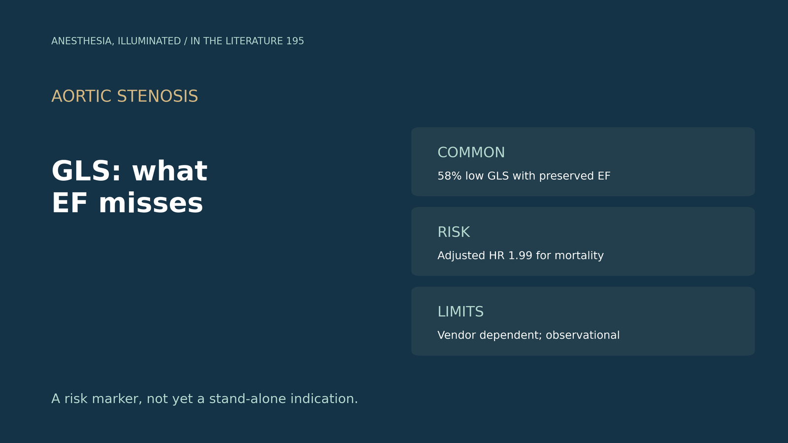
- EF is still the gate. LVEF is the main criterion for selecting asymptomatic patients for surgery, yet GLS is often impaired despite preserved LVEF in severe AS.[1], [2]
- Low GLS carries risk. In severe AS with preserved EF, 58% had a GLS magnitude of 15% or less, with an adjusted mortality HR of 1.99.[1]
- The signal appears before stenosis is severe. In moderate AS, preserved EF with GLS below 16% carried the same risk as EF below 50%.[3]
- Pooled data agree. Across nine studies, impaired GLS raised MACE and mortality, including in patients with LVEF above 50%.[2]
- The evidence is observational. GLS is vendor dependent, one cohort found no prognostic value, and no trial has tested GLS-guided valve replacement.[3], [2], [4]
Before noncardiac surgery, an older patient with asymptomatic severe aortic stenosis has an LVEF of 60%, but the echo report lists a GLS of −13%. Does that strain value reveal myocardial damage that the ejection fraction hides, and how much weight should it carry?[1], [3]
Why EF can look normal
Wang and colleagues note that LVEF remains the main criterion for selecting asymptomatic patients for valve replacement, and that LV deterioration can be cloaked when afterload is reduced.[2] Thellier and colleagues add that GLS is often impaired despite preserved LVEF in severe AS; one of their examples had a GLS magnitude of 12.6% with an LVEF of 55%.[1] GLS is usually reported as a negative number; the studies below use its absolute value, so a smaller number means worse function. A review (Meredith et al., J Am Soc Echocardiogr 2024) notes that nearly one-third of a pooled cohort had impaired strain despite preserved LVEF and concludes that GLS outperforms LVEF in predicting survival.[5]
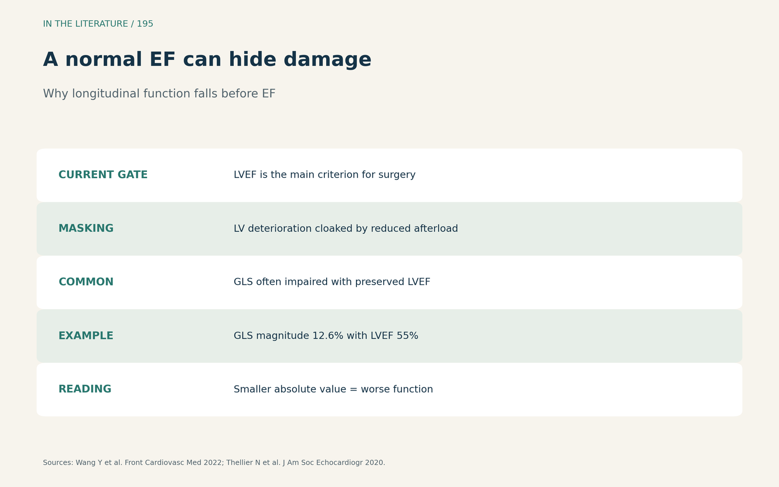
Figure 1. A normal EF can hide longitudinal dysfunction. Synthesized from Wang et al. and Thellier et al.[2], [1] View larger.

What the cohorts show
In Thellier's registry of severe AS with preserved EF and no or mild symptoms, 192 patients (58%) had a GLS magnitude of 15% or less, and 105 patients died over a median of 42 months. Mortality was 52% with impaired GLS versus 19% otherwise; after adjustment that included LVEF below 60%, the HR was 1.99, and in propensity-matched patients it was 2.10. The negative predictive value of GLS above 15% was high.[1] Stassen and colleagues followed 760 patients with moderate AS, of whom 257 (34%) died over 50 months. Patients with LVEF of 50% or more but GLS below 16% had an HR of 2.467, and their outcomes did not differ from patients with LVEF below 50%; asymptomatic patients with low GLS already had 14% mortality at 2 years.[3] Wang's meta-analysis of nine studies found that impaired GLS raised MACE (HR 1.20) and all-cause mortality (HR 1.42), with the association holding when LVEF was above 50% (HR 1.22).[2]
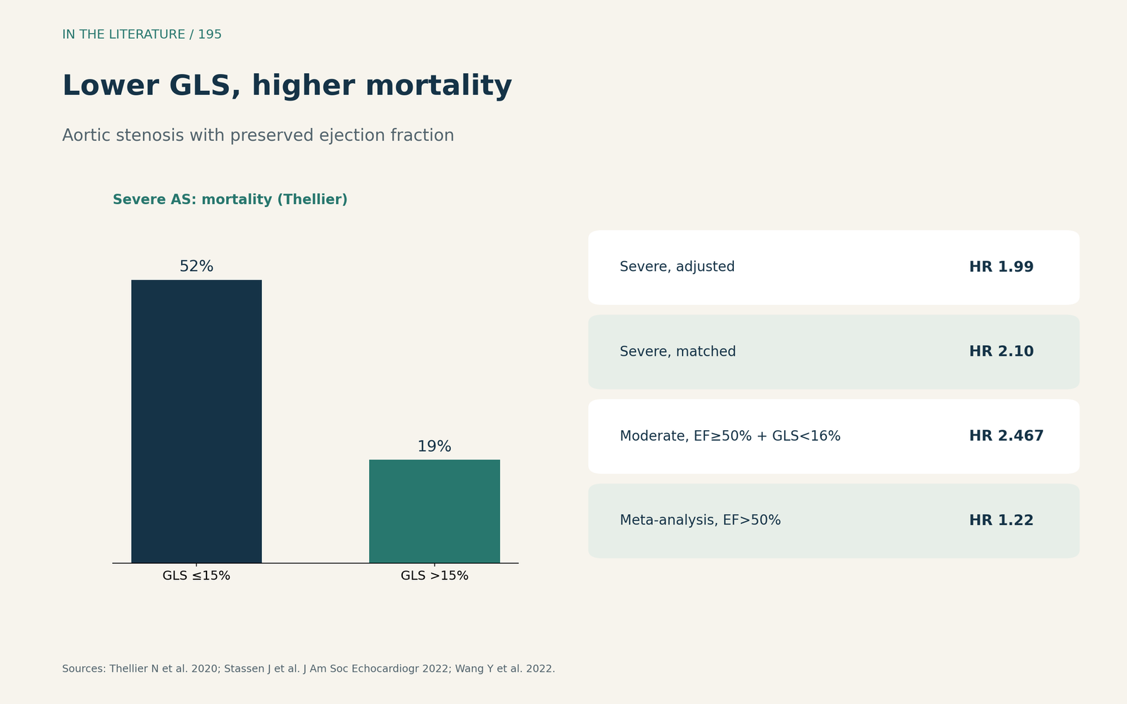
Figure 2. Low GLS, higher mortality, same EF. Redrawn from Thellier et al., Stassen et al. and Wang et al.[1], [3], [2] View larger.

Beyond resting GLS, and the limits
In low-flow, low-gradient AS with reduced EF, Kim and colleagues found resting LVEF of 31.5% versus 31.3% in survivors and nonsurvivors, while the change in GLS during dobutamine stress predicted survival with an AUC of 0.869.[6] Ye and colleagues found that strain detected abnormalities not captured by conventional cardiac damage staging.[7] The limits are real. GLS is vendor dependent and values cannot be compared across platforms.[3] In Tan's well-compensated cohort with a mean LVEF of 64.6%, LV GLS did not predict outcome, whereas left atrial strain did.[4] All of Wang's included studies were observational, and after trim-and-fill for publication bias the MACE HR fell to 1.16.[2]
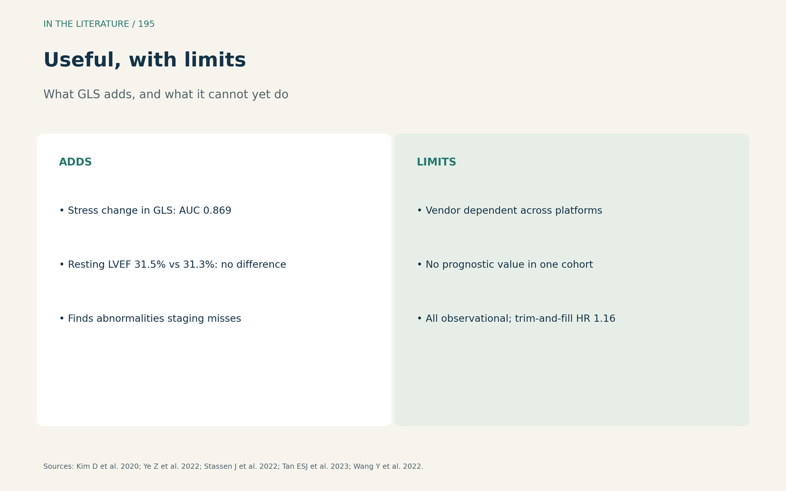
Figure 3. Useful, but not yet a trigger for surgery. Synthesized from Kim et al., Ye et al., Stassen et al., Tan et al. and Wang et al.[6], [7], [3], [4], [2] View larger.

Three takeaways
- A preserved EF does not exclude myocardial damage in aortic stenosis; low GLS often reveals it.[1], [3]
- Impaired GLS is linked to higher mortality, about double in single cohorts, and the link persists with EF above 50%.[1], [2]
- Treat GLS as a risk marker, not a stand-alone indication: values are vendor dependent and evidence is observational.[3], [2]
References
- Thellier N, Altes A, Appert L, et al. Prognostic importance of left ventricular global longitudinal strain in patients with severe aortic stenosis and preserved ejection fraction. J Am Soc Echocardiogr. 2020;33(12):1454–1464. doi:10.1016/j.echo.2020.07.002
- Wang Y, Zhang M, Chen H, Li H. Prognostic value of global longitudinal strain in asymptomatic aortic stenosis: a systematic review and meta-analysis. Front Cardiovasc Med. 2022;9:778027. doi:10.3389/fcvm.2022.778027
- Stassen J, Pio SM, Ewe SH, et al. Left ventricular global longitudinal strain in patients with moderate aortic stenosis. J Am Soc Echocardiogr. 2022;35(8):791–800.e4. doi:10.1016/j.echo.2022.03.008
- Tan ESJ, Jin X, Oon YY, et al. Prognostic value of left atrial strain in aortic stenosis: a competing risk analysis. J Am Soc Echocardiogr. 2023;36(1):29–37.e5. doi:10.1016/j.echo.2022.10.011
- Meredith T, Roy D, Hayward C, et al. Strain assessment in aortic stenosis: pathophysiology and clinical utility. J Am Soc Echocardiogr. 2024;37(1):64–76. doi:10.1016/j.echo.2023.10.001
- Kim D, Hong GR, Ha JW, Shim CY. Association of left ventricular global longitudinal strain during dobutamine stress echocardiography with adverse events in patients with severe low-flow, low-gradient aortic stenosis. J Am Soc Echocardiogr. 2020;33(11):1424–1425. doi:10.1016/j.echo.2020.03.022
- Ye Z, Yang LT, Medina-Inojosa JR, et al. Multichamber strain characterization is a robust prognosticator for both bicuspid and tricuspid aortic stenosis. J Am Soc Echocardiogr. 2022;35(9):956–965. doi:10.1016/j.echo.2022.05.010