ITL-77ad8a00ee8f
Juan Li, MD · Updated November 17, 2026
Watch the video
Key points
- Relative apical sparing of longitudinal strain is a useful red flag for cardiac amyloidosis, not a diagnostic test. Its accuracy depends heavily on who is being tested.[1], [2]
- In an unselected clinical cohort, an apical-sparing ratio of 1 or more had a sensitivity of 66% and a specificity of 59%. That is far below the figures from the original preselected comparison.[1]
- Specificity falls in chronic kidney disease, and other causes of hypertrophy can mimic the pattern. A recent meta-analysis found very large heterogeneity across studies.[2], [3]
- The ratio is not interchangeable across software. In one comparison of two vendors, the same threshold discriminated well with one package and barely better than chance with the other.[4]
- Absence of apical sparing does not exclude amyloidosis. Positive or negative, the pattern should lead to confirmation with nuclear imaging, cardiac magnetic resonance, monoclonal protein testing or biopsy, not to a diagnosis.[1], [5]
The left ventricle is thick, the ejection fraction looks preserved, and the strain bullseye shows a red apex surrounded by pale basal and mid segments, the familiar "cherry on top." For an anesthesiologist reviewing a preoperative echocardiogram, or acquiring strain on intraoperative TEE, the pattern raises the question of cardiac amyloidosis. How much should this one finding change your suspicion, and what can make it misleading?[1]
Where the high accuracy came from
The apical-sparing ratio is calculated as average apical longitudinal strain divided by the sum of the average basal and mid strain; a value of 1 or more is usually called positive. Kyrouac and colleagues summarize the original report as showing 93% sensitivity and 82% specificity. That study compared patients with known amyloidosis against a small, preselected group with hypertrophic cardiomyopathy and aortic stenosis.[1]
A test that looks excellent when known disease is compared with a few chosen controls often performs less well in the patients for whom it is actually used. These are people with unexplained hypertrophy, heart failure or comorbidities that themselves alter regional strain.
Real-world performance is modest
Kyrouac and colleagues identified every transthoracic echocardiogram at their center over almost 4 years that raised concern for amyloidosis. Strain was obtained routinely on all studies. Of 100 patients who completed a full cardiac evaluation, with amyloidosis confirmed by endomyocardial biopsy or diagnostic imaging, an apical-sparing ratio greater than 1.00 had a sensitivity of 66% and a specificity of 59% (area under the curve 0.62). The ejection fraction to global longitudinal strain ratio (EF/GLS) greater than 4.10 was more sensitive (93%) but less specific (38%).[1]
In the biopsy subgroup, 30% of patients with amyloidosis did not have a ratio of 1 or more, whereas 33% of patients without amyloidosis did. The authors suggest that apical sparing may reflect a broader cardiomyopathic process, of which amyloidosis is one important cause. The study was retrospective, single-center and limited to patients in whom the echocardiographer had already raised concern.[1]
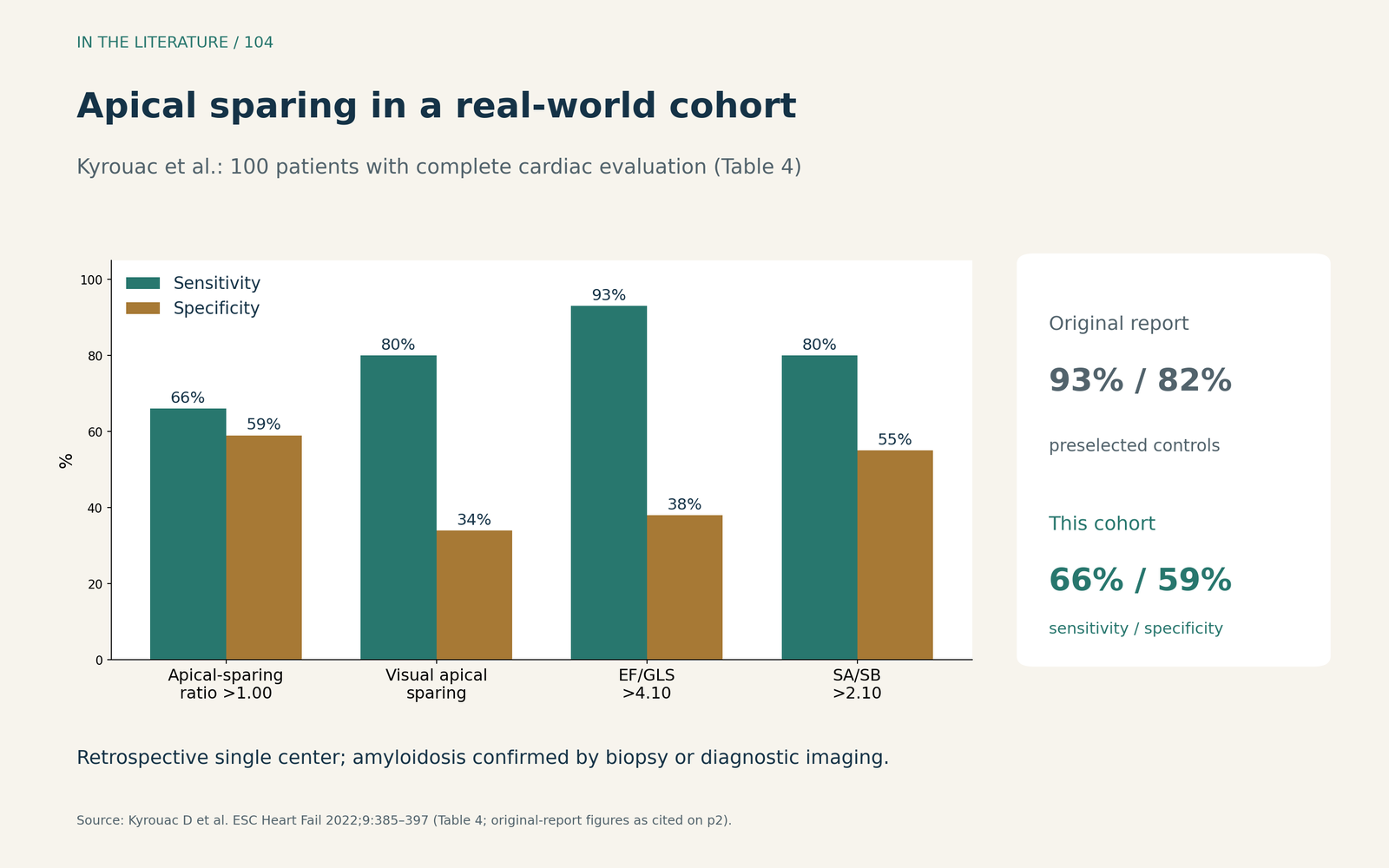
Figure 1. Apical sparing in a real-world cohort. Redrawn from Table 4 of Kyrouac et al. (n = 100 patients with complete cardiac evaluation; amyloidosis confirmed by biopsy or diagnostic imaging). Retrospective single-center data; the figure legend of the source gives 55% specificity for the ratio threshold, whereas Table 4 gives 59%.[1] View larger.

What lowers specificity
Kidney disease. Singh and colleagues studied 63 inpatients referred for technetium pyrophosphate scintigraphy, 24 of whom had cardiac amyloidosis. Specificity of apical sparing was 70% in patients with chronic kidney disease, compared with 94% in those without it. A negative pattern retained a high negative predictive value (94%) even in kidney disease. The study was small and from one center.[3]
Other hypertrophic and cardiomyopathic phenotypes. Sonaglioni and colleagues pooled studies of relative apical sparing across cardiac diseases using echocardiographic and cardiac magnetic resonance strain. Amyloidosis was associated with higher apical sparing than non-amyloid conditions overall, but heterogeneity was very high (I² 96.9%). Effects were smaller in comparisons with hypertrophic cardiomyopathy and advanced cardiomyopathy. Apical sparing was also reported in aortic stenosis and end-stage renal disease. The authors caution that pooled thresholds should not be read as universal diagnostic cut-offs.[2]
Different cohorts, different numbers. In a specialist cohort of 89 patients with amyloidosis compared with 209 with hypertrophic cardiomyopathy, aortic stenosis or hypertensive hypertrophy, Martel and colleagues reported an area under the curve of 0.91 for apical sparing. That is much higher than the 0.62 seen in the unselected cohort. They proposed an ejection fraction to basal strain ratio as an alternative. The contrast illustrates how strongly the comparison group shapes apparent accuracy.[1], [6]
The software and the operator matter
Jiang and colleagues analyzed the same studies of 48 patients with amyloidosis and 52 with other causes of hypertrophy using two strain software packages. Mean apical strain differed systematically between vendors. At the same ratio threshold, discrimination was 0.74 with one package and 0.52 with the other, mainly because sensitivity fell. Visual assessment of the bullseye also differed. The authors concluded that a threshold derived on one software system cannot be assumed to perform the same on another.[4]
For intraoperative use, this adds caution. No study in this folder or our search validated apical-sparing ratios on intraoperative TEE, under general anesthesia or with the loading changes of surgery. Any pattern seen in the operating room should be treated as a prompt for later formal evaluation rather than a measured diagnostic result. This is our inference, not a tested finding.
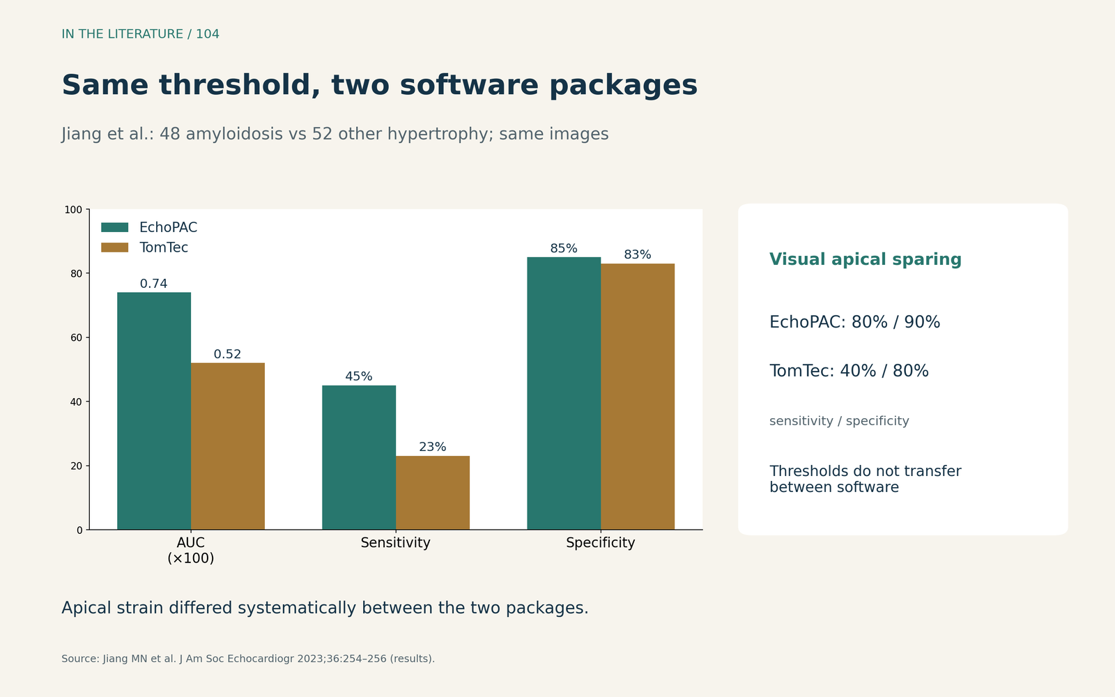
Figure 2. Intervendor variability. Redrawn from the results of Jiang et al. (48 patients with amyloidosis, 52 with other causes of hypertrophy; same images analyzed with both packages).[4] View larger.

Why the apex is spared, and why sometimes it is not
The traditional explanation is a base-to-apex gradient of amyloid deposition. De Gaspari and colleagues examined 27 autopsy hearts with amyloidosis. Overall, amyloid burden was greater in basal and mid segments than at the apex, but the gradient was present in ATTR rather than AL hearts. Among 8 patients with apical sparing on echocardiography, only 2 had a histological base-to-apex gradient. Segmental amyloid burden correlated with segmental strain, but other mechanisms probably contribute.[5]
Clinically, this means apical sparing can appear without the expected deposition pattern and can be absent despite amyloidosis. Both a positive and a negative pattern need interpretation in the context of clinical red flags and confirmatory testing. Kyrouac and colleagues describe that confirmation as cardiac magnetic resonance, technetium pyrophosphate scintigraphy and/or endomyocardial biopsy, together with assessment for monoclonal gammopathy.[1], [5]
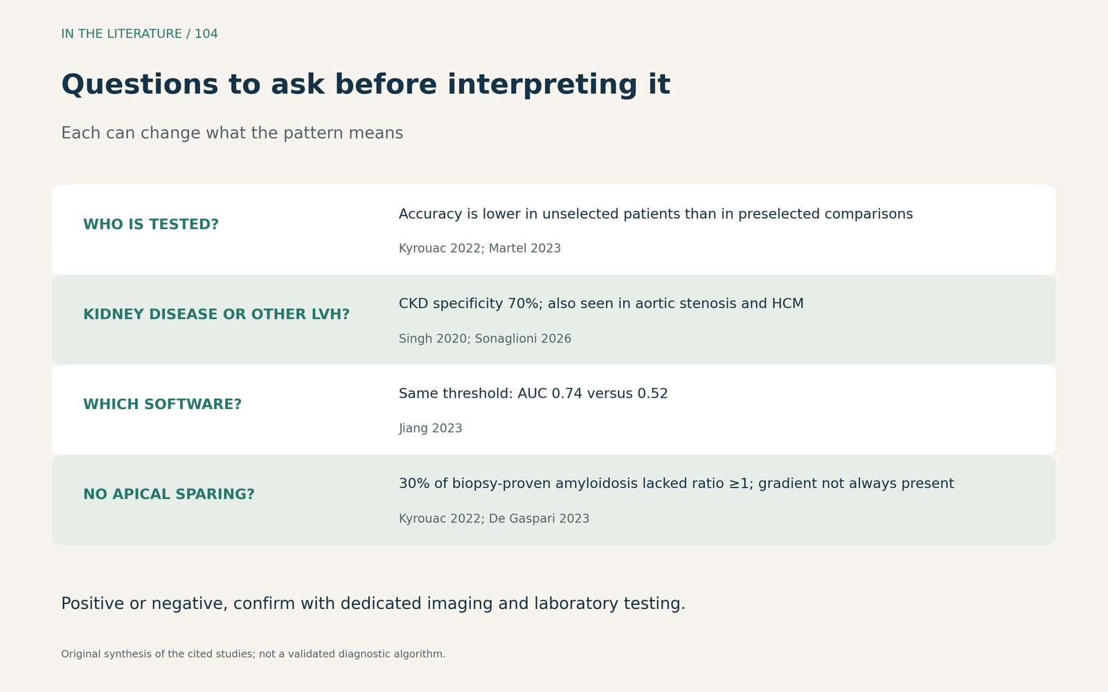
Figure 3. Interpreting the pattern. Original synthesis of the cohort, mechanistic and software studies; not a validated diagnostic algorithm.[1]–[5] View larger.

Three takeaways
- Treat apical sparing as a red flag. In an unselected cohort its sensitivity and specificity were both modest; it should trigger evaluation, not establish the diagnosis.[1]
- Context changes its meaning. Kidney disease, other hypertrophic phenotypes and the comparison population all lower or inflate apparent accuracy.[2], [3], [6]
- Do not transfer thresholds blindly. Ratios differ between software packages, and none has been validated on intraoperative TEE; confirm suspicion with dedicated imaging and laboratory testing.[1], [4]
References
- Kyrouac D, Schiffer W, Lennep B, et al. Echocardiographic and clinical predictors of cardiac amyloidosis: limitations of apical sparing. ESC Heart Fail. 2022;9(1):385–397. doi:10.1002/ehf2.13738
- Sonaglioni A, Gramaglia GF, Nicolosi GL, et al. Diagnostic performance of relative apical sparing across cardiac diseases: a multimodality systematic review and meta-analysis. J Clin Med. 2026;15(5):1685. doi:10.3390/jcm15051685
- Singh V, Soman P, Malhotra S. Reduced diagnostic accuracy of apical-sparing strain abnormality for cardiac amyloidosis in patients with chronic kidney disease. J Am Soc Echocardiogr. 2020;33(7):913–916. doi:10.1016/j.echo.2020.03.012
- Jiang MN, Simkowski J, El Hangouche N, et al. Relative apical sparing of longitudinal strain in cardiac amyloidosis: an intervendor software variability assessment. J Am Soc Echocardiogr. 2023;36(2):254–256. doi:10.1016/j.echo.2022.09.011
- De Gaspari M, Sinigiani G, De Michieli L, et al. Relative apical sparing in cardiac amyloidosis is not always explained by an amyloid gradient. Eur Heart J Cardiovasc Imaging. 2023;24(9):1258–1268. doi:10.1093/ehjci/jead107
- Martel H, Rique A, Piazzai C, et al. Ejection fraction basal strain ratio: a new composite echocardiographic deformation parameter allowing differentiation of cardiac amyloidosis from hypertrophic cardiomyopathies. J Am Soc Echocardiogr. 2023;36(5):555–557. doi:10.1016/j.echo.2022.12.021