ITL-234c05a2b5b8
Juan Li, MD · Updated November 18, 2026
Key points
- The basics are still missed. ARDS recognition ranges from 51.3% to 78.5%, and mortality has stayed at 30% to 40%.[1]
- Advanced tools change decisions. After lung ultrasound, management changed in 47% of ventilated patients.[2]
- Calibrate or mislead. Uncalibrated oesophageal pressure was potentially harmful in 30 to 50% of cases.[3]
- Know EIT's limits. It does not show consolidations, effusions or large bullae.[4]
- Physiology, not survival. EIT-guided PEEP improved oxygenation but not driving pressure or overall survival.[5], [6]
A patient with ARDS has acceptable saturation and a plateau pressure within target, yet is breathing hard against the ventilator. What else should be measured at the bedside?
Why oxygenation and pressure are not enough
ARDS mortality has remained at 30% to 40% over the last ten years, recognition ranges from 51.3% to 78.5% so lung-protective ventilation is often not applied, and oesophageal manometry remains heavily institution-dependent (Welker et al., J Cardiothorac Vasc Anesth 2022).[1] Advanced respiratory monitoring uses several minimally or noninvasive bedside tools: an unmatched ventilation-to-perfusion ratio on EIT was an independent risk factor for mortality, one trial found better survival with EIT-guided PEEP (69% vs 50%), and after lung ultrasound management changed in 47% of ventilated patients, although robust evidence for or against EIT-guided ventilation is lacking (Cammarota et al., Anesthesiology 2023).[2]
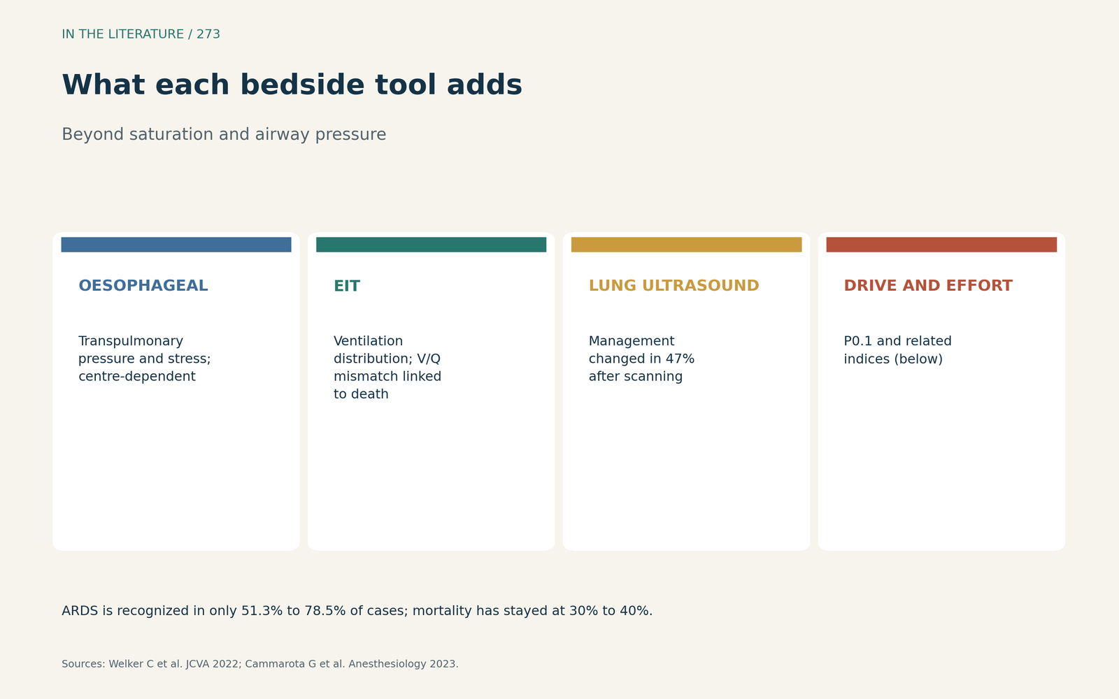
Figure 1. Each tool answers a different question at the bedside. Synthesized from Welker et al. and Cammarota et al.[1], [2] View larger.

Oesophageal pressure and EIT: how to measure well
Without calibration, the oesophageal balloon validation test was passed in only 13 of 30 cases (43.3%), uncalibrated pressure was potentially harmful in 30 to 50% of cases, and calibration is recommended whenever a substantial change occurs (Cammarota et al., Anesthesiology 2020).[3] For EIT, belts placed below the sixth intercostal space let the diaphragm interfere, consolidations, pleural effusions and large bullae are not visualized, and readings should not be relied on during significant ongoing changes in fluid balance (Ribeiro De Santis Santiago et al., Anesthesiology 2023).[4]
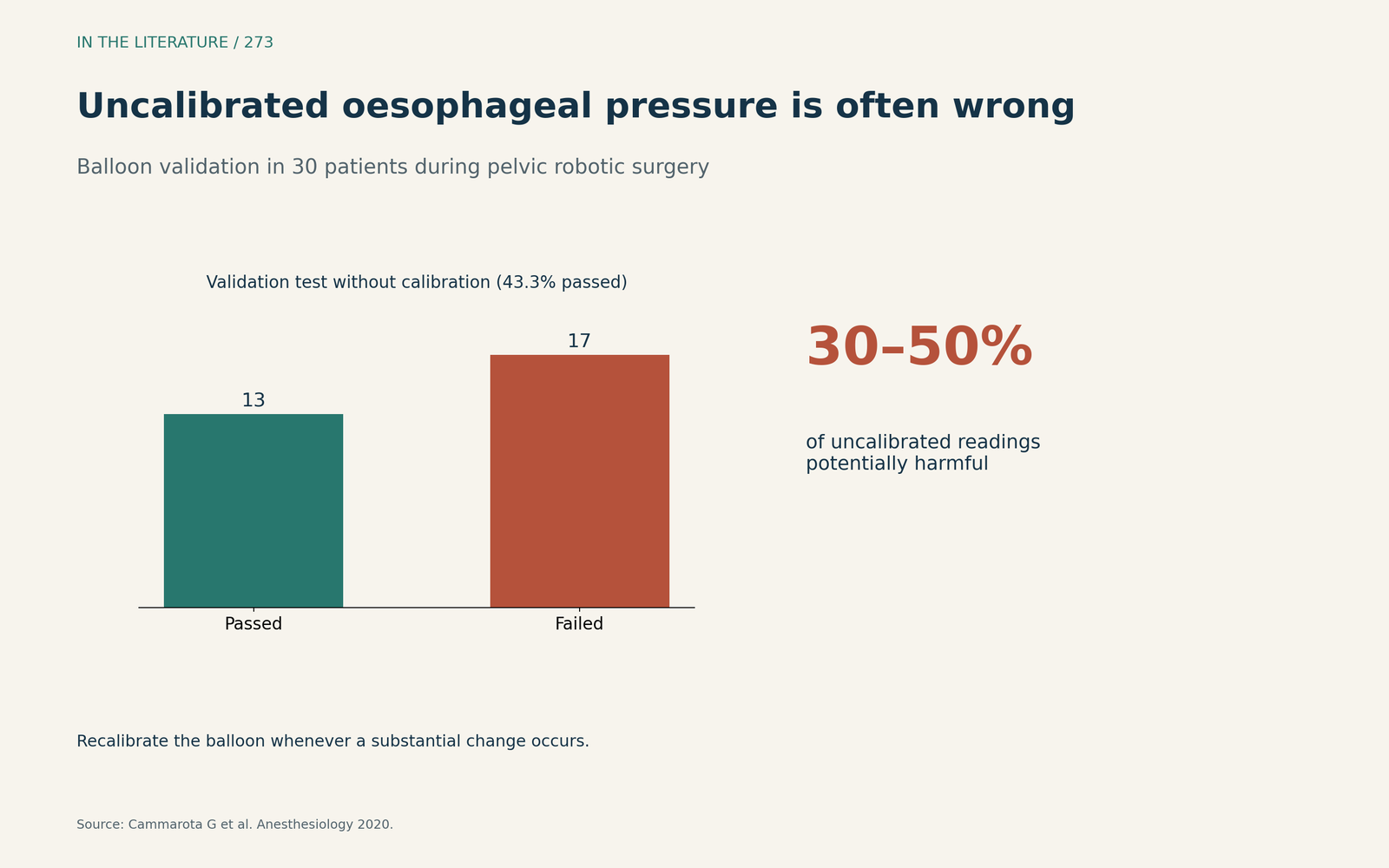
Figure 2. Most uncalibrated balloon readings failed validation. Data from Cammarota et al.[3] View larger.

What monitoring-guided ventilation achieves
A meta-analysis of 9 studies with 356 adults with ARDS (Rauseo et al., Ann Intensive Care 2026) found that EIT-guided PEEP improved the PaO2/FiO2 ratio, with no significant difference in driving pressure, and all non-randomized studies were at serious risk of bias.[5] Excessive respiratory effort was independently associated with higher ICU mortality, the EIT-guided approach did not improve overall survival, a P0.1 between 1.0 and 4.0 cm H2O is generally considered normal, and EIT assesses only a single transverse slice of the lung (Cornejo et al., J Clin Med 2026).[6]
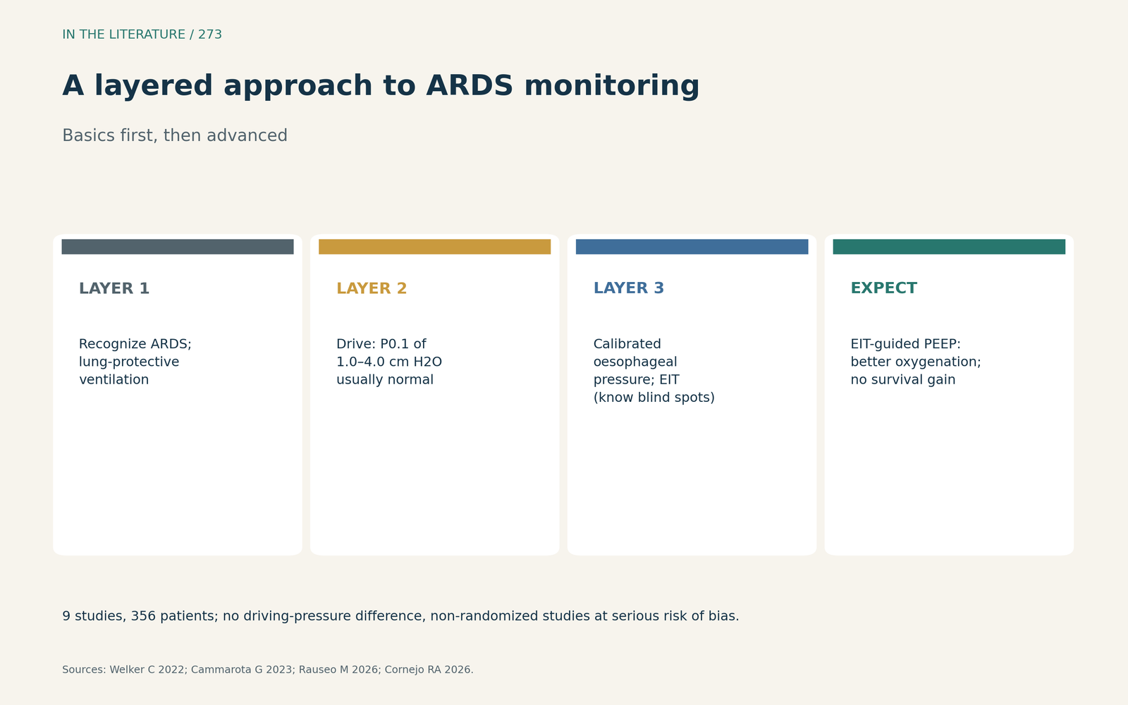
Figure 3. Layer advanced measures on top of the basics. Synthesized from Welker et al., Cammarota et al., Rauseo et al. and Cornejo et al.[1], [2], [5], [6] View larger.

Three takeaways
- First get the basics right: recognize ARDS and apply lung-protective ventilation.[1]
- Add effort and stress measures such as P0.1 and oesophageal pressure, but calibrate the balloon and know EIT's blind spots.[3], [4], [6]
- Expect better physiology, not proven survival: EIT-guided PEEP improves oxygenation only.[2], [5], [6]
References
- Welker C, Huang J, Núñez Gil IJ, et al. 2021 acute respiratory distress syndrome update, with coronavirus disease 2019 focus. J Cardiothorac Vasc Anesth. 2022;36(4):1188–1195. doi:10.1053/j.jvca.2021.02.053
- Cammarota G, Simonte R, Longhini F, et al. Advanced point-of-care bedside monitoring for acute respiratory failure. Anesthesiology. 2023;138(3):317–334. doi:10.1097/ALN.0000000000004480
- Cammarota G, Lauro G, Santangelo E, et al. Mechanical ventilation guided by uncalibrated esophageal pressure may be potentially harmful. Anesthesiology. 2020;133(1):145–153. doi:10.1097/ALN.0000000000003327
- Ribeiro De Santis Santiago R, Xin Y, Gaulton TG, et al. Lung imaging acquisition with electrical impedance tomography: tackling common pitfalls. Anesthesiology. 2023;139(3):329–341. doi:10.1097/ALN.0000000000004613
- Rauseo M, Azzolina D, Scaramuzzo G, et al. Personalized positive-end expiratory pressure using electrical impedance tomography in ARDS patients: a systematic review and meta-analysis. Ann Intensive Care. 2026;16:100049. doi:10.1016/j.aicoj.2026.100049
- Cornejo RA, Morais CCA, Arellano DH, et al. Protective ventilation during controlled and partial ventilatory support in ARDS: clinical-physiological background and monitoring. J Clin Med. 2026;15(5):1830. doi:10.3390/jcm15051830