
ITL-a9605f978ecd
Watch the video

Key points
- Training can thicken the left ventricular wall to 13–15 mm, and a wall thicker than 15 mm should raise suspicion of disease. Hypertrophic cardiomyopathy (HCM) is diagnosed at 15 mm or more, or 13 mm or more with a family history or a disease-causing variant, so the two overlap.[1], [2]
- Echocardiography looks at the pattern, not just the number. Athlete's heart shows balanced dilation of all four chambers and uniform thickening; a global longitudinal strain less negative than −15% is a red flag. Normal diastolic function does not exclude HCM.[1], [2]
- The ECG helps. Isolated voltage criteria for left ventricular hypertrophy are rare in HCM, whereas lateral or inferolateral T-wave inversion calls for cardiac MRI.[3], [2]
- Cardiac MRI adds tissue characterization. Late gadolinium enhancement, prolonged T1 and increased extracellular volume favor HCM, but many athletes with HCM have no enhancement.[4], [2]
- Detraining and exercise testing are imperfect tie-breakers. Detraining as a diagnostic test has never been formally tested, hypertrophy can regress in HCM too, and peak oxygen uptake alone does not reliably separate the two.[5], [2], [6]
A young competitive rower is scheduled for surgery, and the preoperative echocardiogram reports a septum of 14 mm. Is this an athlete's heart or hypertrophic cardiomyopathy, and which findings help decide?[1], [2]
Where the two overlap
The 2020 American Society of Echocardiography (ASE) recommendations for competitive athletes note that exercise-induced remodeling can produce wall thickness of 13–15 mm, typically 11–13 mm in white athletes and up to 15 mm in Black athletes, and that a wall thicker than 15 mm should raise suspicion of disease.[1] The 2022 ASE recommendations for HCM define the disease at 15 mm or more, or 13 mm or more when there is a family history or a known disease-causing variant, and add that walls thicker than 12 mm are rare in white athletes.[2]
The international recommendations for ECG interpretation in athletes describe the grey zone as a maximal wall thickness of 13 to 16 mm in men.[3] Sex matters: La Gerche and colleagues conclude that in female athletes, concentric hypertrophy or a wall thicker than 12 mm is very rare and should raise suspicion of disease.[5] In a cardiac MRI study by Czimbalmos and colleagues, 47.5% of male elite athletes but only 4.1% of female athletes had wall thickness in the grey zone.[4]
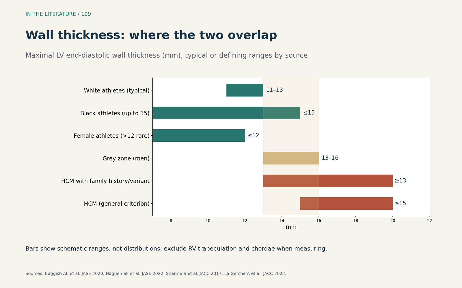
Figure 1. Where the two overlap. Schematic ranges compiled from the ASE recommendations, the international ECG recommendations and La Gerche et al.; measured walls should exclude RV trabeculation and chordae.[1], [2], [3], [5] View larger.

What echocardiography should look at
The pattern helps. The 2022 ASE document describes athlete's heart as balanced dilation of all four chambers with wall thickness typically below 15 mm, and physiological thickening as uniform, with less than 2 mm difference between adjacent segments.[2] The 2020 recommendations advise imaging the apex carefully in multiple planes to avoid missing apical HCM, and state that a global longitudinal strain less negative than −15% should raise consideration of disease.[1]
Some apparent clues are unreliable. Normal diastolic function does not exclude HCM, and normal diastolic function and exercise capacity can also be seen in athletes with HCM.[1], [2] Czimbalmos and colleagues also observed pronounced asymmetry in some healthy, highly trained athletes.[4]
What the ECG adds
The ECG often separates the two before imaging. Sharma and colleagues note that isolated voltage criteria for left ventricular hypertrophy occur alone in less than 2% of patients with HCM, and recommend cardiac MRI as a routine test when lateral or inferolateral T-wave inversion is present.[3] The ECG is not perfect: between 5% and 10% of people with HCM have a normal ECG.[2]
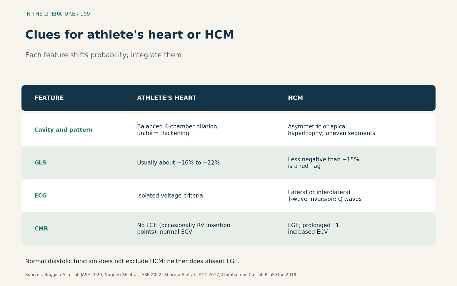
Figure 2. Clues for athlete's heart or HCM. Synthesized from the ASE recommendations, the international ECG recommendations and Czimbalmos et al.; each feature shifts probability rather than proving a diagnosis.[1], [2], [3], [4] View larger.

What cardiac MRI adds
Cardiac MRI measures wall thickness more completely and characterizes the tissue. In athlete's heart, late gadolinium enhancement is absent except occasionally at the right ventricular insertion points, and extracellular volume is normal; prolonged T1 and increased extracellular volume favor HCM.[2] A negative scan does not settle the question. In the study by Czimbalmos and colleagues, 75% of non-athletes with HCM but only 40% of athletes with HCM showed late enhancement, and the authors stress that absent fibrosis does not rule out the disease.[4]
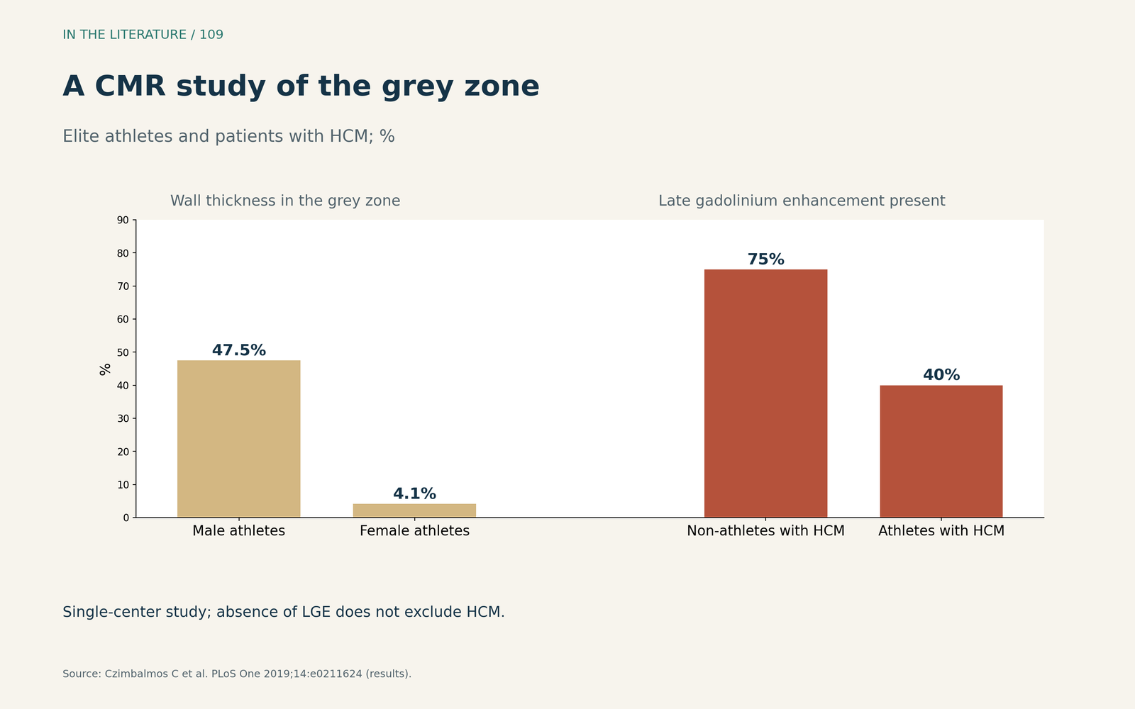
Figure 3. A cardiac MRI study of the grey zone. Redrawn from the results of Czimbalmos et al. (single center).[4] View larger.

Detraining and exercise testing
Detraining is often proposed: physiological remodeling should regress, whereas HCM should persist. La Gerche and colleagues point out that this theory has never been tested, and that caution is needed if detraining is used as a diagnostic test.[5] The ASE notes limited normative data on the expected rate of regression and that regression may also occur in detrained athletes with HCM.[2] In athletes with HCM studied by Czimbalmos and colleagues, 3 months without training produced little or no regression.[4]
Cardiopulmonary exercise testing helps less than often assumed. A 2026 narrative review by Caloian and colleagues concluded that peak oxygen uptake alone was an unreliable discriminator, although ventilatory efficiency was worse in HCM than in fitness-matched athletes.[6]
Three takeaways
- Know the grey zone. About 13–16 mm in men overlaps HCM; above 15 mm, or above 12 mm in women, favors disease.[1], [3], [5]
- Combine pattern, ECG, strain and cardiac MRI. Balanced dilation and uniform thickening favor training; inferolateral T-wave inversion, abnormal strain, late enhancement and raised extracellular volume favor HCM.[1], [2], [3]
- Do not rely on one tie-breaker. Absent enhancement, normal diastolic function, regression with detraining or a high peak oxygen uptake do not exclude HCM.[2], [4], [6]
References
- Baggish AL, Battle RW, Beaver TA, et al. Recommendations on the use of multimodality cardiovascular imaging in young adult competitive athletes: a report from the American Society of Echocardiography in collaboration with the Society of Cardiovascular Computed Tomography and the Society for Cardiovascular Magnetic Resonance. J Am Soc Echocardiogr. 2020;33(5):523–549. doi:10.1016/j.echo.2020.02.009
- Nagueh SF, Phelan D, Abraham T, et al. Recommendations for multimodality cardiovascular imaging of patients with hypertrophic cardiomyopathy: an update from the American Society of Echocardiography, in collaboration with the American Society of Nuclear Cardiology, the Society for Cardiovascular Magnetic Resonance, and the Society of Cardiovascular Computed Tomography. J Am Soc Echocardiogr. 2022;35(6):533–569. doi:10.1016/j.echo.2022.03.012
- Sharma S, Drezner JA, Baggish A, et al. International recommendations for electrocardiographic interpretation in athletes. J Am Coll Cardiol. 2017;69(8):1057–1075. doi:10.1016/j.jacc.2017.01.015
- Czimbalmos C, Csecs I, Toth A, et al. The demanding grey zone: sport indices by cardiac magnetic resonance imaging differentiate hypertrophic cardiomyopathy from athlete's heart. PLoS One. 2019;14(2):e0211624. doi:10.1371/journal.pone.0211624
- La Gerche A, Wasfy MM, Brosnan MJ, et al. The athlete's heart—challenges and controversies: JACC Focus Seminar 4/4. J Am Coll Cardiol. 2022;80(14):1346–1362. doi:10.1016/j.jacc.2022.07.014
- Caloian B, Caloian CS, Tomoaia R, et al. Cardiopulmonary exercise testing in the differential diagnosis between athlete's heart and cardiac pathology: current evidence and diagnostic challenges. Diagnostics (Basel). 2026;16(16):2573. doi:10.3390/diagnostics16162573