ITL-6f2052f70763
Juan Li, MD · Updated November 18, 2026
Watch the video
Key points
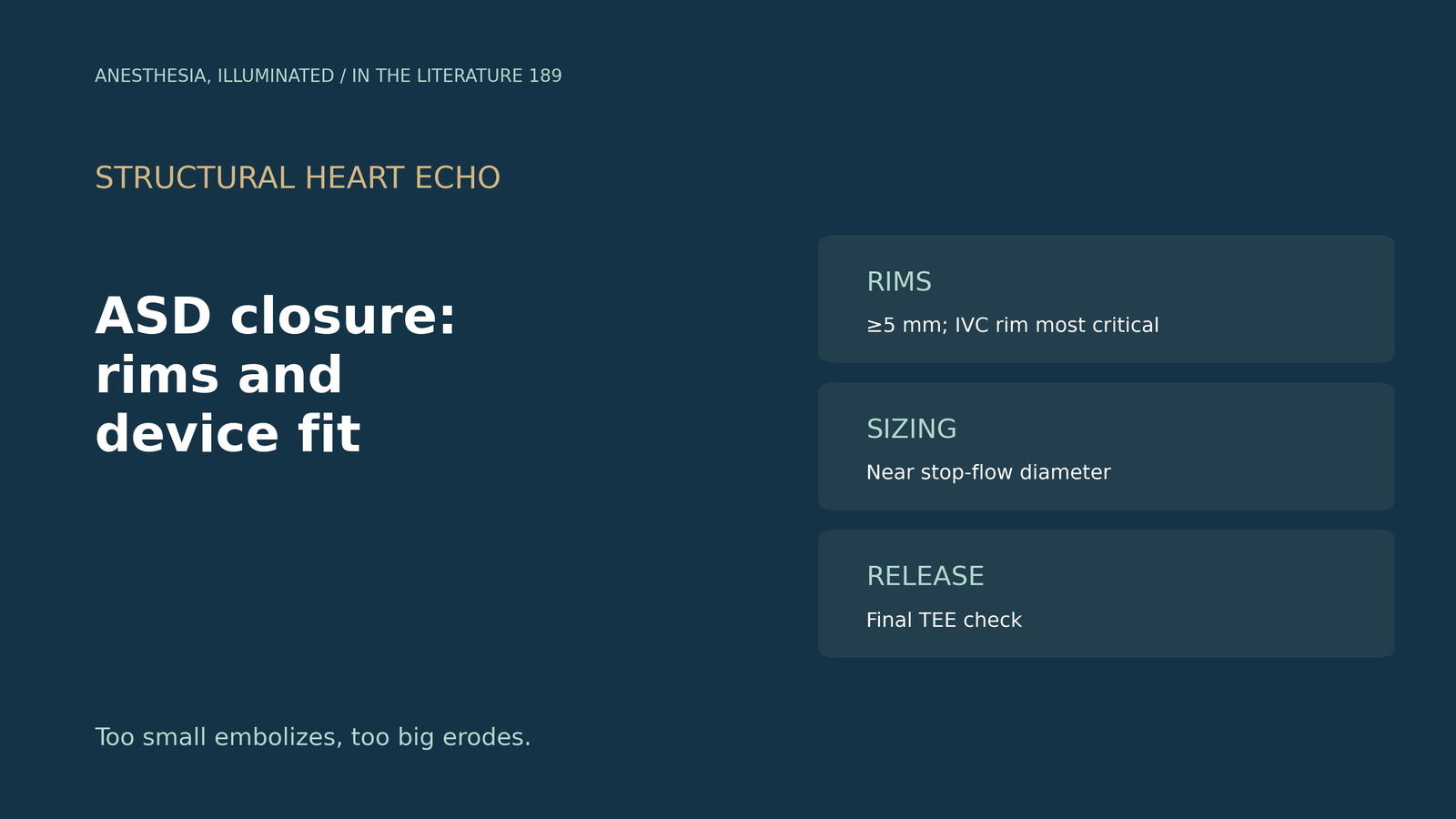
- Rims decide candidacy. Device closure requires adequate septal rims, defined as at least 5 mm long; an absent aortic rim is traditionally tolerated.[1]
- Not all rims are equal. Posteroinferior or IVC rim deficiency increases embolization risk, and erosion is associated with oversizing.[2]
- Size close to the stop-flow diameter. The device should be no more than 2 mm larger than the balloon stop-flow diameter, and oversizing should be avoided when the aortic rim is deficient.[3]
- The septum sets an upper limit. An occluder longer than the septum can impinge on the AV valve.[4]
- Check before release. With ICE guidance alone, 3 of 53 devices dropped off, and a final TEE check before release is considered indispensable.[5]
During TEE for transcatheter closure of a large secundum atrial septal defect, the aortic rim looks very short and the inferior rim is hard to see. Is the defect suitable for a device, and how should the device be sized?[1], [2]
Judging the rims
Rana notes that device closure requires adequate rims of septal tissue, defined as at least 5 mm long. Traditional teaching allows an absent aortic rim, but high-risk features include absent or limited aortic and superior vena cava rims.[1] Turner and colleagues add that selection depends on defect location and rim adequacy, that devices can close defects up to 40 mm, and that posteroinferior or IVC rim deficiency increases embolization risk to the extent that it may preclude closure.[2] Jung and Choi note that aortic rim deficiency is the most common rim deficiency and that the IVC rim is hard to see on TEE, so ICE is preferable.[3]
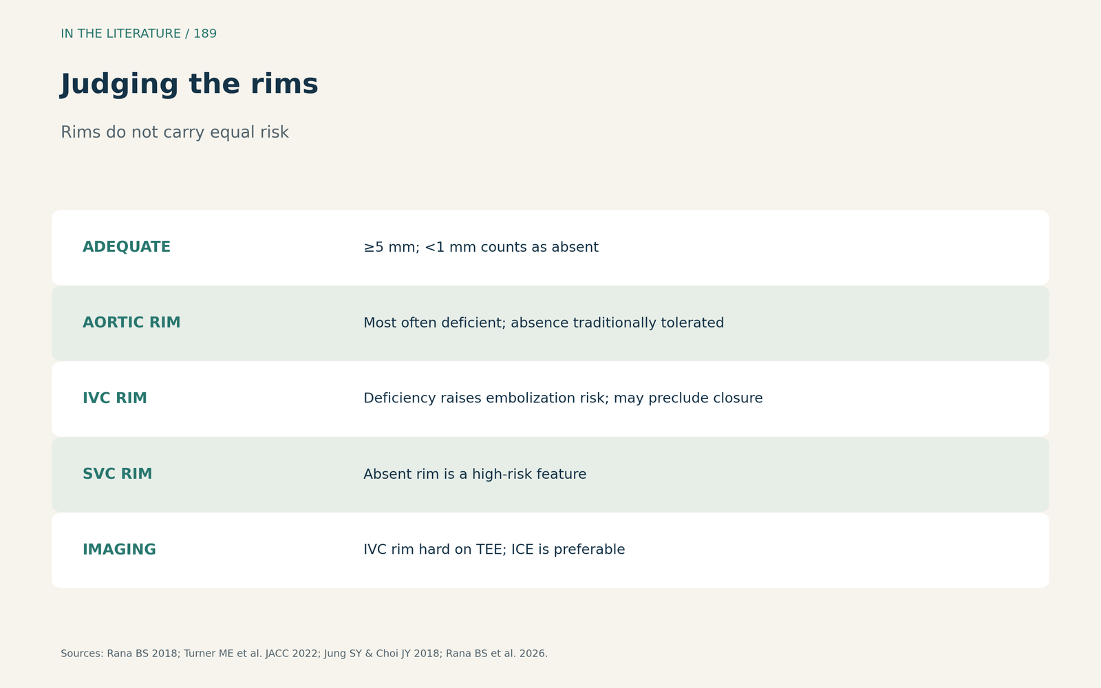
Figure 1. Not all rims carry the same risk. Synthesized from Rana, Turner et al., Jung and Choi, and Rana et al.[1], [2], [3], [6] View larger.

Sizing the device
Jung and Choi recommend a device no more than 2 mm larger than the stop-flow diameter and advise avoiding an oversized device when the aortic rim is deficient.[3] Without balloon sizing, Rana uses the measured defect size plus 20%, or plus 25% in some anatomies.[1] Turner and colleagues associate embolization with undersizing and erosion with oversizing.[2] In Huang's series, occluders were 0 to 6 mm larger than the maximum defect diameter, but an occluder longer than the septum can impinge on the AV valve.[4] A case report (McGrail et al., J Cardiothorac Vasc Anesth 2022) adds that ASD devices are sized after a balloon dilation and occlusion test, whereas balloon dilation of a PFO is avoided, so the two must be told apart first.[7]
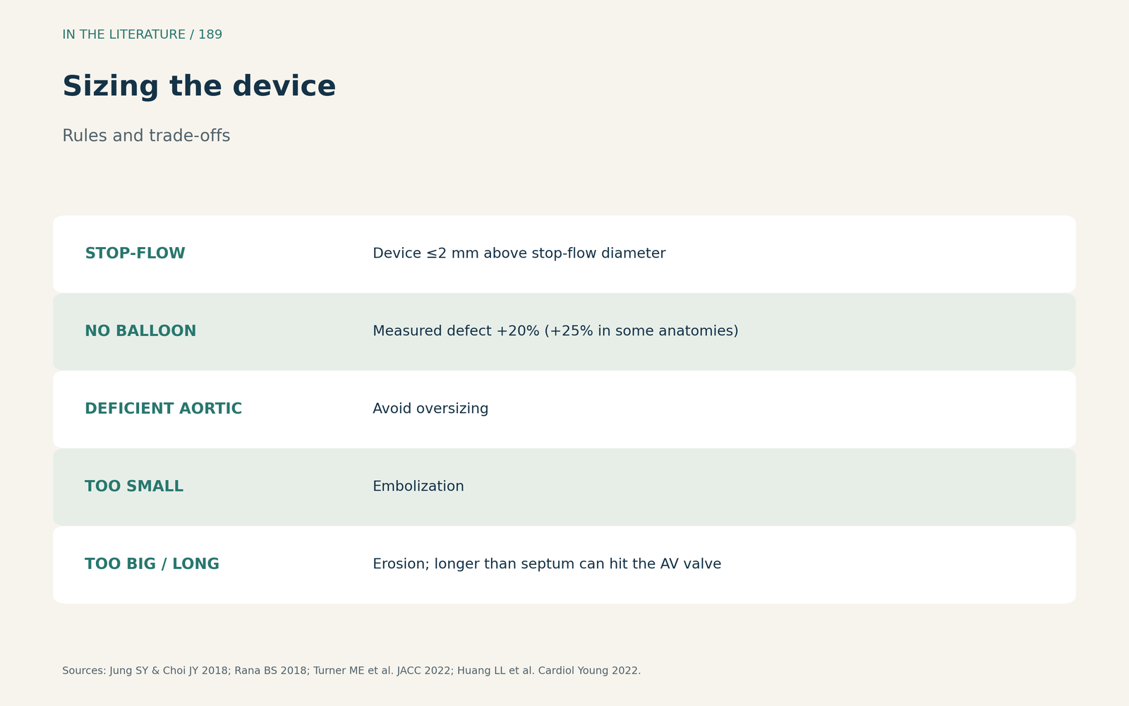
Figure 2. Too small embolizes, too big erodes. Synthesized from Jung and Choi, Rana, Turner et al. and Huang et al.[3], [1], [2], [4] View larger.

Imaging guidance and safety
Yamano and colleagues defined a hypermobile rim as excursion above 10 mm; with ICE guidance alone, 3 of 53 devices dropped off, and they consider a final TEE check before release indispensable.[5] Rana and colleagues classify rims as sufficient at 5 mm or more and absent below 1 mm, report that device erosion is rare at 0.1 to 0.3%, mostly with a deficient aortic rim and oversizing, and that good case selection keeps complications at 1% or less.[6] A case report (Mendez-Ruiz et al., CASE 2023) shows the cost of a marginal fit: a 26.8 mm defect with a 3.1 mm posterior rim and absent aortic rim was closed with a 46 mm occluder that later migrated; risk factors for migration include a large defect, rim deficiency and device under- or oversizing.[8]
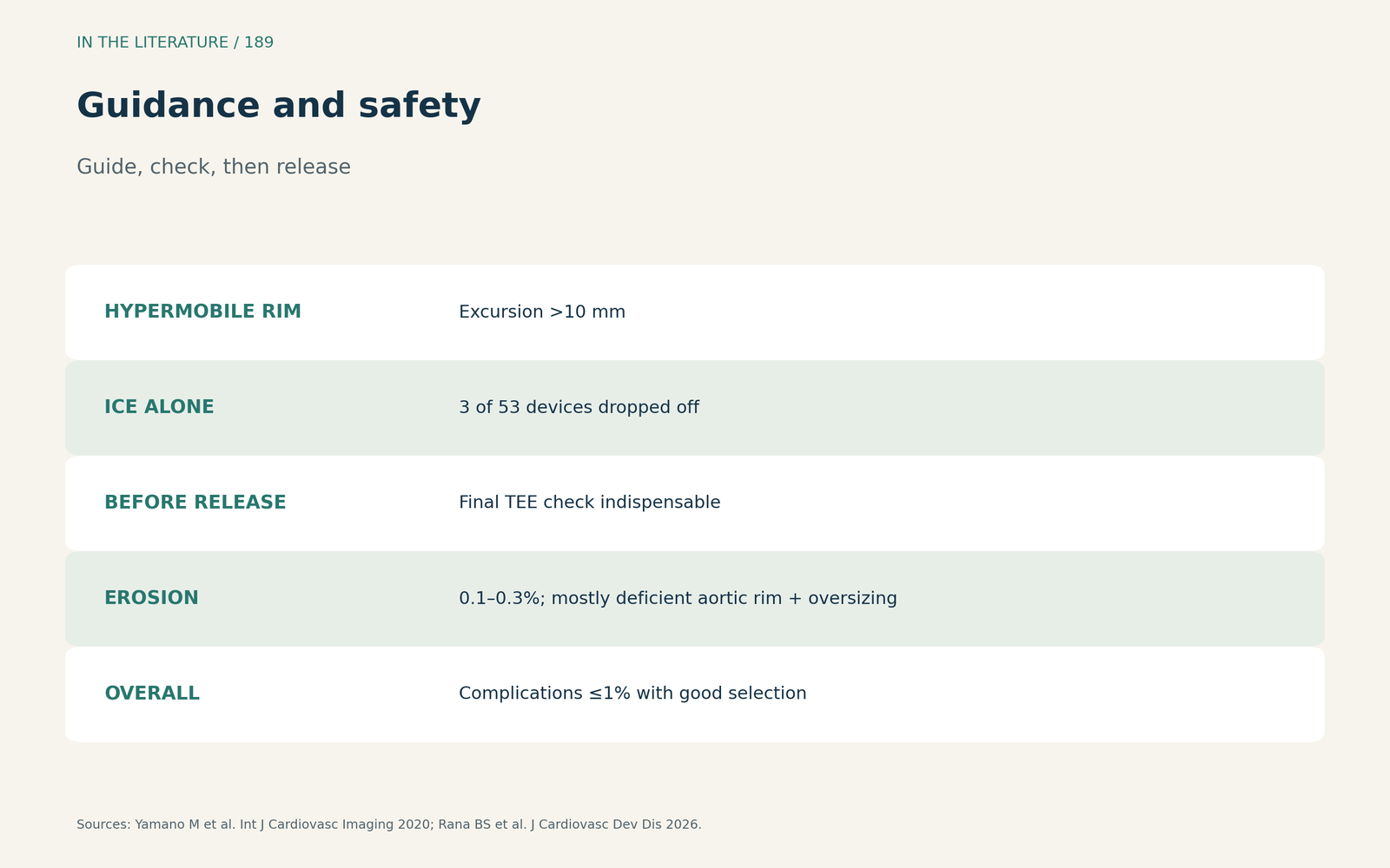
Figure 3. Guide, check, then release. Synthesized from Yamano et al. and Rana et al.[5], [6] View larger.

Three takeaways
- Measure every rim; 5 mm is the usual floor, and the IVC rim matters most for embolization.[1], [2]
- Size near the stop-flow diameter and avoid oversizing with a deficient aortic rim.[3]
- Confirm position on TEE before release.[5]
References
- Rana BS. Echocardiography guidance of atrial septal defect closure. J Thorac Dis. 2018;10(Suppl 24):S2899–S2908. doi:10.21037/jtd.2018.07.126
- Turner ME, Bouhout I, Petit CJ, Kalfa D. Transcatheter closure of atrial and ventricular septal defects: JACC focus seminar. J Am Coll Cardiol. 2022;79(22):2247–2258. doi:10.1016/j.jacc.2021.08.082
- Jung SY, Choi JY. Transcatheter closure of atrial septal defect: principles and available devices. J Thorac Dis. 2018;10(Suppl 24):S2909–S2922. doi:10.21037/jtd.2018.02.19
- Huang LL, Wu J, Chen M, et al. Transcatheter closure of atrial septal defect with deficient posterior-inferior or inferior vena cava rim under echocardiography only: a feasibility and safety analysis. Cardiol Young. 2022;32(4):589–596. doi:10.1017/S104795112100264X
- Yamano M, Yamano T, Nakamura T, et al. Appropriate selection of echocardiographic guidance for transcatheter atrial septal defect closure. Int J Cardiovasc Imaging. 2020;36(5):855–863. doi:10.1007/s10554-020-01778-9
- Rana BS, Clapp B, Malik IS, et al. The pivotal role of advanced echocardiography in transcatheter closure of challenging secundum atrial septal defect anatomies: an expert-based review. J Cardiovasc Dev Dis. 2026;13(6):261. doi:10.3390/jcdd13060261
- McGrail D, Sehgal S, Tuttle MK, et al. The many faces of the interatrial septum: a diagnostic dilemma and considerations for defect closure device selection. J Cardiothorac Vasc Anesth. 2022;36(8):3156–3162. doi:10.1053/j.jvca.2022.01.034
- Mendez-Ruiz A, Dangl M, Peñalver J, Colombo R. Migration of an atrial septal defect occluder in a pregnant patient. CASE (Phila). 2023;7(11):445–448. doi:10.1016/j.case.2023.08.002