
ITL-62450d1d10fe
Juan Li · In the Literature · Updated November 16, 2026
Watch the video

Key points
- BASILICA addresses selected leaflet-mediated mechanisms of coronary obstruction; identifying a low coronary ostium alone does not establish suitability.
- A split leaflet must move apart enough to leave a flow pathway. A transcatheter-valve skirt or commissural post can still obstruct that pathway.
- Technical success and clinical safety are different endpoints. Successful traversal and laceration do not mean freedom from stroke or coronary obstruction.
- Proper lengthwise laceration can preserve leaflet coaptation before TAVR. Severe aortic regurgitation is not inevitable, but hemodynamic deterioration still requires immediate assessment.
- Plan the coronary protection and rescue strategy before starting, and reassess coronary perfusion after implantation.
First identify what will block the artery
When CT predicts coronary obstruction during transcatheter aortic valve replacement (TAVR), the useful question is: will splitting the threatened leaflet create a usable route for coronary blood flow? BASILICA intentionally lacerates that leaflet before valve implantation so its halves can move apart when displaced by the new valve.
The 2026 working-group position statement distinguishes two principal leaflet-mediated targets: direct coverage of the coronary ostium and sinus sequestration, in which displaced leaflets prevent blood from entering the sinus at the sinotubular junction. BASILICA can generally mitigate these mechanisms when anatomy and subsequent leaflet movement permit. It does not remove a bulky calcific mass, embolic material, an obstructing valve skirt, or a commissural post. [1]
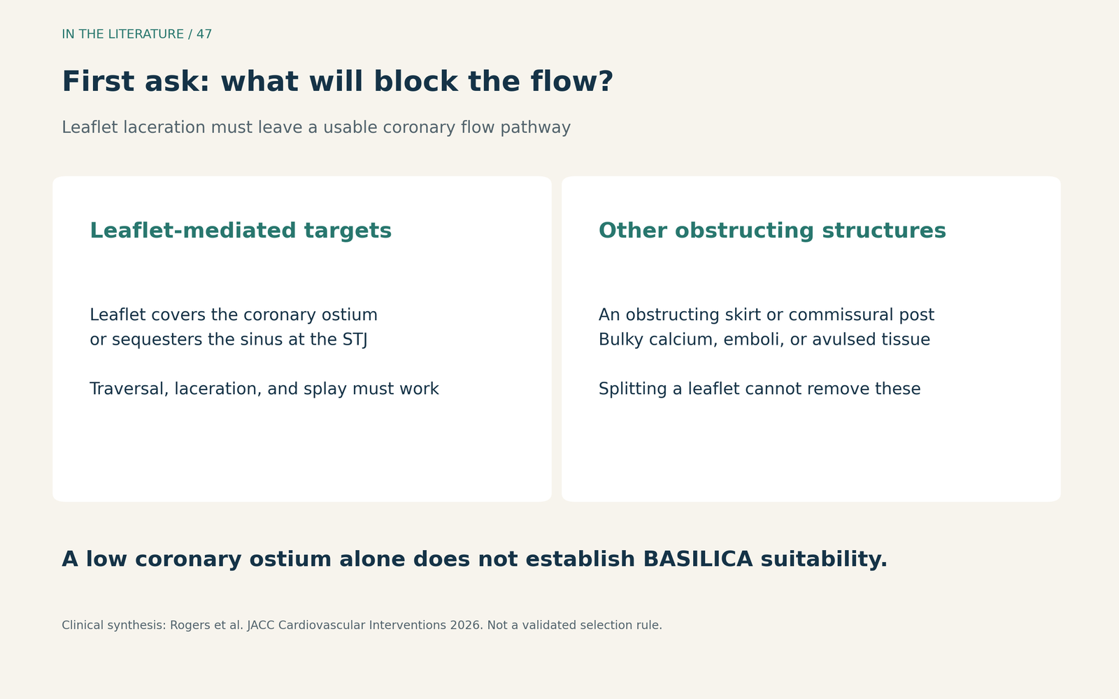
Figure 1. A mechanism-based discussion aid. Suitability depends on the planned valve and the available flow pathway, not simply on completing a cut. [1] View figure

Read the CT as a geometry problem
Review leaflet height against coronary and sinotubular-junction heights, virtual valve-to-coronary and valve-to-sinotubular-junction distances, leaflet calcium, and the planned valve position. These measurements interact; a single clearance value is insufficient. Redo-TAVR also requires assessment of the existing frame, the height of the leaflets pinned open by the new valve, and commissural alignment. [1]
The original BASILICA trial illustrates a practical constraint: its two traversal failures involved confluent calcium at the intended leaflet-base target. A large total calcium burden and the absence of a usable basal crossing point are different findings. Planning must identify both a traversable target and a laceration likely to open toward the threatened coronary artery. [2]
What the original trial actually established
Khan and colleagues’ 2019 prospective, single-arm feasibility trial enrolled 30 patients at high or prohibitive surgical risk. Patient-level procedural success occurred in 28/30 (93%); traversal and laceration succeeded in 35/37 intended leaflets (95%). The patient and leaflet denominators answer different questions. [2]
No coronary obstruction occurred, but this cannot be read as 100% protection from BASILICA alone. Coronary stents were positioned in both traversal failures, and one was deployed prophylactically after TAVR. The trial had no untreated control group. At 30 days, 21/30 patients (70%) met the VARC-2 early-safety endpoint, and 3/30 (10%) had stroke, including one disabling stroke. Those outcomes establish the need to separate procedural feasibility from clinical safety; they do not quantify a causal stroke excess or risk reduction attributable to BASILICA. [2]
Why “100% technical success” needs its definition
The 2025 TELLTALE prospective multicenter single-arm study evaluated a purpose-built electrosurgical system in 90 selected patients. Its 100% technical-success rate meant successful leaflet traversal, laceration, and system retrieval. Freedom from stroke and coronary obstruction was not part of that technical endpoint. [3]
The in-hospital safety endpoint was met in 96%, while three strokes and one coronary obstruction still occurred. The report identifies valve-commissural-post obstruction despite successful leaflet splitting. Thus, a successful cut does not settle the final coronary-perfusion assessment. [3]
TELLTALE excluded patients requiring protection of both coronary arteries. Its results should not be extended directly to double-leaflet BASILICA, and the 100% figure should not be compared with the original trial’s 93% as though the endpoints and populations were interchangeable. Neither study randomized BASILICA against another protection strategy. [2, 3]
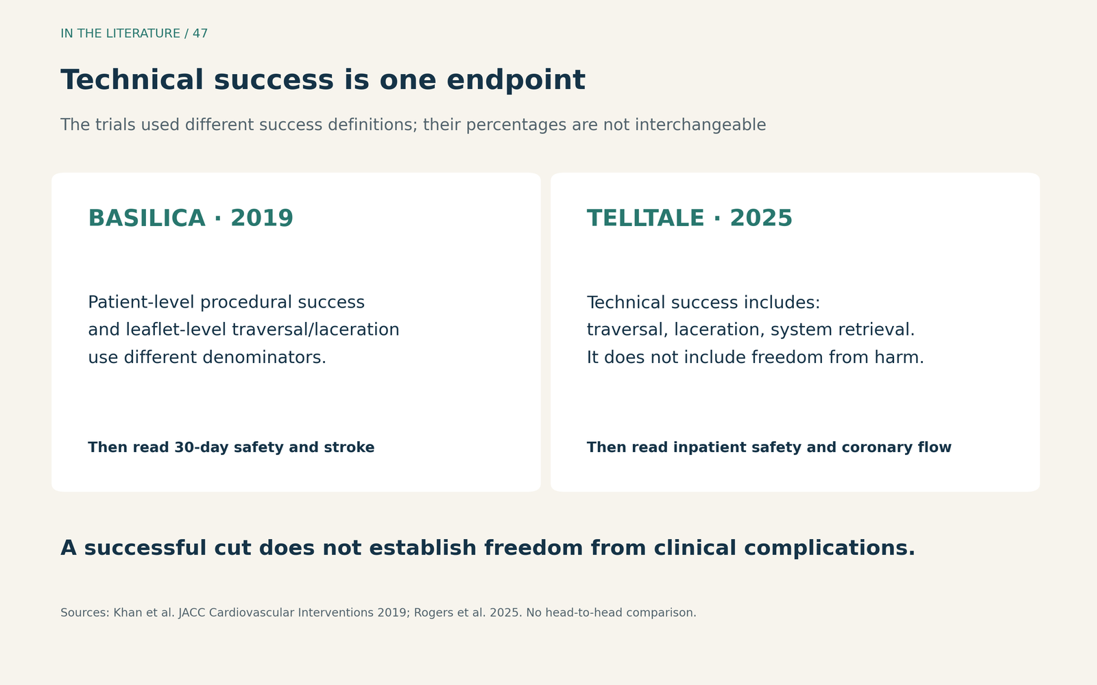
Figure 2. Read the endpoint before interpreting the success rate. The two trials used different definitions; this is an explanation of study interpretation, not a comparative outcome chart. [2, 3] View figure

What the anesthesia and imaging teams need to anticipate
The 2026 statement explains that a clean lengthwise split can retain diastolic coaptation. Do not assume every successful BASILICA produces severe acute aortic regurgitation. Leaflet avulsion is different: uncontrolled tearing can produce severe regurgitation or obstructive prolapse. Preparedness remains necessary even when the intended mechanism preserves stability. [1]
In the original trial, transient hypotension after laceration occurred in 2/30 patients (7%) and resolved promptly with TAVR. This small descriptive result supports readiness for hemodynamic support and valve implantation; it does not define a universally safe waiting interval. [2]
Protsyk and colleagues’ structured echocardiographic review describes how multiplane transesophageal echocardiography (TEE) complements fluoroscopy during alignment and traversal. After valve implantation, assess coronary perfusion and look for new left- or right-ventricular wall-motion abnormalities. Interpret imaging alongside blood pressure and the ECG; persistent concern warrants further coronary evaluation. These are imaging and procedural recommendations, not evidence that every BASILICA requires one particular anesthetic technique. [1, 4]
Before starting, the team should be able to explain the predicted obstruction mechanism, the expected route for flow after laceration, and how persistent obstruction or sudden instability will be treated. This is a practical synthesis of the cited sources. The final confirmation is adequate perfusion after the new valve is in place. [1–4]
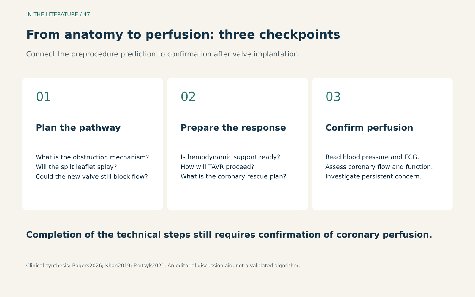
Figure 3. Three checkpoints for the team discussion. This editorial aid supports a patient-specific plan; it is not a validated selection algorithm. [1, 2, 4] View figure

Three takeaways
- Match the treatment to the obstruction mechanism. BASILICA can address displaced leaflets; a skirt, post, calcium mass, or embolus may require a different strategy.
- Separate the cut from the outcome. Technical success does not establish freedom from coronary obstruction, stroke, or other clinical complications.
- Confirm flow and preserve a rescue route. Anticipate instability, reassess perfusion after implantation, and keep the coronary response plan actionable.
References
- Rogers T, Halaby RN, Greenbaum AB, et al. Aortic Valve Leaflet Modification: A Working Group Position Statement on Best Practices and Step-by-Step Guide. JACC Cardiovasc Interv. 2026;19(11):1347–1377. doi:10.1016/j.jcin.2026.03.033.
- Khan JM, Greenbaum AB, Babaliaros VC, et al. The BASILICA Trial: Prospective Multicenter Investigation of Intentional Leaflet Laceration to Prevent TAVR Coronary Obstruction. JACC Cardiovasc Interv. 2019;12(13):1240–1252. doi:10.1016/j.jcin.2019.03.035.
- Rogers T, Khan JM, Laham R, et al. Electrosurgical Leaflet Modification to Prevent Coronary Obstruction During Transcatheter Aortic Valve Replacement in Failing Native and Bioprosthetic Valves. JACC Cardiovasc Interv. 2025;18(19):2355–2368. doi:10.1016/j.jcin.2025.07.032.
- Protsyk V, Meineri M, Kitamura M, et al. Echocardiographic Guidance of Intentional Leaflet Laceration prior to Transcatheter Aortic Valve Replacement: A Structured Approach to the Bioprosthetic or Native Aortic Scallop Intentional Laceration to Prevent Iatrogenic Coronary Artery Obstruction Procedure. J Am Soc Echocardiogr. 2021;34(6):676–689. doi:10.1016/j.echo.2021.01.015.