ITL-a631425d838d
Juan Li, MD · Updated November 17, 2026
Watch the video
Key points
- Assess the right ventricle with clinical findings, imaging and invasive hemodynamics together before durable LVAD implantation.[1], [2]
- Interpret PAPi and other RV indices in the loading and support conditions in which they were measured; one cutoff cannot capture the whole risk.[2]
- EUROMACS and STOP-RVF can organize risk assessment, but their validation populations and endpoints matter when applying an estimate to an individual patient.[3], [4]
- A model containing cardiopulmonary bypass time cannot provide purely preoperative prediction. A 2026 validation study makes this timing distinction explicit.[5]
- Persistent RV dysfunction after optimization should prompt an LVAD-team discussion of planned right-heart support; a score alone does not mandate an RVAD.[1], [3]
A patient being considered for a durable LVAD has a dilated, weak right ventricle and a low pulmonary artery pulsatility index (PAPi). The immediate clinical task is to determine how much of this picture reflects modifiable loading conditions, how much dysfunction persists despite treatment, and how those findings should change the implant plan. A calculator can contribute to that discussion, but it cannot conduct it.[1], [2]
Define the event you are trying to anticipate
“RV failure after LVAD” covers several outcomes. Failure to separate from bypass requiring immediate right-sided mechanical support differs from prolonged postoperative inotrope dependence, and both differ from late right-heart failure. The timing and severity definition should be stated when discussing a patient's risk.[2]
For example, STOP-RVF predicted a treatment-based early outcome: inotropes for more than 14 days and/or right-sided mechanical circulatory support within 30 days. Such an endpoint is clinically important, but it also reflects local support and weaning practices. An estimate for that outcome is not automatically an estimate for isolated RVAD use, mortality or late RV failure.[4]
Read the RV in its current physiologic state
The ISHLT guidance recommends integrating invasive assessment with imaging focused on RV function and tricuspid-valve integrity. At the bedside, organize the information around three related questions.[1], [2]
- What does the ventricle look like? Review RV size, systolic function and tricuspid regurgitation together. TAPSE, fractional area change or strain can contribute, but each needs image-quality and loading context.
- What load is it working against, and what flow is it producing? Read right atrial pressure, pulmonary artery pressures, wedge pressure and cardiac output together. PAPi is pulmonary artery pulse pressure divided by right atrial pressure; its components deserve attention as well as the ratio.
- How is the patient tolerating the circulation? Consider congestion, perfusion, clinical acuity, inotrope or temporary-support requirements, and renal and hepatic function.[1], [2]
Record the treatment state alongside the measurements. A study obtained during substantial inotropic or mechanical support answers a different physiologic question from an unsupported measurement. Preload, afterload and contractile support can all alter the apparent RV performance. Reassessment after optimization is therefore useful, provided the changes in treatment conditions remain visible in the interpretation.[1], [2]
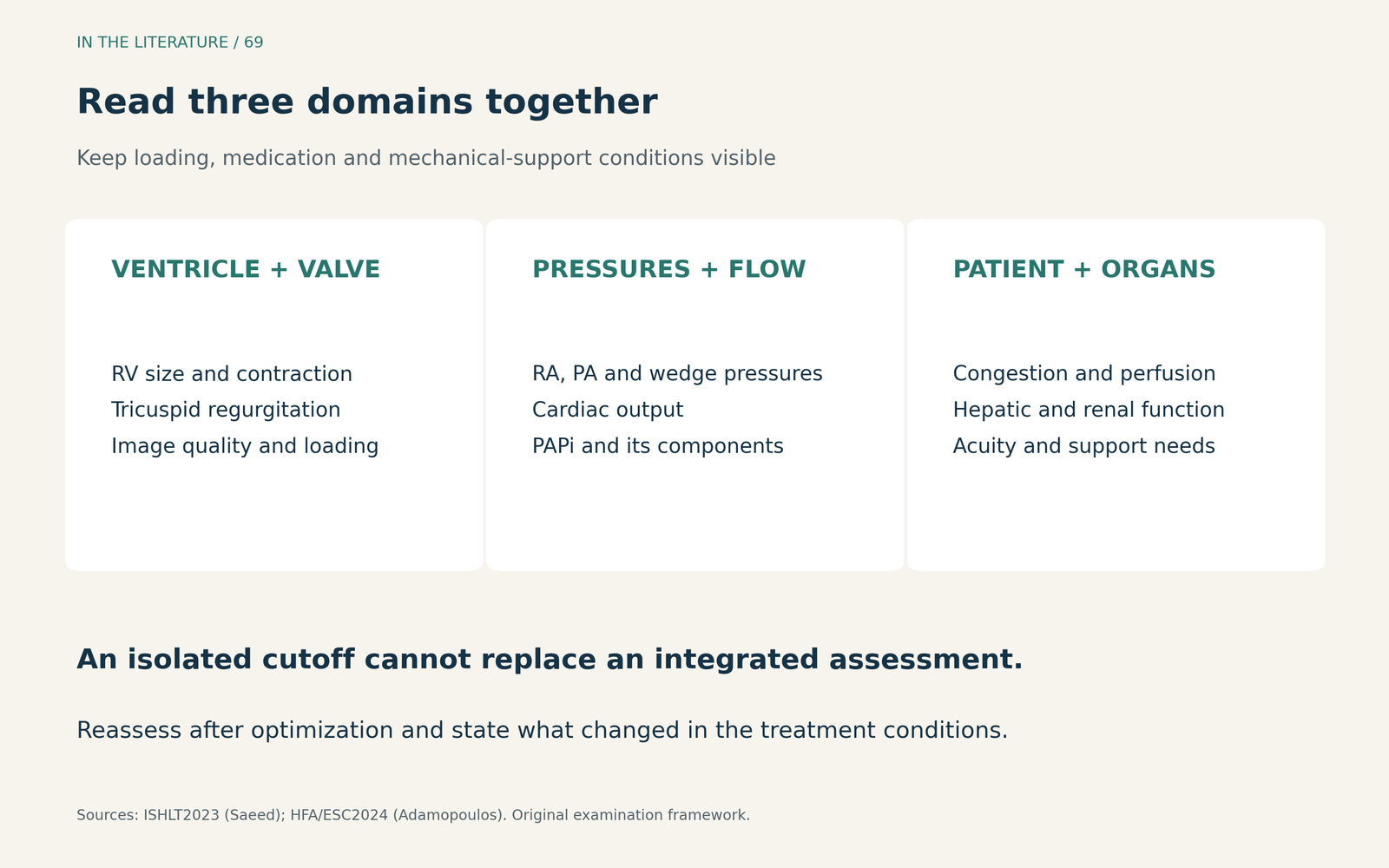
Figure 1. Combine the ventricle, the circulation and the patient. This is an examination framework, not a new prediction score.[1], [2] View larger.

Use a score with its population and validation in view
EUROMACS combines five preoperative domains: INTERMACS profile, inotropic support, severe RV dysfunction, right atrial-to-wedge pressure ratio and hemoglobin. Its original 2,988-patient registry was randomly divided into 2,000 derivation and 988 validation patients. The validation C index was 0.67. That was a holdout sample from the same registry, and only 8% of the overall cohort had HeartMate 3.[3]
STOP-RVF adds evidence from independent external validation. It was developed in 798 patients from five institutions and tested in 327 patients at a separate sixth institution. The external C statistic was 0.73 (95% CI, 0.67–0.80). This supports useful, imperfect discrimination. However, enrollment extended from 2008 to 2019 across several pump platforms; performance in today's exclusively HeartMate 3 population cannot simply be assumed.[4]
These results do not constitute a head-to-head ranking. Discrimination describes how well a model separates patients who experience the outcome from those who do not; calibration asks whether its estimated probabilities match the observed risks. Neither establishes that an intervention triggered by the score improves outcomes. The original EUROMACS investigators explicitly cautioned against using their model alone for clinical decisions.[3], [4]
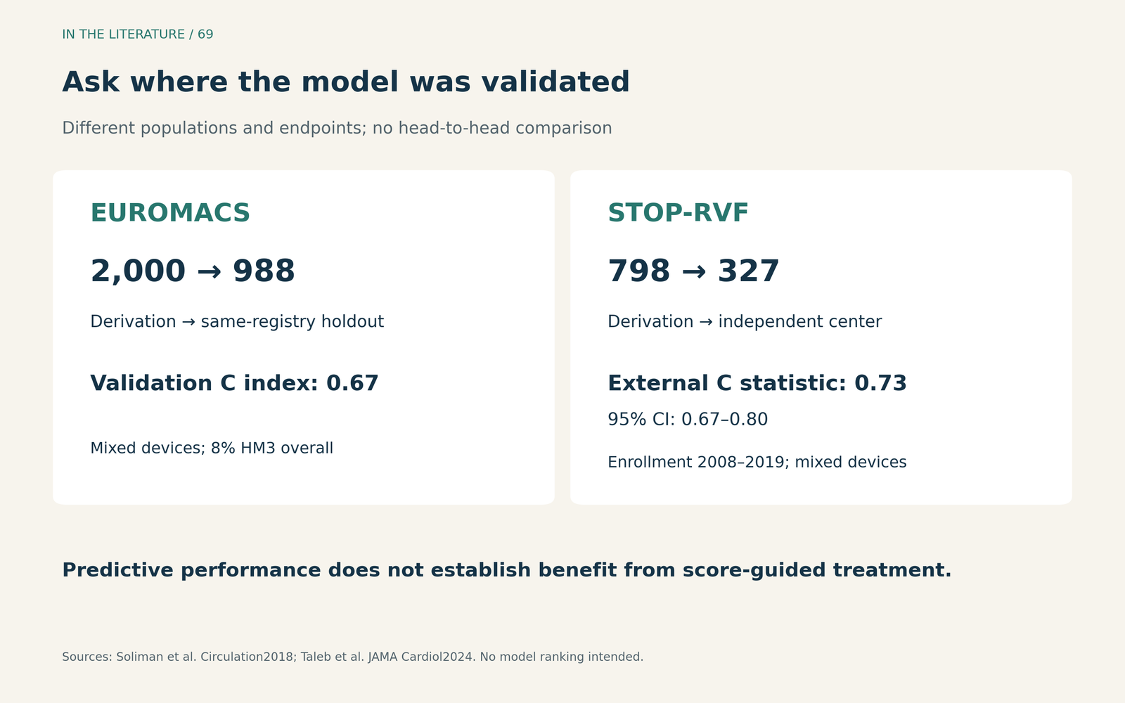
Figure 2. Identify what “validated” means before relying on a score. Different populations and outcome definitions prevent a simple comparison of the two C statistics.[3], [4] View larger.

What the 2026 evidence changes in the discussion
Sales and colleagues reassessed established scores in a single-center cohort. They enrolled 105 patients and analyzed 103 after excluding two intraoperative deaths attributed to causes other than right-heart failure; 21 developed moderate-to-severe RHF. Crucially, they evaluated the postoperative EUROMACS version separately because it includes bypass time. Its performance cannot be presented as information available before implantation.[5]
Their exploratory five-variable model also illustrates why a promising result is not yet a bedside rule. It was fitted in just 65 complete cases with 12 events. The apparent AUC of 0.854 fell to 0.773 after bootstrap correction for optimism. That internal correction is useful, but it does not replace testing in an independent cohort. The practical lesson is to check the measurement time, missing data and validation method before adopting a new prediction tool.[5]
Make the preoperative discussion produce a plan
For a patient with concerning RV findings, the useful next step is hemodynamically guided optimization and reassessment. Address congestion, perfusion and the factors impairing RV performance with the advanced-heart-failure, surgical and anesthesia teams. The ISHLT guidance advises considering planned biventricular support when RV function remains suboptimal after optimization. This is a clinical recommendation supported by limited evidence, not a score-triggered mandate.[1]
A practical way to apply that guidance is to leave the preimplant discussion with three documented conclusions: which abnormalities appear modifiable, what residual RV concern remains under the current support conditions, and what right-heart support strategy the team has agreed upon if isolated LV support is insufficient. This proposed discussion structure translates the guidance into a handoff; it is not a separately validated algorithm.[1], [2]
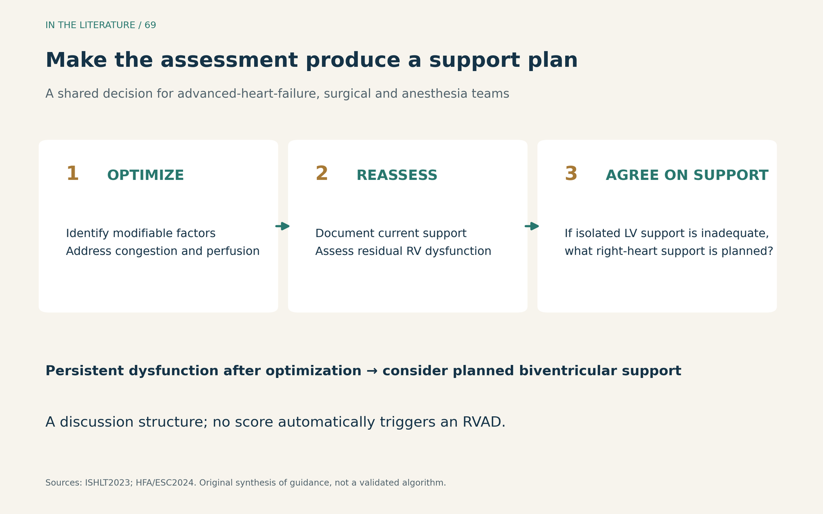
Figure 3. Use risk assessment to prepare a response. Persistent dysfunction after optimization warrants a team decision; no numeric score in this diagram automatically selects an RVAD.[1], [2] View larger.

Three takeaways
- Bring the measurement conditions to the discussion. RV imaging, pressures and flow are most informative when loading and support are documented together.
- Ask what the score actually predicts. Match the endpoint, pump population, timing of inputs and validation setting to the decision in front of you.
- Finish with a support plan. Reassess after optimization and discuss persistent RV dysfunction before implantation; risk prediction alone does not establish a treatment benefit.[1], [2], [3], [4]
References
- Saeed D, Feldman D, Banayosy AE, et al. The 2023 International Society for Heart and Lung Transplantation Guidelines for Mechanical Circulatory Support: A 10-Year Update. J Heart Lung Transplant. 2023;42(7):e1-e222. doi:10.1016/j.healun.2022.12.004
- Adamopoulos S, Bonios M, Ben Gal T, et al. Right heart failure with left ventricular assist devices: Preoperative, perioperative and postoperative management strategies. A clinical consensus statement of the Heart Failure Association (HFA) of the ESC. Eur J Heart Fail. 2024;26(11):2304–2322. doi:10.1002/ejhf.3323
- Soliman OII, Akin S, Muslem R, et al. Derivation and Validation of a Novel Right-Sided Heart Failure Model After Implantation of Continuous Flow Left Ventricular Assist Devices: The EUROMACS (European Registry for Patients with Mechanical Circulatory Support) Right-Sided Heart Failure Risk Score. Circulation. 2018;137(9):891–906. doi:10.1161/CIRCULATIONAHA.117.030543
- Taleb I, Kyriakopoulos CP, Fong R, et al. Machine Learning Multicenter Risk Model to Predict Right Ventricular Failure After Mechanical Circulatory Support: The STOP-RVF Score. JAMA Cardiol. 2024;9(3):272–282. doi:10.1001/jamacardio.2023.5372
- Sales MC, Zayat R, Hatam N, et al. Predicting the right! Validation of right heart failure predictive risk models after primary durable ventricular assist device implantation. PLoS One. 2026;21(9):e0357025. doi:10.1371/journal.pone.0357025