ITL-2f861437674d
Juan Li, MD · Updated November 17, 2026
Watch the video
Key points
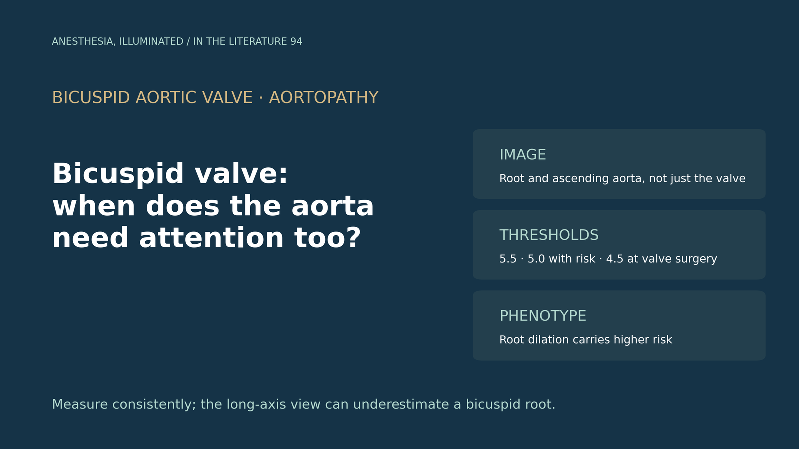
- Every patient with a bicuspid aortic valve (BAV) needs the aortic root and ascending aorta imaged, not only the valve; CT or MRI is indicated when echocardiography cannot fully assess them.[1], [2]
- Replacement is recommended at 5.5 cm. Thresholds of 5.0 cm apply with added risk factors, and the 2024 ESC guideline recommends surgery at 5.0 cm for the root phenotype.[1], [2]
- When the valve is being operated on, replacing a root or ascending aorta of 4.5 cm or more at the same time is reasonable.[1], [2]
- Dissection is uncommon in monitored BAV, but root dilation carried about twice the risk in one large cohort. Aortic events after isolated valve replacement were rare when the aorta was below 4.5 cm.[3], [4], [5]
- Measurement matters: the standard echo view can underestimate the bicuspid root by several millimetres. Screening first-degree relatives finds BAV in about 7%.[1], [6], [7]
A patient with a bicuspid aortic valve is scheduled for aortic valve replacement. Intraoperative transesophageal echocardiography (TEE) shows an ascending aorta of 4.7 cm and a root of 4.4 cm. The surgeon asks whether the aorta should be replaced as well. When does a bicuspid valve mean the aorta needs monitoring or surgery, and how much do the measurements and phenotype matter?[1], [2]
Image the whole aorta, not just the valve
The 2022 American College of Cardiology/American Heart Association (ACC/AHA) aortic disease guideline recommends transthoracic echocardiography (TTE) for every patient with a BAV to assess the valve, aortic root and ascending aorta (Class 1). CT or MRI is indicated when echocardiography cannot adequately assess the diameter and morphology of the aorta. Patients with a root or ascending aorta of 4.0 cm or more need lifelong surveillance at an interval dependent on diameter and growth, even after valve surgery.[1]
How the aorta is measured changes the number. The guideline recommends inner-edge to inner-edge measurement on electrocardiogram-gated CT or MRI, the root recorded as the maximum sinus-to-sinus diameter, leading-edge to leading-edge measurement on echocardiography, and indexing to height or body surface area in the report.[1] In 85 patients with BAV, Milleron and colleagues found that the largest root diameter runs roughly perpendicular to the valve opening, so the standard parasternal long-axis view underestimated the maximal root diameter by a mean of 6 mm in valves with a vertical opening. Their findings support short-axis, 3D or CT assessment of the root.[6]
When to replace the aorta
The ACC/AHA and 2024 European Society of Cardiology (ESC) recommendations for BAV aortopathy are broadly similar:[1], [2]
- 5.5 cm or more: replacement of the root, ascending aorta or both is recommended (ACC/AHA Class 1).
- 5.0–5.4 cm with an additional risk factor: replacement is reasonable (ACC/AHA Class 2a). Risk factors include a family history of dissection, growth of 0.3 cm/year or more, coarctation and the root phenotype; an aortic cross-sectional area to height ratio of 10 cm²/m or more is a separate Class 2a criterion.
- Root phenotype of 5.0 cm or more: surgery is recommended in the 2024 ESC guideline (Class 1), which also lists age under 50 years, ascending aortic length over 11 cm and resistant hypertension among factors favoring earlier surgery.
- 4.5 cm or more at the time of valve surgery: concomitant replacement of the root or ascending aorta is reasonable (ACC/AHA and ESC Class 2a).
All the "reasonable" options assume an experienced surgical team. Every BAV aortopathy recommendation rests on non-randomized data or expert opinion.[1]
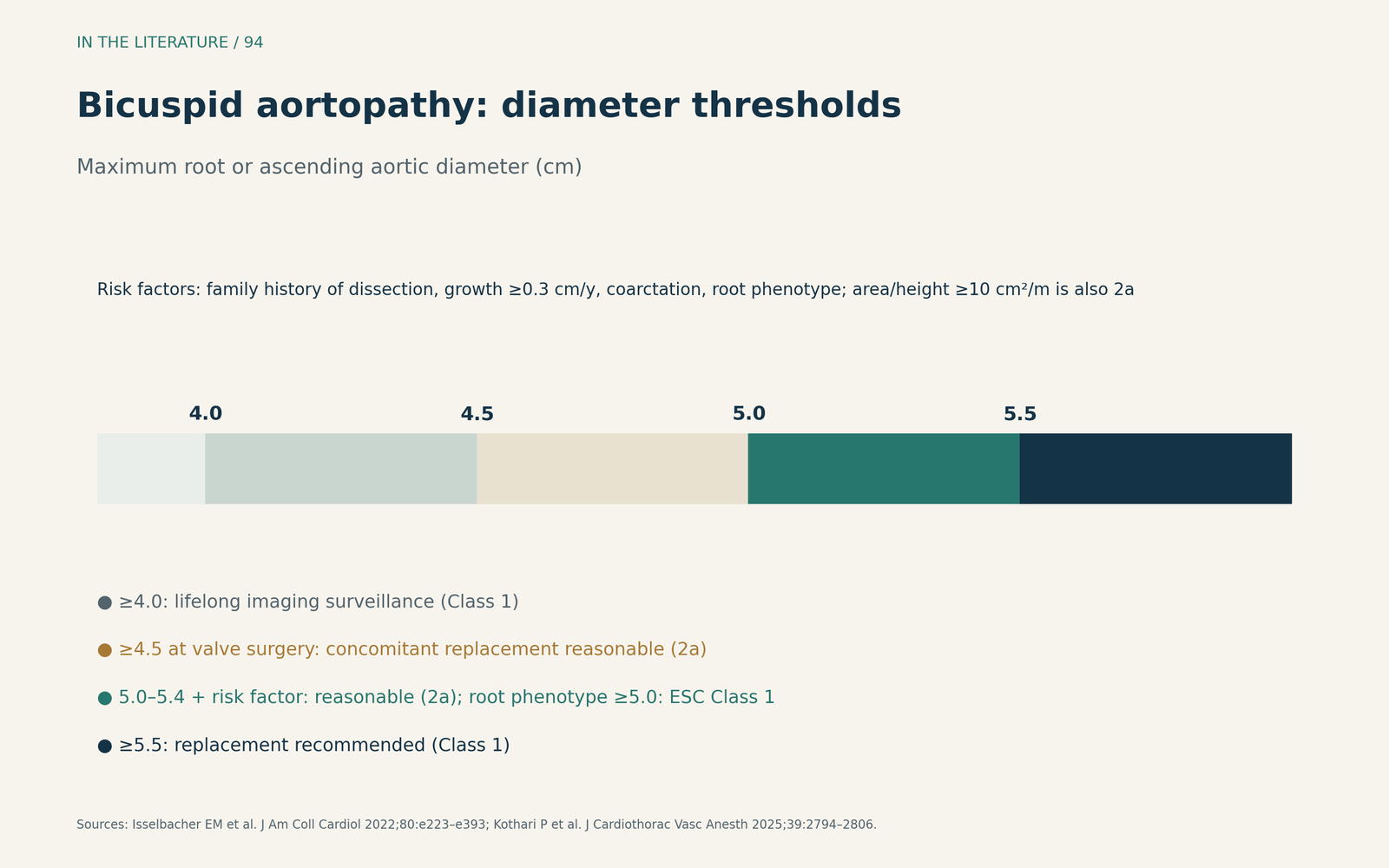
Figure 1. Size, phenotype and context. Thresholds summarized from the 2022 ACC/AHA aortic disease guideline and the 2024 ESC guideline as reviewed by Kothari and colleagues.[1], [2] View larger.

How large is the risk?
Dissection is uncommon in monitored patients. A meta-analysis by Wilson-Smith and colleagues of 11 studies and 4,330 unoperated patients found a pooled incidence of BAV-related aortic dissection of 0.6% over a median of 9 years.[3]
Phenotype appears to matter. In 1,582 patients with BAV and an aorta of 40 mm or more, Gao and colleagues found an annual dissection incidence of 0.43% overall, 0.78% with root dilation versus 0.30% without, and a more than two-fold adjusted risk with root dilation. Spline analysis suggested risk inflection points at 45 mm for the root and 50 mm for the ascending aorta. The study was single-center with only 24 dissections, so these inflection points need confirmation.[4]
After isolated aortic valve replacement for BAV with an ascending aorta below 45 mm, Hachiro and colleagues reported that 3 of 181 patients (1.7%) needed later aortic surgery over a median of 6 years. All three had diameters of about 44.5 mm at valve replacement, and no patient died of aortic disease.[5] The small number of events limits conclusions about borderline diameters.
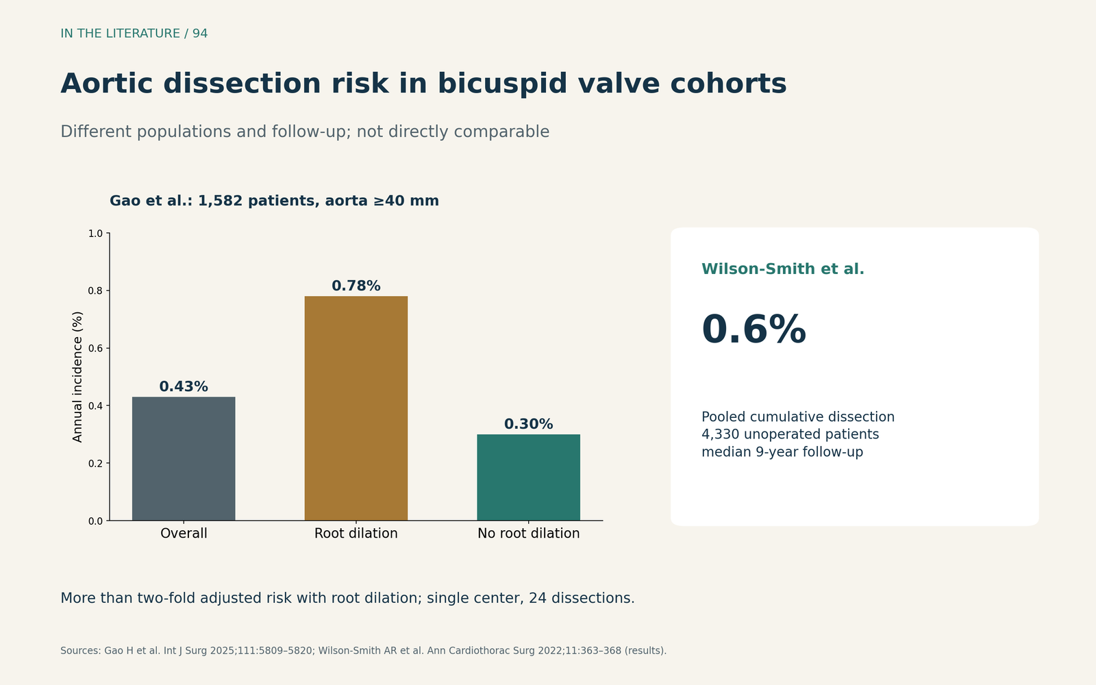
Figure 2. Dissection is uncommon but higher with root dilation. Redrawn from the results of Gao et al. (annual incidence by root dilation) and Wilson-Smith et al. (pooled cumulative incidence); different populations and follow-up.[3], [4] View larger.

Family screening
Bicuspid aortopathy runs in families. The ACC/AHA guideline recommends TTE screening of first-degree relatives when the patient's aorta is dilated (Class 1) and considers it reasonable for all BAV patients (Class 2a).[1] In a meta-analysis of 6,054 relatives, Bray and colleagues found BAV in 7.3% of screened relatives and aortic dilatation in 9.4%; the estimated number needed to screen to find one affected relative was 20 (95% CI 5–120). No data yet show that screening improves outcomes.[7]
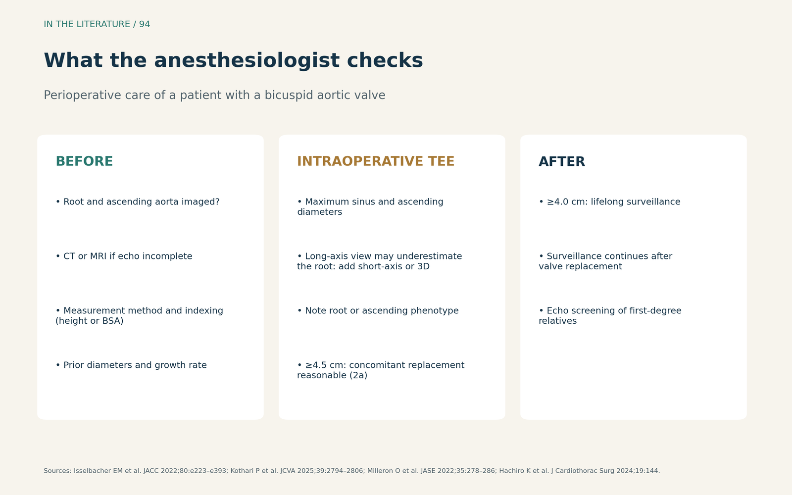
Figure 3. A perioperative checklist for bicuspid aortopathy. Synthesized from the 2022 ACC/AHA guideline, the 2024 ESC guideline review, Milleron et al. and Hachiro et al.; not a validated protocol.[1], [2], [5], [6] View larger.

Three takeaways
- A bicuspid valve is an aortic diagnosis too. Image the root and ascending aorta, measure consistently and index to body size; use CT or MRI when echocardiography is incomplete.[1], [6]
- Know the thresholds and the phenotype. Replacement at 5.5 cm, at 5.0 cm with risk factors or a root phenotype, and at 4.5 cm or more when the valve is already being operated on.[1], [2], [4]
- Measure carefully at valve surgery. The intraoperative diameter can change the operation, and the long-axis view may underestimate a bicuspid root.[1], [5], [6]
References
- Isselbacher EM, Preventza O, Hamilton Black J 3rd, et al. 2022 ACC/AHA guideline for the diagnosis and management of aortic disease: a report of the American Heart Association/American College of Cardiology Joint Committee on Clinical Practice Guidelines. J Am Coll Cardiol. 2022;80(24):e223–e393. doi:10.1016/j.jacc.2022.08.004
- Kothari P, Bodmer NJ, Vanneman MW, et al. Highlights and perioperative implications from the 2024 European Society of Cardiology guidelines for the management of peripheral arterial and aortic diseases. J Cardiothorac Vasc Anesth. 2025;39(10):2794–2806. doi:10.1053/j.jvca.2025.03.022
- Wilson-Smith AR, Eranki A, Muston B, et al. Incidence of bicuspid valve related aortic dissection: a systematic review and meta-analysis. Ann Cardiothorac Surg. 2022;11(4):363–368. doi:10.21037/acs-2022-bav-21
- Gao H, Liu X, Wang Y, et al. Aortic root dilation in bicuspid aortic valve disease: impact on dissection risk and evaluation of guideline-based surgical thresholds—a retrospective cohort study. Int J Surg. 2025;111(9):5809–5820. doi:10.1097/JS9.0000000000002657
- Hachiro K, Takashima N, Suzuki T. Later aorta operation after aortic valve replacement for bicuspid aortic valve. J Cardiothorac Surg. 2024;19(1):144. doi:10.1186/s13019-024-02638-6
- Milleron O, Masi P, Eliahou L, et al. Aortic root anatomy is related to the bicuspid aortic valve phenotype. J Am Soc Echocardiogr. 2022;35(3):278–286. doi:10.1016/j.echo.2021.11.012
- Bray JJH, Freer R, Pitcher A, Kharbanda R. Family screening for bicuspid aortic valve and aortic dilatation: a meta-analysis. Eur Heart J. 2023;44(33):3152–3164. doi:10.1093/eurheartj/ehad320