ITL-e1700d07ea96
Juan Li, MD · Updated November 18, 2026
Key points
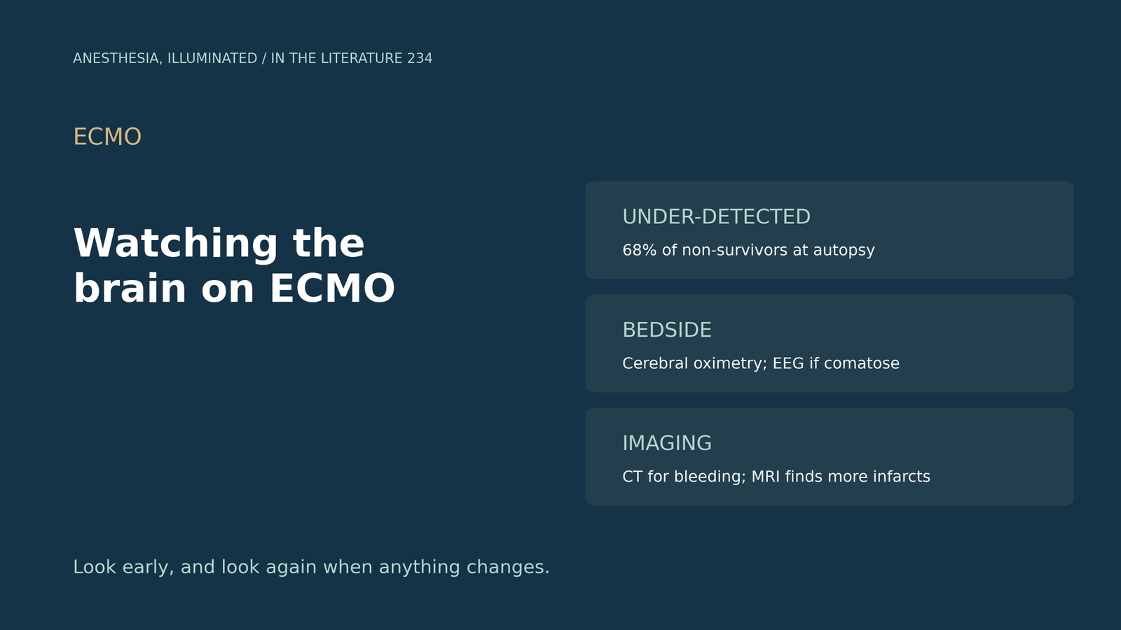
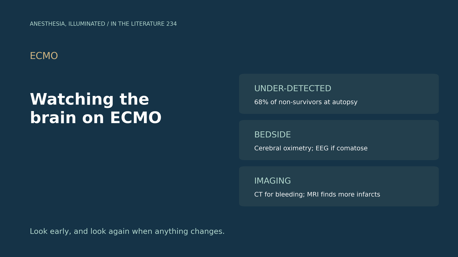
- Common and missed. At autopsy, 68% of ECMO non-survivors had acute brain injury, and incidence is likely higher than clinical detection.[1]
- It matters. Acute brain injury increases in-hospital mortality by a factor of 2 to 3.[1]
- Guideline tools: continuous cerebral oximetry, intermittent EEG and SSEP in comatose patients, and non-contrast head CT to rule out ICH when stroke is suspected.[1]
- Imaging gaps. More than 30% of patients never get a head CT during ECMO; portable MRI found injury in 44% of patients scanned.[1], [2]
- No standard protocol. No included study imaged patients with a standardized protocol, so risk-factor data are limited.[3]
A sedated patient on venovenous ECMO is on heparin and has a low platelet count. The pupils look unchanged, but the neurologic exam is limited. How should the brain be watched, and when should it be imaged?[1]
How often the brain is injured
The ELSO consensus guidelines (Cho et al., Crit Care 2024) report that acute brain injury increases in-hospital mortality by a factor of 2 to 3; at autopsy, 68% of ECMO non-survivors had developed it, and MRI found cerebral infarction or hemorrhage in 37 to 52% of adult ECMO survivors.[1] In a COVID cohort under standardized neuromonitoring (Kannapadi et al., J Cardiothorac Vasc Anesth 2021), 25% of VV-ECMO patients had neurologic events, the median time from cannulation to ICH was three days, and ICHs were managed by discontinuing heparin upon CT detection.[4] Injury is not limited to the brain: spinal cord infarction developed in 5.3% of patients on prolonged femoral VA-ECMO, and recognition was delayed in 71%, partly due to critical illness polyneuropathy.[5]
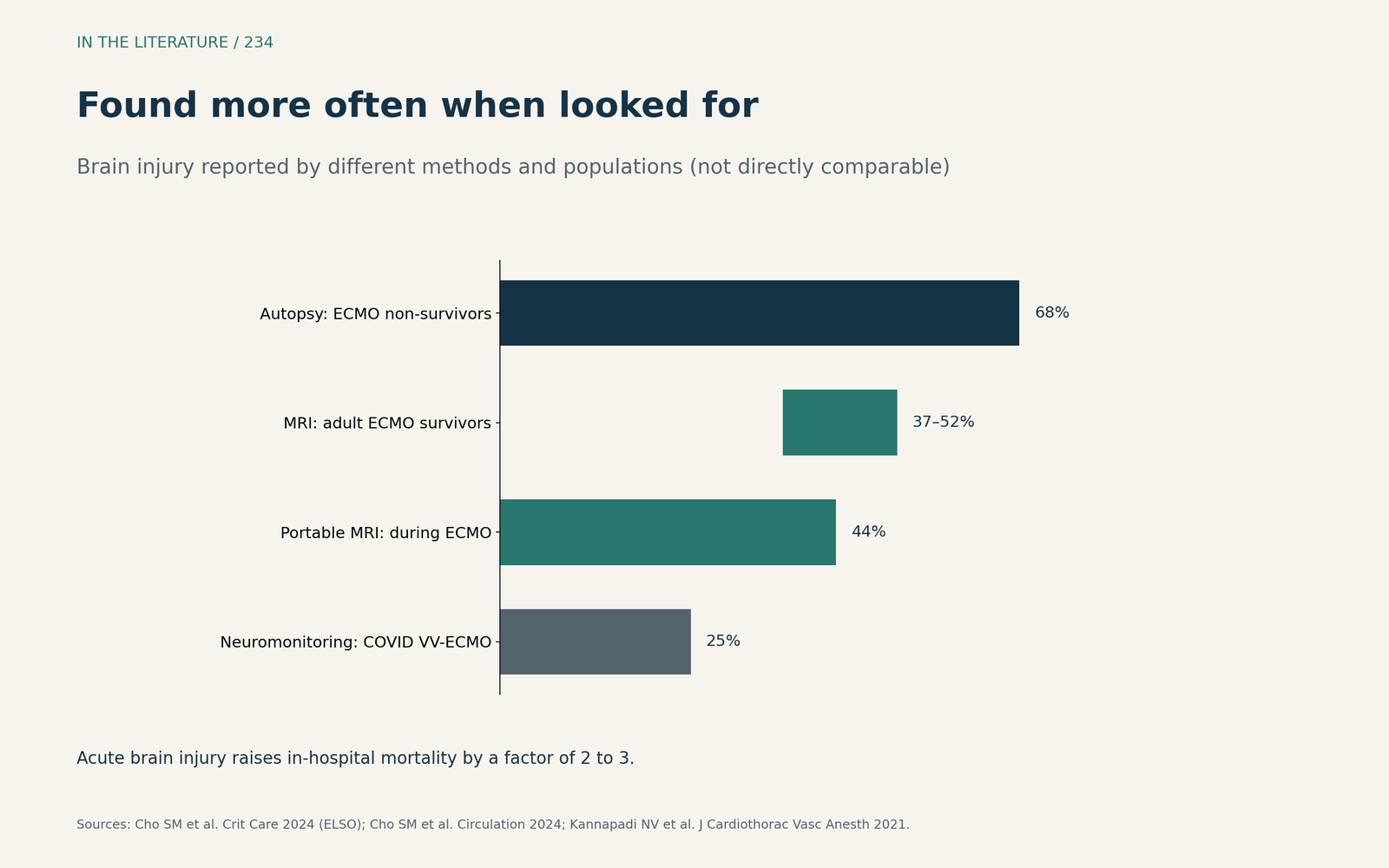
Figure 1. Found more often when looked for. Data from Cho et al. (ELSO 2024), the SAFE MRI ECMO study and Kannapadi et al.[1], [2], [4] View larger.

What to monitor
The ELSO guidelines recommend continuous cerebral oximetry to follow trends and detect brain injury early, and intermittent EEG and SSEP, particularly in comatose patients.[1] Non-contrast head CT is used to rule out ICH when stroke is suspected, and studies have reported routine head CT within 24 h of cannulation to be beneficial; yet more than 30% of patients do not receive a head CT during ECMO support.[1]
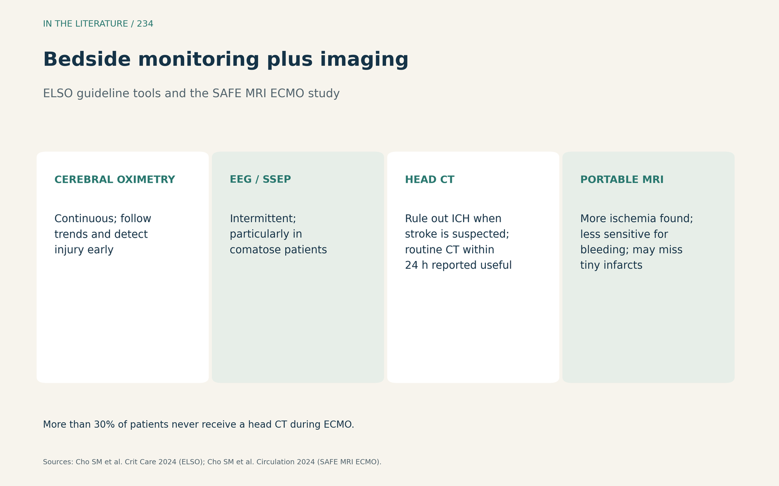
Figure 2. Bedside monitoring plus imaging. Synthesized from Cho et al. (ELSO 2024) and the SAFE MRI ECMO study.[1], [2] View larger.

CT or bedside MRI?
In the multicenter SAFE MRI ECMO study (Cho et al., Circulation 2024), portable MRI found acute brain injury in 44% of 50 patients.[2] Among patients with both scans within 24 hours, portable MRI detected all 8 ischemic events and head CT only 4; for hemorrhage, portable MRI detected only 1 of 2 events that head CT detected.[2] Head CT has limited sensitivity for hyperacute to acute ischemic injury, while portable MRI may miss tiny punctate infarcts seen on conventional MRI.[2]
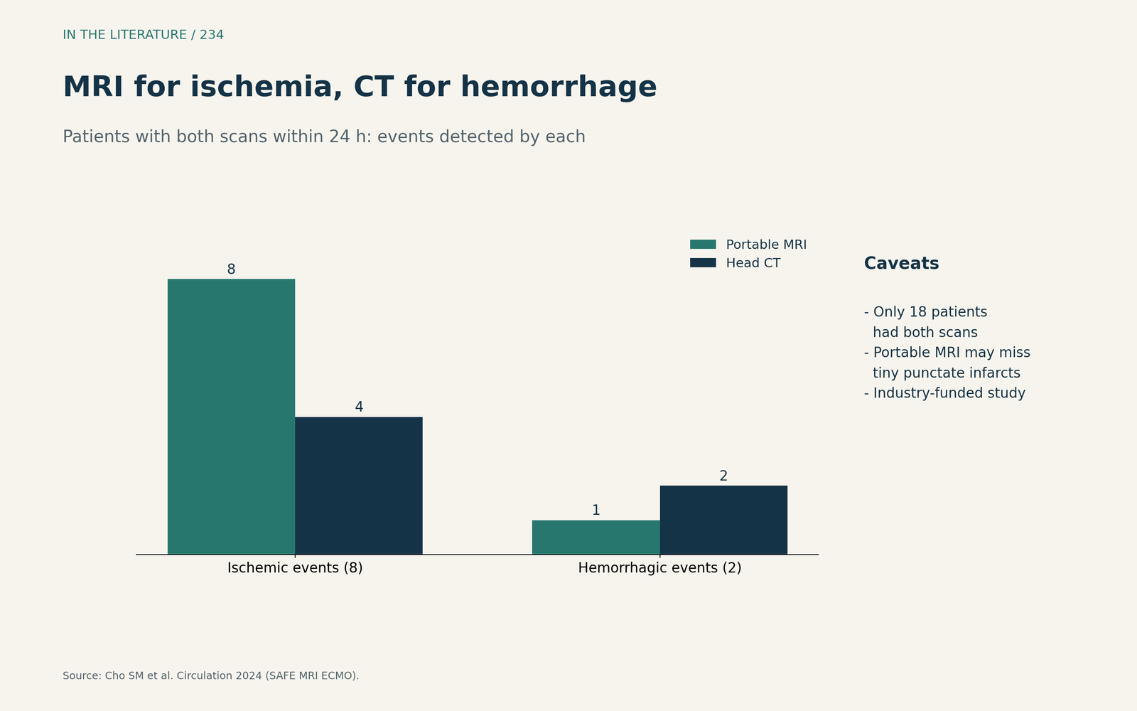
Figure 3. MRI for ischemia, CT for hemorrhage. Redrawn from the SAFE MRI ECMO study.[2] View larger.

Who is at risk
In a systematic review of VV-ECMO (Thiara et al., Crit Care Med 2025), lower pH before VV-ECMO was associated with increased ICH risk (moderate certainty), and no predictors of ischemic stroke were identified in two or more studies; no included study had a standardized imaging protocol.[3] Dong et al. (Front Med 2026) pooled 29 studies with 62,656 patients; each additional day of ECMO support was associated with a 2% increase in stroke risk, but the findings should be interpreted as hypothesis-generating rather than definitive.[6]
Three takeaways
- Assume brain injury is under-detected; it is common and raises mortality.[1]
- Use cerebral oximetry and EEG at the bedside, and image early and whenever stroke is suspected.[1]
- CT rules out hemorrhage; MRI, including bedside MRI, finds more ischemic injury.[2]
References
- Cho SM, Hwang J, Chiarini G, et al. Neurological monitoring and management for adult extracorporeal membrane oxygenation patients: Extracorporeal Life Support Organization consensus guidelines. Crit Care. 2024;28(1):296. doi:10.1186/s13054-024-05082-z
- Cho SM, Khanduja S, Wilcox C, et al. Clinical use of bedside portable ultra-low-field brain magnetic resonance imaging in patients on extracorporeal membrane oxygenation: results from the multicenter SAFE MRI ECMO study. Circulation. 2024;150(24):1955–1965. doi:10.1161/CIRCULATIONAHA.124.069187
- Thiara S, Willms AJ, Tran A, et al. Prognostic factors associated with intracranial hemorrhage and ischemic stroke during venovenous extracorporeal membrane oxygenation: a systematic review. Crit Care Med. 2025;53(2):e400–e409. doi:10.1097/CCM.0000000000006520
- Kannapadi NV, Jami M, Premraj L, et al. Neurologic injury in patients with COVID-19 who receive VV-ECMO therapy: a cohort study. J Cardiothorac Vasc Anesth. 2021;35(11):3456–3461. doi:10.1053/j.jvca.2021.05.017
- Pasrija C, Kon ZN, Mazzeffi MA, et al. Spinal cord infarction with prolonged femoral venoarterial extracorporeal membrane oxygenation. J Cardiothorac Vasc Anesth. 2023;37(5):758–766. doi:10.1053/j.jvca.2022.12.025
- Dong M, Gu P, Cai L, et al. Risk factors for neurological complications in adult ECMO patients: a systematic review and meta-analysis. Front Med (Lausanne). 2026;12:1726621. doi:10.3389/fmed.2025.1726621