ITL-28cfa4f37c78
Juan Li, MD · Updated November 17, 2026
Watch the video
Key points
- The right heart is hit first. In a meta-analysis, 32% of patients with carcinoid syndrome had cardiac involvement on echo; 79% of these had tricuspid abnormalities, and moderate-severe tricuspid regurgitation was the most common lesion, in 70%.[1]
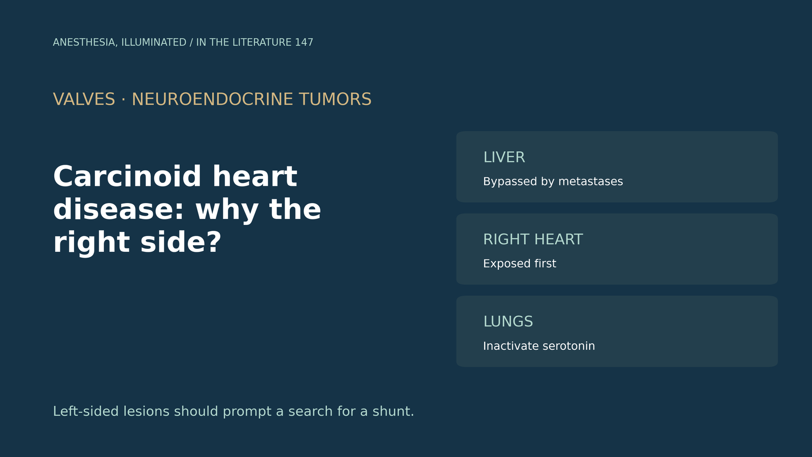
- The liver and the lungs act as filters. Monoamine oxidase in the liver and lung inactivates serotonin; once metastatic disease releases vasoactive substances into the systemic veins, the right-sided valves are exposed before the lungs clear them.[2]
- Left-sided disease is the exception. Significant mitral and aortic regurgitation were found in 11% and 10%, and left-sided lesions are reported in up to 15% of patients.[1], [2]
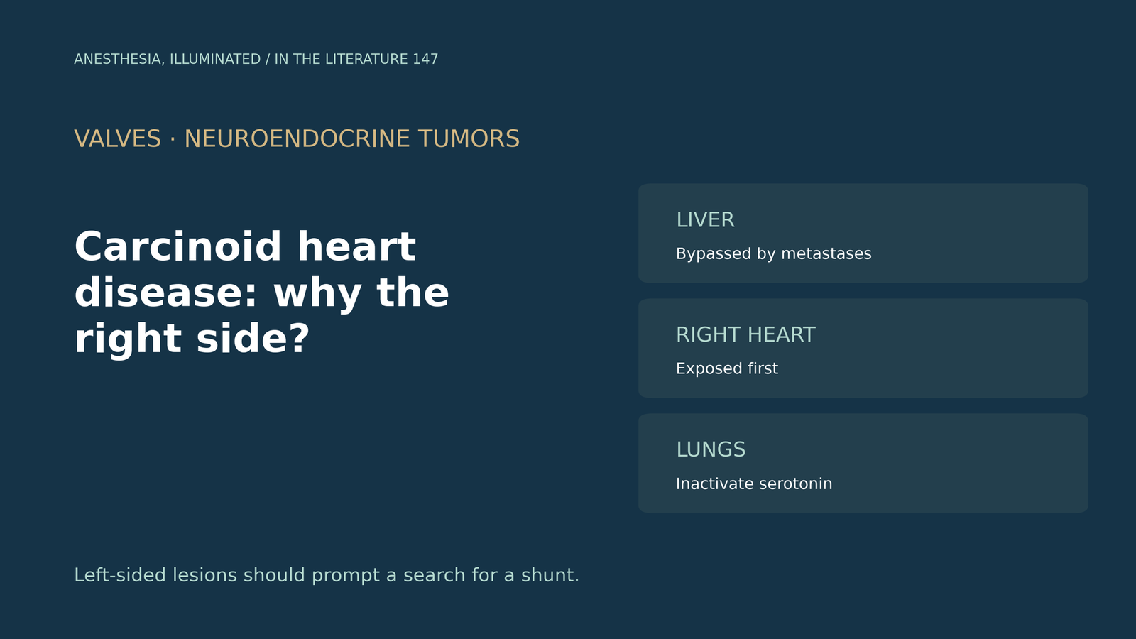
- Left-sided lesions should prompt a search for a patent foramen ovale, which is more common in carcinoid heart disease (about 59%) than in the general population (25%).[3], [4]
- For the anesthesiologist, the right ventricle needs protection, and TEE shows the pulmonary valve poorly.[5]
A patient with a metastatic neuroendocrine tumor comes for tricuspid and pulmonary valve replacement. Pre-bypass TEE shows thickened, fixed tricuspid leaflets and a mildly thickened mitral valve. Why are the right-sided valves affected, and what does left-sided involvement mean?[1], [3]
The right-sided pattern
Namkoong and colleagues pooled 36 studies. Of patients with carcinoid syndrome, 32% had cardiac involvement on echo; 79% of these had tricuspid abnormalities, moderate-severe tricuspid regurgitation was seen in 70%, and pulmonary stenosis in 43%. Significant mitral regurgitation (11%) and aortic regurgitation (10%) were less common, although heterogeneity between studies was substantial.[1] Stavagna and colleagues cite tricuspid involvement in up to 90% of cases, followed by the pulmonary (69%), mitral (29%) and aortic (27%) valves; plaque-like fibrotic deposits leave the valves thickened, retracted and fixed.[3]
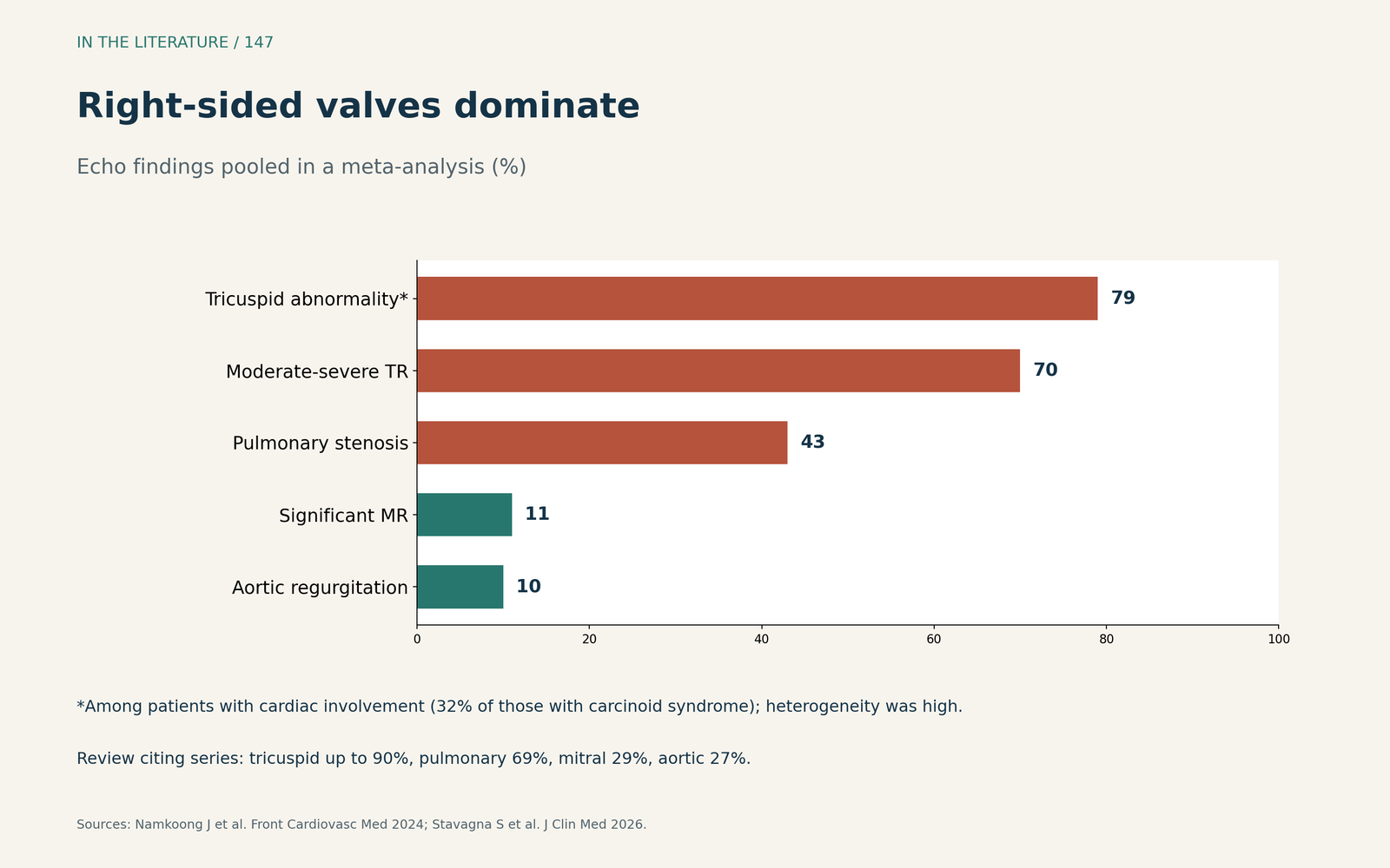
Figure 1. Right-sided valves dominate. Redrawn from Namkoong et al. and Stavagna et al.[1], [3] View larger.

Why the left heart is usually spared
Willauer and colleagues explain that monoamine oxidase in the liver, lung and brain converts serotonin to its inactive metabolite; tumor confined to the gut is cleared by the liver, but metastatic disease releases vasoactive substances into the systemic circulation. These reach the right-sided valves first, and left-sided lesions are less common because the lung inactivates serotonin.[2] Namkoong and colleagues and Stavagna and colleagues give the same explanation: the substances are broken down in the pulmonary circulation before reaching the left heart. This is stated as background physiology rather than tested in these studies.[1], [3]
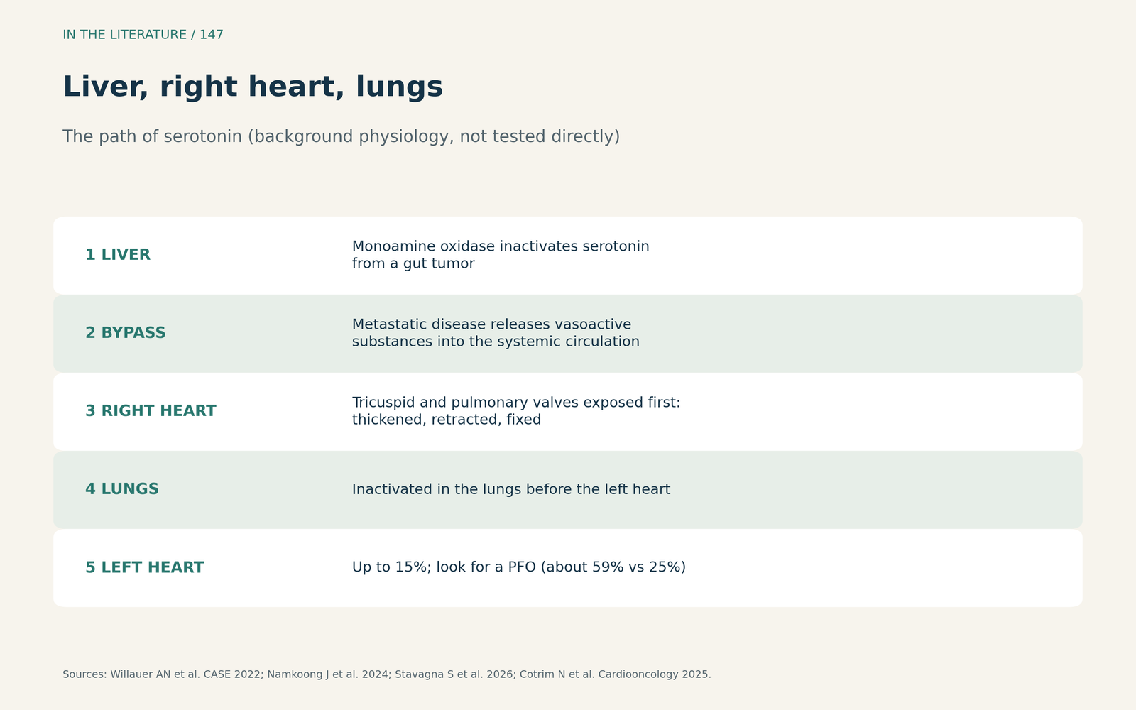
Figure 2. Liver, right heart, lungs. Synthesized from Willauer et al., Namkoong et al., Stavagna et al. and Cotrim et al.[2], [1], [3], [4] View larger.

When the left heart is involved
Left-sided lesions are present in up to 15% of patients and are generally less severe.[2] Stavagna and colleagues advise that left-sided disease should prompt a search for a patent foramen ovale, whose prevalence is about 59% in carcinoid heart disease versus 25% in the general population.[3] Cotrim and colleagues describe a patient in whom left-sided valve involvement was likely related to right-to-left shunting through a patent foramen ovale, and warn that isolated closure in the presence of severe tricuspid regurgitation can increase right ventricular afterload.[4]
What it means in the operating room
In a case conference, Alvarez-Villegas and colleagues note that left-sided valves are affected rarely and in the presence of intracardiac shunts, that the pulmonary valve is not well visualized on TEE, and that cautious protection of the right ventricle is mandatory.[5] With severe tricuspid regurgitation, transthoracic indices of right ventricular function overestimate performance, and the right ventricle and atrium behave as a single chamber.[2], [3] Prognosis remains poor: 3-year survival was 31% with carcinoid heart disease versus 69% without, and there is not yet clear evidence that surgery lowers mortality today.[1]
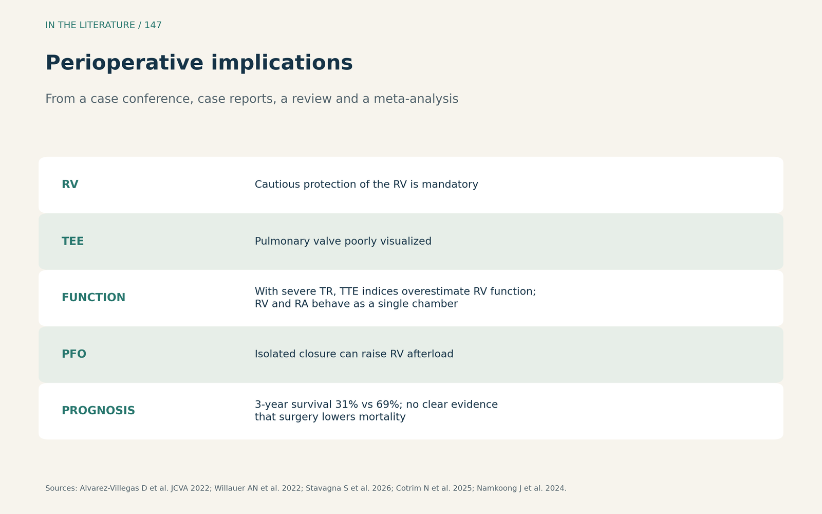
Figure 3. Perioperative implications. Synthesized from Alvarez-Villegas et al., Willauer et al., Stavagna et al., Cotrim et al. and Namkoong et al.[5], [2], [3], [4], [1] View larger.

Three takeaways
- Right first, lungs second. Vasoactive substances that escape the liver reach the right-sided valves before the lungs inactivate them.[1], [2]
- Left-sided lesions mean a bypass. Look for a patent foramen ovale and plan closure with the valve surgery in mind.[3], [4]
- Protect the right ventricle. Expect a fixed tricuspid valve, a poorly seen pulmonary valve and overestimated right ventricular function.[5], [2]
References
- Namkoong J, Andraweera PH, Pathirana M, et al. A systematic review and meta-analysis of the diagnosis and surgical management of carcinoid heart disease. Front Cardiovasc Med. 2024;11:1353612. doi:10.3389/fcvm.2024.1353612
- Willauer AN, Hajjar R, Estes CW, Laipply KR. Delayed presentation of carcinoid heart disease and subsequent modified healthcare delivery in the COVID-19 era. CASE (Phila). 2022;6(3):128–133. doi:10.1016/j.case.2022.01.006
- Stavagna S, Manzi G, Angotti D, et al. Carcinoid heart disease: the role of echocardiography in raising the first suspicion. J Clin Med. 2026;15(5):1978. doi:10.3390/jcm15051978
- Cotrim N, Carvalheiro R, Teixeira B, et al. Concomitant carcinoid heart disease and patent foramen ovale: a case report and review of literature. Cardiooncology. 2025;11(1):80. doi:10.1186/s40959-025-00380-z
- Alvarez-Villegas D, Giménez-Milà M, Sbraga F, et al. Dealing with the right side: carcinoid heart disease. J Cardiothorac Vasc Anesth. 2022;36(8):2793–2802. doi:10.1053/j.jvca.2021.10.030