ITL-d4c6fb3c5bc6
Juan Li, MD · Updated November 18, 2026
Watch the video
Key points
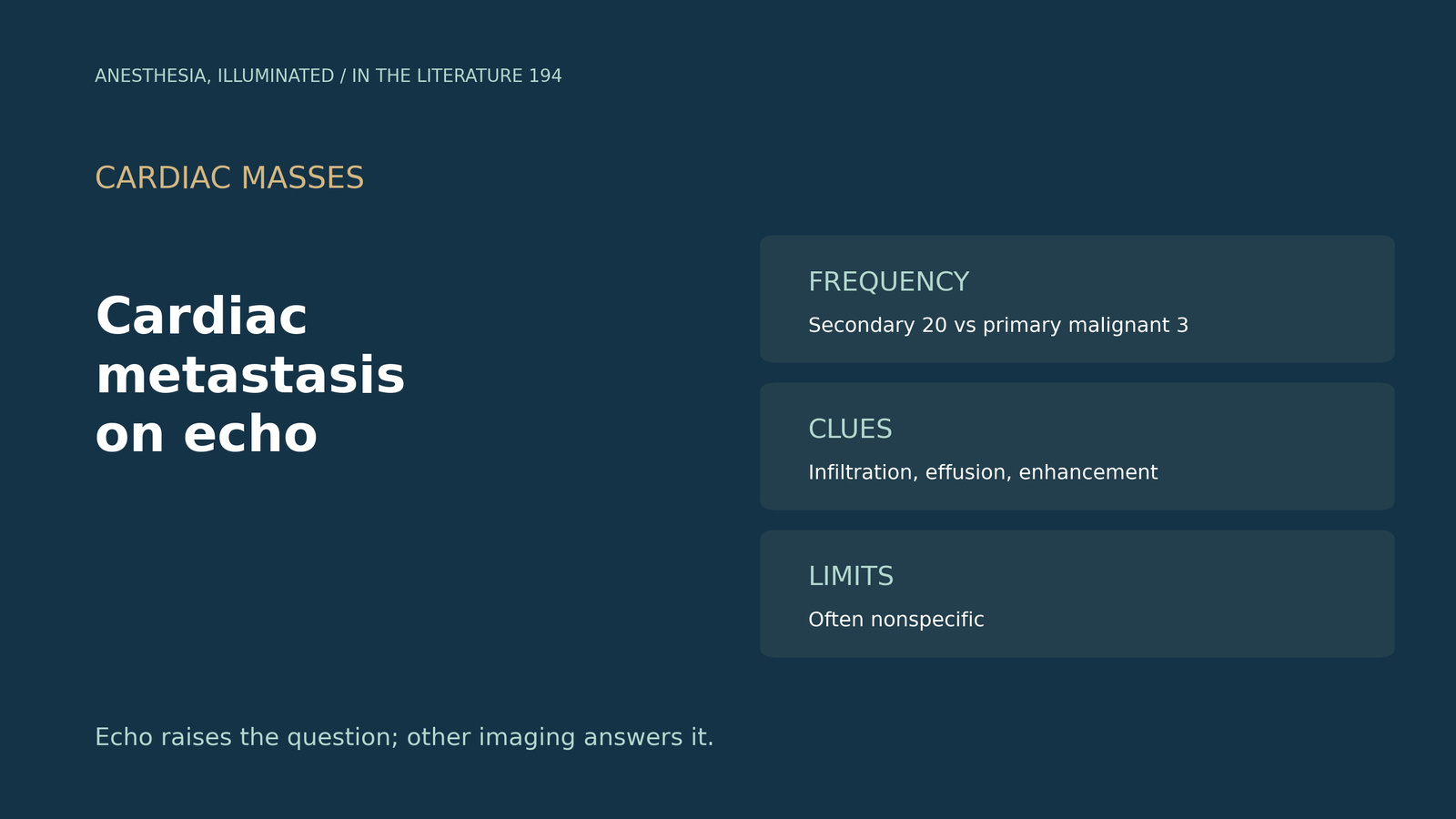
- Secondary beats primary. In one 12-year series, secondary cardiac tumours outnumbered primary malignant tumours 20 to 3, with lymphoma and lung cancer the most frequent primaries.[1]
- The route predicts the site. Lymphatic spread often produces pericardial and epicardial tumour; two-thirds of 622 autopsy cases involved the pericardium.[1], [2]
- Contrast helps separate tumour from clot. Masses with neovascularity enhance relative to myocardium, while thrombi and myxomas show little or no enhancement.[2], [3]
- Look for infiltration, not only a mass. Ill-defined echodensities can thicken the RV wall and depress its function.[4]
- Echo is the start, not the end. It is the initial modality, but its findings can be nonspecific.[3], [5]
During pre-induction TEE in a patient with known cancer, the right ventricular free wall looks thick and irregular, and a small pericardial effusion is present. Could this be cardiac metastasis, and what on echocardiography should raise or lower that suspicion?[1], [2]
How common it is and where it goes
Bugra and colleagues reviewed the records of 64,862 patients over 12 years in the I-MASS series. Metastases or invasions made up 15.75% of cardiac masses (20 patients), secondary tumours outnumbered primary malignant tumours 20 to 3, and lymphoma and lung cancer were the most frequent primaries. Each mass was assessed for location, mobility, echogenicity and pedicle, and the authors note that lymphatic spread often produces pericardial and epicardial tumour.[1] Dudorova and colleagues cite an autopsy series of 622 cardiac metastases in which two-thirds involved the pericardium, and they emphasize that mural thrombi account for most ventricular masses.[2] Richardson and colleagues note that renal cell carcinoma spreads into the IVC in up to 15% of cases and describe a mobile mass extending from the IVC into the right atrium.[6]
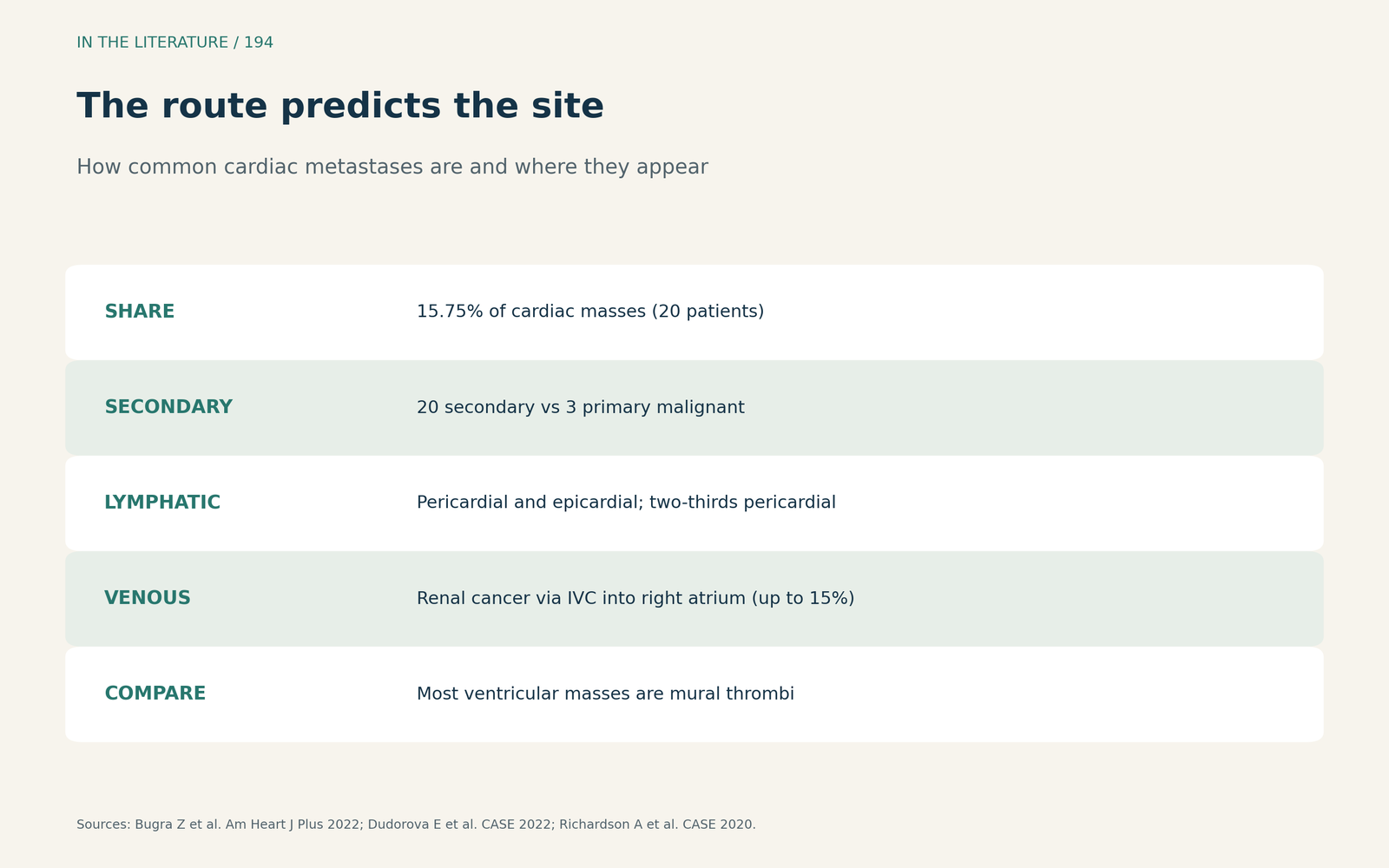
Figure 1. The route of spread predicts the site. Synthesized from Bugra et al., Dudorova et al. and Richardson et al.[1], [2], [6] View larger.

Echo clues that favour tumour
TTE is the initial test of choice for cardiac tumours, and thrombi and myxomas show little or no contrast enhancement.[3] Dudorova and colleagues add that contrast perfusion echocardiography helps separate malignant from benign masses, because masses with neovascularity enhance relative to myocardium.[2] Metastasis does not always form a discrete mass: Xu and colleagues describe ill-defined echodensities that thickened the RV wall, RV fractional area change that fell to 36% from 66%, and profound anterior ST elevation caused by the metastasis.[4]
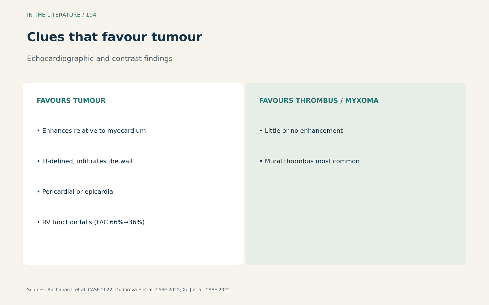
Figure 2. Enhancement and infiltration point to tumour. Synthesized from Buchanan et al., Dudorova et al. and Xu et al.[3], [2], [4] View larger.

When echo is not enough
In Lara-Sampayo and colleagues' systematic review of melanoma, cardiac metastases are found at autopsy in 50 to 65% of patients, and echocardiography served as the initial diagnostic modality. Their index case showed infiltration but no discrete intracardiac mass, and they warn that echo findings can be nonspecific.[5] All of this evidence comes from single-centre series, case reports and small reviews, so no echo pattern has a defined accuracy for metastasis.[1], [5] Cystic masses are a particular trap: an ultrasound enhancing agent may help show that a lesion is avascular (Mir et al., CASE 2021),[7] walled-off cysts that do not communicate with the blood pool will not enhance with contrast, unlike tumours, while CT cannot reliably distinguish malignant tissue from nonmalignant fluid (Maidman et al., CASE 2023),[8] and neoplasms, though typically solid, often have cystic components or necrosis (Chalhoub et al., CASE 2023).[9]
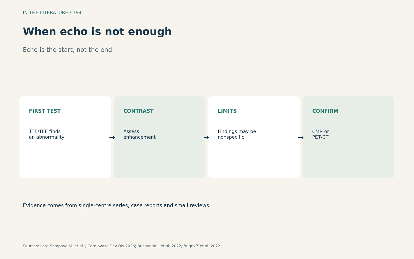
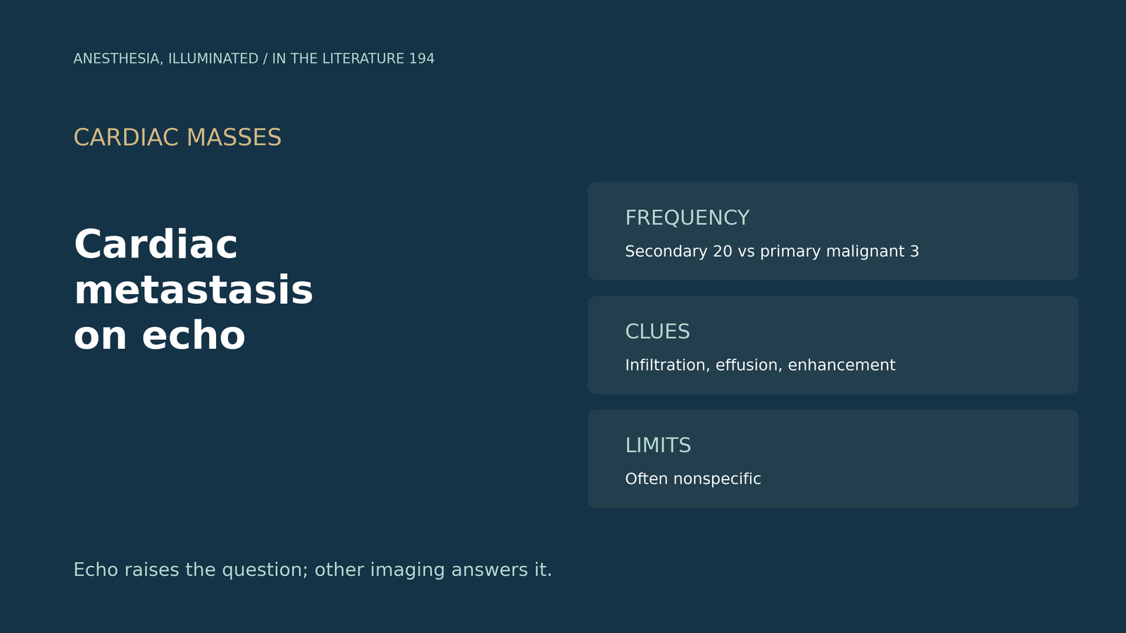
Figure 3. Echo raises the question; other imaging answers it. Synthesized from Lara-Sampayo et al., Buchanan et al. and Bugra et al.[5], [3], [1] View larger.

Three takeaways
- In a patient with cancer, a new cardiac mass is more likely secondary than a primary malignant tumour.[1]
- Contrast enhancement favours tumour; thrombi and myxomas enhance little.[2], [3]
- Wall thickening or effusion without a discrete mass can still be metastasis; confirm with other imaging.[4], [5]
References
- Bugra Z, Emet S, Umman B, et al. Intracardiac masses: single center experience within 12 years: I-MASS study. Am Heart J Plus. 2022;13:100081. doi:10.1016/j.ahjo.2021.100081
- Dudorova E, Lavelle M, Lahey R, Ravalli S. Left ventricular metastases from penile squamous cell carcinoma. CASE (Phila). 2022;6(2):67–72. doi:10.1016/j.case.2022.01.009
- Buchanan L, Dangl M, Syed A, et al. Metastatic carcinoma to the right heart: the complementary utility of contrast-enhanced echocardiography and cardiac positron emission tomography/computed tomography. CASE (Phila). 2022;6(1):3–7. doi:10.1016/j.case.2021.09.012
- Xu J, Cui F, Wang D, et al. Profound anterior ST-segment elevation in a patient with lung cancer and echocardiographic evidence of right ventricle metastasis. CASE (Phila). 2022;6(2):59–62. doi:10.1016/j.case.2021.11.001
- Lara-Sampayo KL, Ibarrola-Peña JC, de la Pena-Tamez M, et al. Multimodality imaging in cardiac metastasis of cutaneous melanoma: case report and systematic review. J Cardiovasc Dev Dis. 2026;13(2):84. doi:10.3390/jcdd13020084
- Richardson A, Ganji M, Omman R, et al. Renal cell carcinoma with right ventricular metastatic spread: an unusual finding with poor prognosis. CASE (Phila). 2020;4(5):374–376. doi:10.1016/j.case.2020.04.012
- Mir H, McClure A, Thampinathan B, et al. Echocardiographic features of cardiac echinococcal infection. CASE (Phila). 2021;5(1):26–32. doi:10.1016/j.case.2020.10.002
- Maidman SD, Bamira D, Vainrib AF, Ro R, Saric M. Cysts around the heart: differential diagnosis and multimodality imaging strategies for paracardiac cysts. CASE (Phila). 2023;7(9):365–376. doi:10.1016/j.case.2023.05.006
- Chalhoub G, Kamel A, Levsky J, Schenone A, Garcia MJ. A large cyst in an unlikely location: the interventricular septum. CASE (Phila). 2023;7(10):401–404. doi:10.1016/j.case.2023.06.001