ITL-47c2b6b4faf0
Juan Li, MD · Updated November 17, 2026
Watch the video
Key points
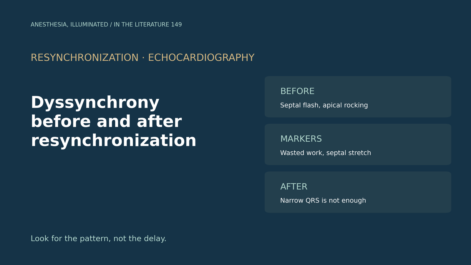
- Timing delays fell out of favor. The 2008 American Society of Echocardiography consensus listed indices such as a 33 ms Yu index, but reproducibility of dyssynchrony analysis needed improvement, and about 25% to 35% of recipients did not respond.[1]
- Look for the pattern, not the delay. Septal flash results from early septal contraction before delayed lateral contraction; visual assessment needs high-quality multi-view images, and scar or foreshortening can hide or mimic it.[2]
- Myocardial work adds information. Only 37% of patients with low global wasted work responded, yet the authors say such patients should not be excluded from therapy.[3]
- Combine electrical and mechanical markers. With high QRS area plus septal rebound stretch, 90% responded, versus 54% with QRS area alone.[4]
- After implantation, a narrowed QRS does not guarantee response, activation mapping remains a pilot tool, and intraoperative TEE can reveal pacing-induced dyssynchrony that causes functional mitral regurgitation.[5], [6]
After aortic valve replacement, epicardial pacing is started and TEE shows new moderate mitral regurgitation with normal-looking leaflets. In another patient, a biventricular device was placed last month. How should mechanical dyssynchrony be recognized before and after resynchronization?[6], [2]
From timing indices to contraction patterns
The 2008 consensus described tissue Doppler and speckle-tracking timing indices, including a 12-site standard deviation cut-off of 33 ms. It noted that M-mode should not be used in isolation and that, after the PROSPECT study, future work was needed to improve reproducibility; about 25% to 35% of recipients did not respond favorably.[1]
Diogo and colleagues describe a visual approach. Septal flash is caused by early contraction of the interventricular septum, which precedes the delayed contraction of the lateral wall; apical rocking reflects the same sequence. A high-quality, multi-view acquisition is essential, reduced septal contractility or scar attenuates septal flash, and foreshortening may amplify apical rocking. A randomized trial of this visual approach is ongoing.[2]
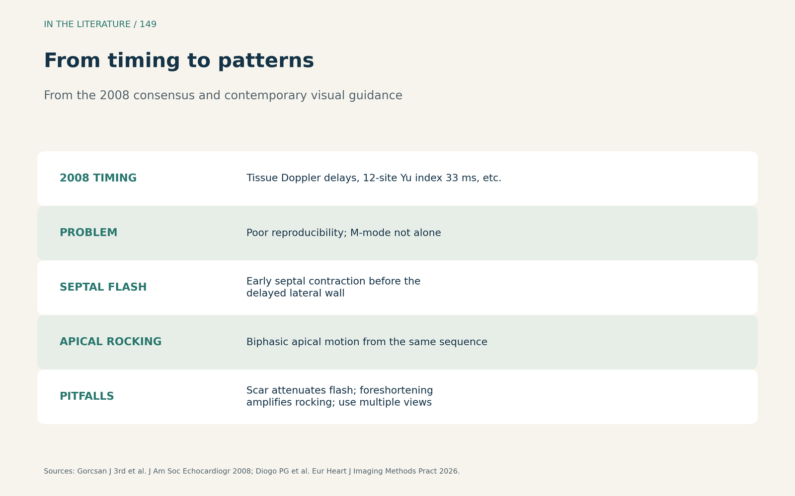
Figure 1. From timing to patterns. Synthesized from the ASE consensus and Diogo et al.[1], [2] View larger.

Quantitative markers before implantation
Riolet and colleagues measured myocardial work from pressure-strain loops: low preoperative global wasted work was associated with absence of response, and only 37% of such patients responded. Immediate changes in wasted work after implantation were not prognostic, and the authors state that patients with low wasted work should not be excluded from therapy.[3]
In the multicenter MARC study of 240 patients, Wouters and colleagues found sustained remodeling in 114 (57%). Despite a mean QRS duration of 180 ms, 39% did not respond. With both high QRS area and septal rebound stretch, 90% responded, compared with 54% with high QRS area alone; the authors acknowledge that echo dyssynchrony analysis still has no place in contemporary practice.[4]
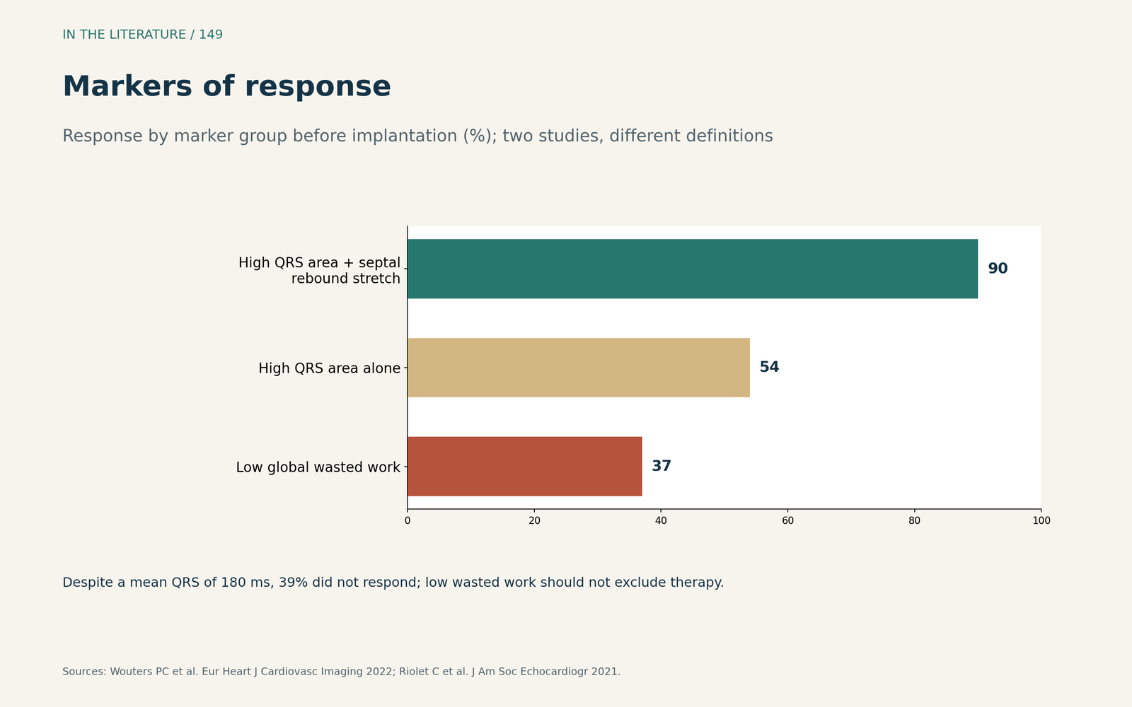
Figure 2. Markers of response. Redrawn from Riolet et al. and Wouters et al.[3], [4] View larger.

After implantation and in the operating room
Melki and colleagues note that one third of patients do not improve despite a narrowed QRS. Their electromechanical wave imaging quantified the percentage of the left ventricle activated within 120 ms of QRS onset within 24 hours of implantation, but it was a single-center pilot.[5]
Nelson and colleagues describe new severe functional mitral regurgitation after non-mitral cardiac surgery with normal leaflets and normal ventricular function. Epicardial pacing produced left ventricular dyssynchrony; native conduction reduced it, and regurgitation fell. They recommend trying to improve conduction before returning to bypass for the mitral valve.[6]
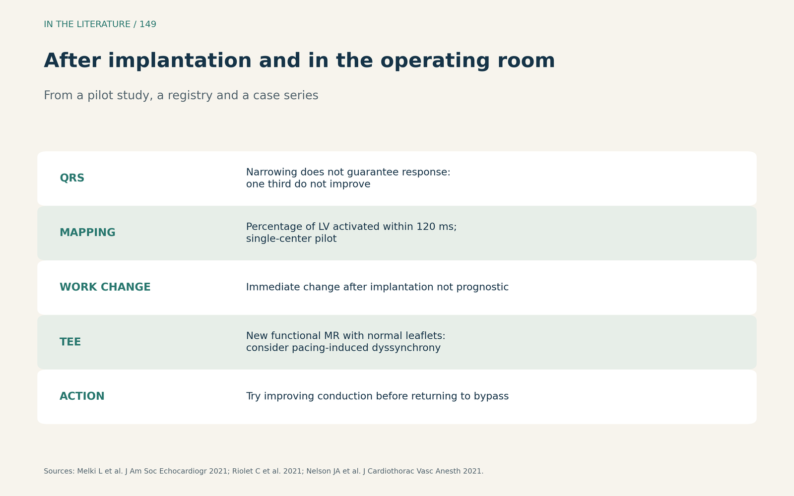
Figure 3. After implantation and in the operating room. Synthesized from Melki et al., Riolet et al. and Nelson et al.[5], [3], [6] View larger.

Three takeaways
- Look for the pattern. Septal flash and apical rocking on good multi-view images matter more than timing delays.[1], [2]
- Use markers to inform, not exclude. Wasted work and septal stretch refine prediction but should not deny eligible patients therapy.[3], [4]
- Think of pacing in the operating room. New functional mitral regurgitation with normal leaflets may be pacing-induced dyssynchrony.[6]
References
- Gorcsan J 3rd, Abraham T, Agler DA, et al. Echocardiography for cardiac resynchronization therapy: recommendations for performance and reporting--a report from the American Society of Echocardiography Dyssynchrony Writing Group endorsed by the Heart Rhythm Society. J Am Soc Echocardiogr. 2008;21(3):191–213. doi:10.1016/j.echo.2008.01.003
- Diogo PG, Demesure K, Puvrez A, et al. How to visually diagnose mechanical dyssynchrony in cardiac resynchronization therapy candidates using echocardiography. Eur Heart J Imaging Methods Pract. 2026;4(1):qyaf158. doi:10.1093/ehjimp/qyaf158
- Riolet C, Menet A, Mailliet A, et al. Clinical significance of global wasted work in patients with heart failure receiving cardiac resynchronization therapy. J Am Soc Echocardiogr. 2021;34(9):976–986. doi:10.1016/j.echo.2021.06.008
- Wouters PC, van Everdingen WM, Vernooy K, et al. Does mechanical dyssynchrony in addition to QRS area ensure sustained response to cardiac resynchronization therapy? Eur Heart J Cardiovasc Imaging. 2022;23(12):1628–1635. doi:10.1093/ehjci/jeab264
- Melki L, Wang DY, Grubb CS, et al. Cardiac resynchronization therapy response assessment with electromechanical activation mapping within 24 hours of device implantation: a pilot study. J Am Soc Echocardiogr. 2021;34(7):757–766.e8. doi:10.1016/j.echo.2021.02.012
- Nelson JA, Espinosa R, Michelena H, Rehfeldt K. Acute severe functional mitral regurgitation after non-mitral valve cardiac surgery--left ventricular dyssynchrony as a potential mechanism. J Cardiothorac Vasc Anesth. 2021;35(5):1292–1298. doi:10.1053/j.jvca.2020.08.045