ITL-a39e0451c500
Juan Li, MD · Updated November 17, 2026
Watch the video
Key points
- The classic target is uncertain. In 268 arrests, a maximal LV point estimated nearer the midsternum was linked to worse ROSC and survival, challenging the maximal-LV target.[1]
- The outflow tract matters in swine. Compression over the LVOT collapsed it in 82% vs 10% of compressions, and mid-LV compression raised coronary perfusion pressure by 15.6 mmHg.[2]
- Clinicians already adjust. 96.7% of surveyed clinicians had moved the compression site during TEE-guided CPR, mostly leftward (61.0%); the lower-half-of-sternum recommendation is weak.[3]
- TEE can show the problem. In one case, compressions over the LVOT and aortic valve were moved toward the LV apex under TEE guidance.[4]
- Outcomes are unproven. The swine study did not assess survival, and clinical trials of TEE in arrest are lacking.[2], [4]
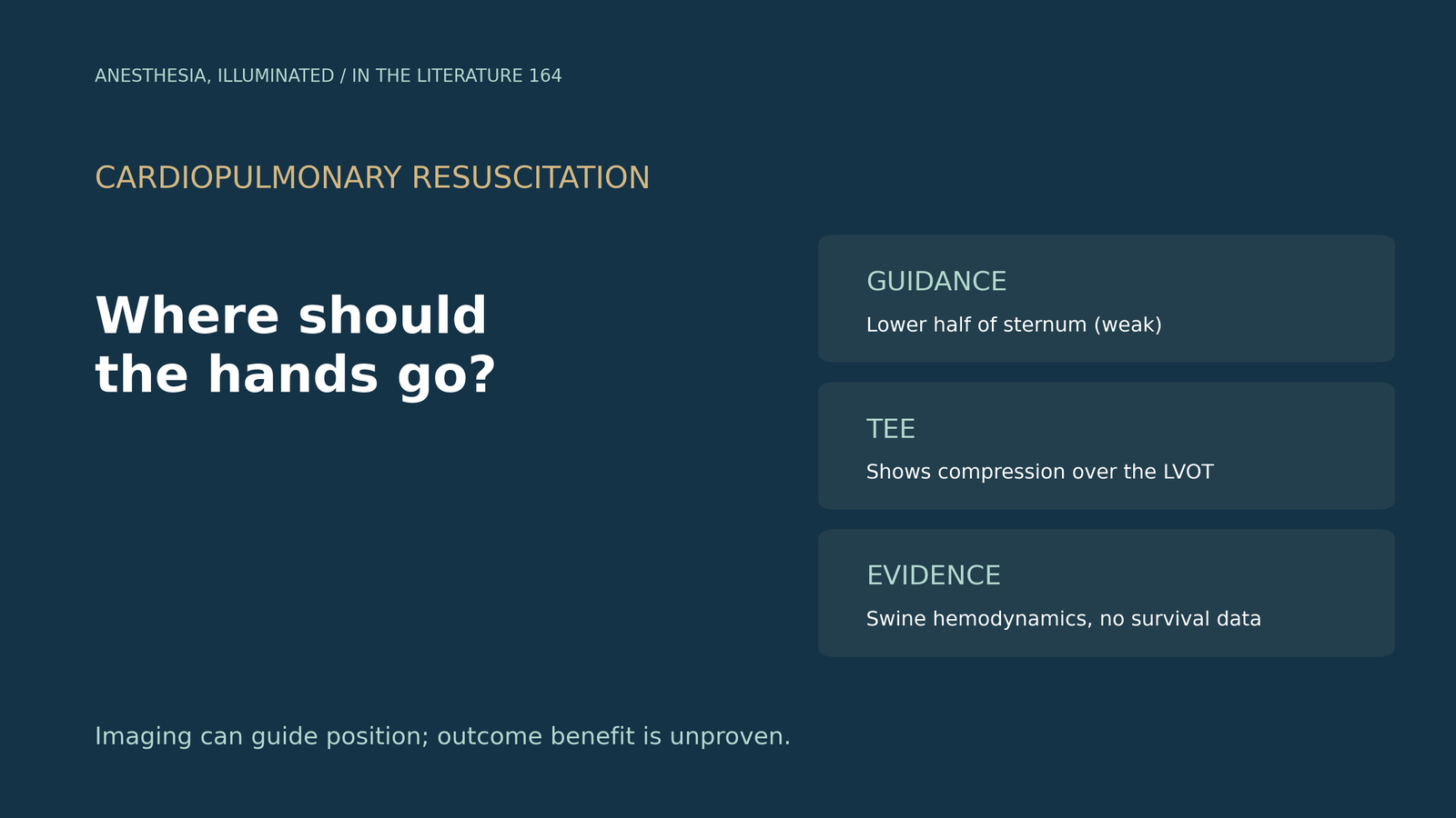
During an intraoperative cardiac arrest with a TEE probe in place, the mid-oesophageal long-axis view shows the compressions squeezing the aortic root rather than the ventricle. Should the hands move, and what is the evidence?[2], [4]
Is the maximal LV diameter the right target?
Park and colleagues estimated, from old chest radiographs, the point over the maximal LV diameter in out-of-hospital arrests. Of 268 patients, 123 (45.9%) achieved ROSC and 40 (14.9%) survived to discharge; patients whose point lay closer to the midsternum had worse ROSC and survival, which the authors say challenges the maximal-LV target. They note that the personalized point could hardly be located during actual CPR, suggest that LV and RV stroke volumes should be balanced, and acknowledge that the retrospective design cannot show causality and that selection bias is possible.[1]
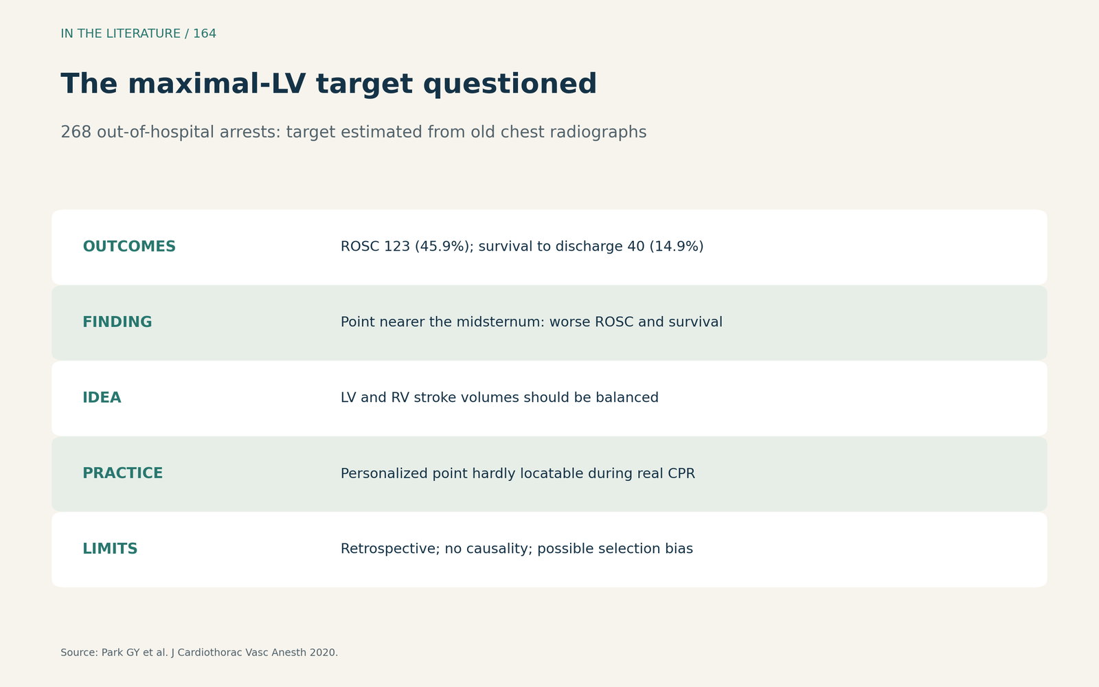
Figure 1. The maximal-LV target questioned. Redrawn from Park et al.[1] View larger.

What TEE showed in swine
Standard guidance places compressions over the lower half of the sternum.[2] In Teran's randomized swine model, the LVOT compression site lay about 2 cm caudal to the aortic annulus; LVOT collapse occurred in 82% vs 10% of compressions, mid-LV compression raised coronary perfusion pressure by 15.6 mmHg, and end-tidal CO2 averaged 30.9 mmHg with mid-LV compression. The authors argue that compression location is modifiable, like rate and depth, but did not assess ROSC or survival, and two pilot animals were added to the final cohort.[2]
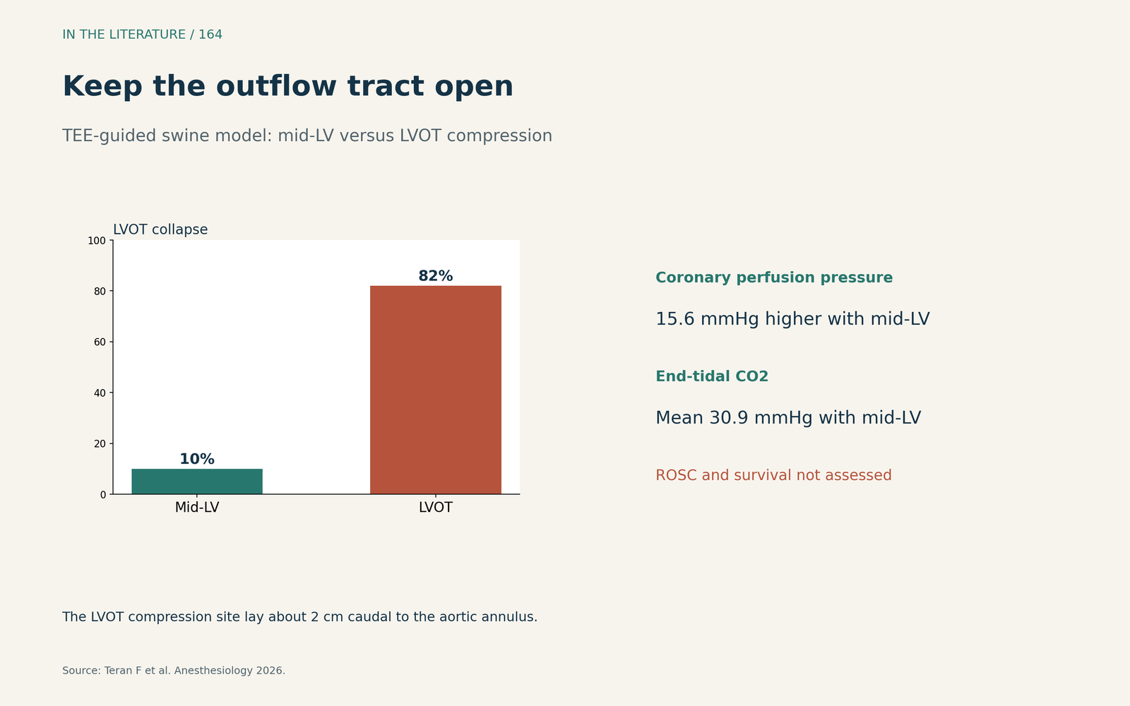
Figure 2. Keep the outflow tract open. Redrawn from Teran et al.[2] View larger.

What clinicians do
Marshall and colleagues note that the lower-half-of-sternum recommendation is weak, based on very-low-certainty evidence. In their survey, 96.7% of clinicians had moved the compression site during TEE-guided CPR, mostly leftward (61.0%) and then caudally (26.2%); injury effects of moving the site are unknown, and recall bias limits the data.[3] Amador describes TEE showing compressions over the LVOT and aortic valve that were then moved toward the LV apex, while noting that clinical trials of TEE in arrest are lacking.[4]
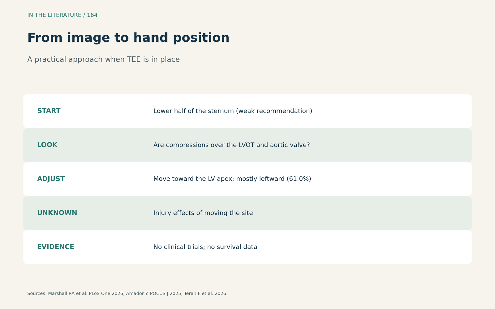
Figure 3. From image to hand position. Synthesized from Marshall et al., Amador and Teran et al.[3], [4], [2] View larger.

Three takeaways
- Location may matter. Compressions over the outflow tract can impair CPR hemodynamics, at least in swine.[2]
- If TEE is in place, look. It can show compressions over the aortic root, and the site can be adjusted.[3], [4]
- Do not overclaim. No human study shows better survival from imaging-guided hand position.[1], [2]
References
- Park GY, Oh WS, Chon SB, Kim S. The maximum diameter of the left ventricle may not be the optimum target for chest compression during cardiopulmonary resuscitation: a preliminary, observational study challenging the traditional assumption. J Cardiothorac Vasc Anesth. 2020;34(2):383–391. doi:10.1053/j.jvca.2019.07.005
- Teran F, Owyang CG, Martin-Flores M, et al. Outflow tract patency and ventricular compression as determinants of cardiopulmonary resuscitation hemodynamics in a transesophageal echocardiography–guided swine model of cardiac arrest. Anesthesiology. 2026;145(3):638–649. doi:10.1097/ALN.0000000000006175
- Marshall RA, DeVito-Porter K, Dallaire G, et al. Transesophageal echocardiography during cardiopulmonary resuscitation is associated with alternate areas of compression: analysis of healthcare provider experiences with potential implications for conventional CPR. PLoS One. 2026;21(1):e0339974. doi:10.1371/journal.pone.0339974
- Amador Y. Point of care transesophageal echocardiogram-guided CPR: area of maximal compression. POCUS J. 2025;10(1):61–63. doi:10.24908/pocusj.v10i01.18111