ITL-9a274e0681a1
Juan Li, MD · Updated November 18, 2026
Watch the video
Key points
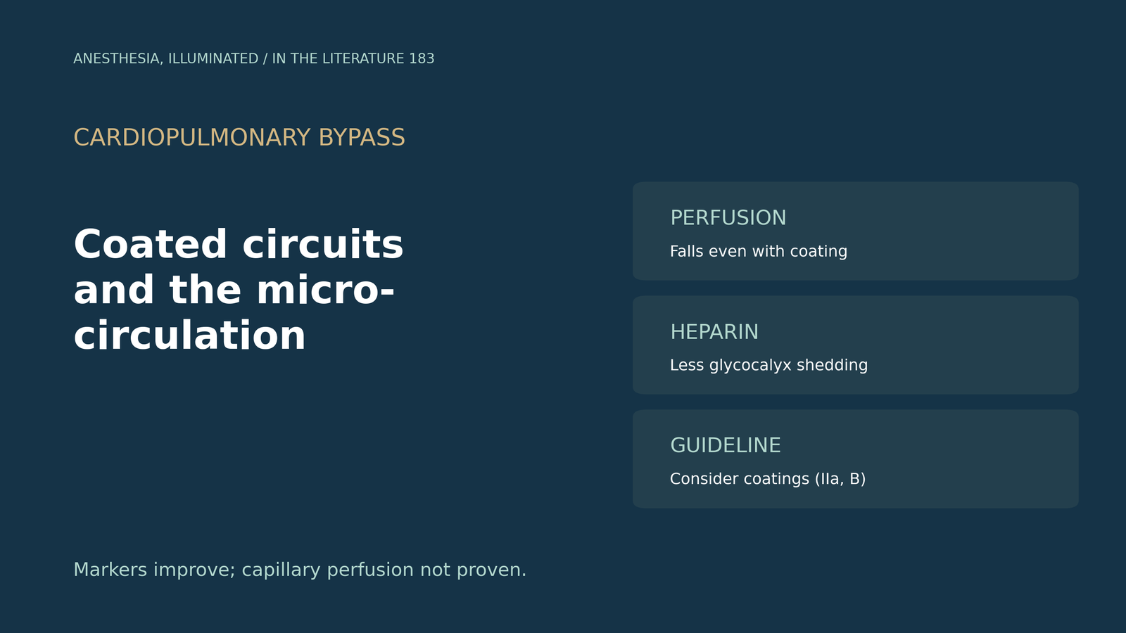
- Coating does not prevent the fall. With a phosphorylcholine-coated circuit, the proportion of perfused sublingual vessels fell on bypass and stayed impaired for three postoperative days.[1]
- The problem is perfusion, not vessel loss. Bypass mainly reduces the fraction of perfused microvessels, as reported in two thirds of sublingual imaging studies.[2]
- Heparin may spare the glycocalyx. Heparin coating has been linked to less complement activation, lower syndecan-1 and smaller rises in perfused boundary region.[2]
- Phosphorylcholine alone does not stop activation. Complement rose despite a phosphorylcholine circuit (C3a 8.8-fold), and coated circuits did not attenuate inflammation in a small trial.[3], [4]
- Guidelines are cautious. Biocompatible coatings "should be considered" (Class IIa, Level B), based on about 5 hours shorter ICU stay and no difference in deaths.[5]
Before coronary surgery, the perfusion team asks whether a heparin-coated or a phosphorylcholine-coated circuit would better protect the patient's microcirculation. Does either coating actually improve capillary perfusion?[1], [2]
What happens to the microcirculation on bypass
Dekker and colleagues imaged the sublingual microcirculation in 17 patients having on-pump coronary surgery with a phosphorylcholine-coated circuit; mean non-pulsatile bypass time was 103 minutes. The proportion of perfused vessels fell on bypass and did not recover after transfusion of salvaged blood, and the perfusion disturbances persisted for the first three postoperative days. Syndecan-1 rose after weaning, consistent with glycocalyx shedding. All measurements were sublingual, and there was no comparison circuit.[1] Zheng and colleagues' review notes that bypass reduced sublingual perfusion measures in two thirds of imaging studies and that it mainly reduces the fraction of microvessels that are perfused.[2]
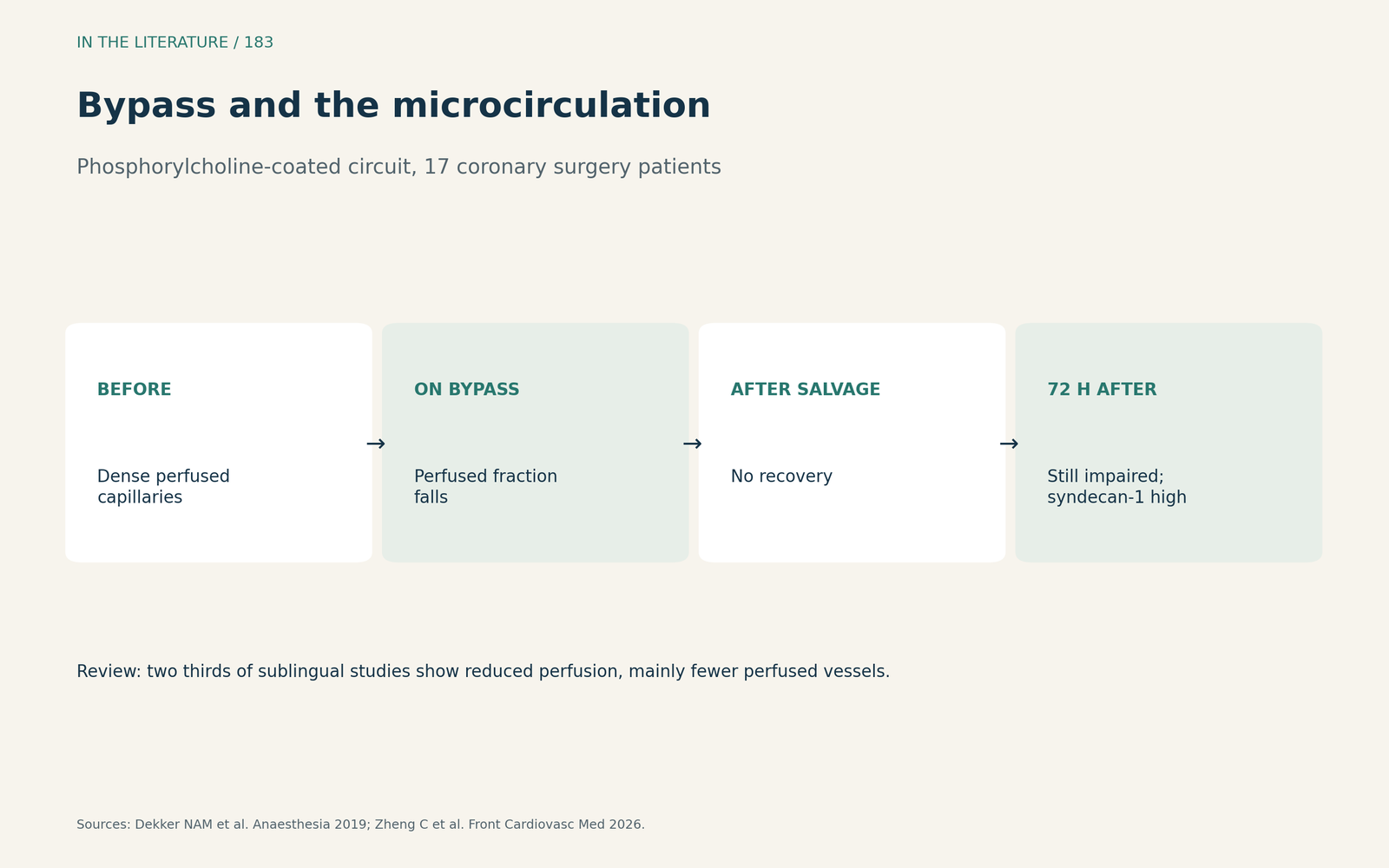
Figure 1. Perfusion falls and stays low. Synthesized from Dekker et al. and Zheng et al.[1], [2] View larger.

What coatings change
According to Zheng and colleagues, heparin-coated circuits have been shown to attenuate complement activation, and a clinical study found lower syndecan-1 and smaller rises in perfused boundary region with heparin coating, consistent with partial preservation of the glycocalyx. They conclude that clinical evidence for protecting the microcirculation remains inconsistent.[2] Bierer and colleagues ran 45 ex vivo primes in a phosphorylcholine-coated circuit and found significant complement activation despite modern biocompatible technology, with C3a rising 8.8-fold after adjustment; the in vivo reaction could differ.[3] In Hatemi's study of 32 coronary surgery patients allocated to coated or uncoated circuits, coated circuits did not attenuate the inflammatory response.[4]
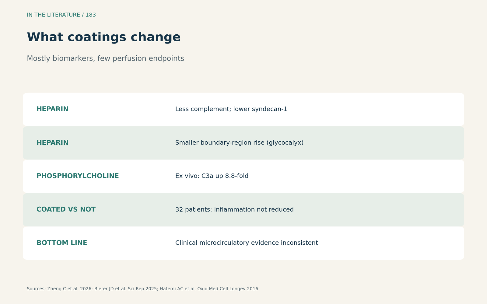
Figure 2. Markers move, perfusion evidence is thin. Synthesized from Zheng et al., Bierer et al. and Hatemi et al.[2], [3], [4] View larger.

What the guideline says
The 2024 EACTS/EACTAIC/EBCP guideline states that biocompatible surface treatment to reduce complications should be considered (Class IIa, Level B). In the meta-analysis it cites, coatings shortened ICU stay by about 5 hours but did not reduce deaths (OR 0.78), and 78% of the trials used heparin-bonded surfaces only. The guideline summarizes that biocompatible surfaces may reduce postoperative inflammation; it gives no microcirculatory endpoint.[5]
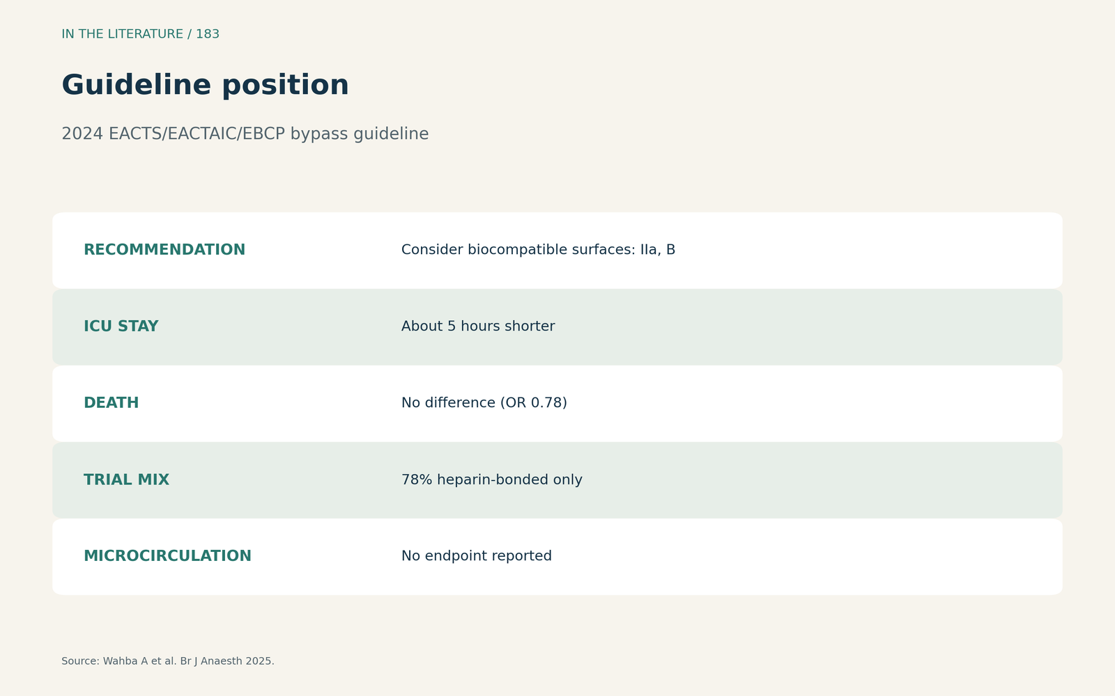
Figure 3. Consider coatings, but expect modest effects. Synthesized from Wahba et al.[5] View larger.

Three takeaways
- Coated or not, bypass impairs the microcirculation, and the impairment can last days.[1]
- Heparin coating may spare the glycocalyx more than phosphorylcholine, but proof of better perfusion is lacking.[2], [3]
- Coatings are reasonable for modest clinical benefits, not as microcirculatory protection.[5]
References
- Dekker NAM, Veerhoek D, Koning NJ, et al. Postoperative microcirculatory perfusion and endothelial glycocalyx shedding following cardiac surgery with cardiopulmonary bypass. Anaesthesia. 2019;74(5):609–618. doi:10.1111/anae.14577
- Zheng C, Muir K, Kaushik H, et al. Microcirculation dysfunction and cardioprotection in cardiac surgery with cardiopulmonary bypass: mechanisms, monitoring, and therapeutic strategies. Front Cardiovasc Med. 2026;13:1843167. doi:10.3389/fcvm.2026.1843167
- Bierer JD, Stanzel R, Henderson M, et al. Complement activation by the artificial surface of cardiopulmonary bypass is a persistent clinical problem. Sci Rep. 2025;15(1):27643. doi:10.1038/s41598-025-11157-w
- Hatemi AC, Çeviker K, Tongut A, Özgöl İ, Mert M, Kaya A. Oxidant status following cardiac surgery with phosphorylcholine-coated extracorporeal circulation systems. Oxid Med Cell Longev. 2016;2016:3932092. doi:10.1155/2016/3932092
- Wahba A, Kunst G, De Somer F, et al. 2024 EACTS/EACTAIC/EBCP guidelines on cardiopulmonary bypass in adult cardiac surgery. Br J Anaesth. 2025;134(4):917–1008. doi:10.1016/j.bja.2025.01.015