ITL-91119735c521
Juan Li, MD · Updated November 17, 2026
Watch the video
Key points
- Both conditions cause heart failure with a preserved ejection fraction, but constriction is potentially curable by pericardiectomy and restriction is not. In experienced hands, echocardiography is often enough to make the diagnosis.[1], [2]
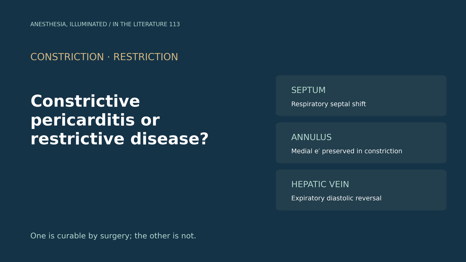
- Three echocardiographic findings favor constriction: respiratory septal shift, a preserved medial mitral annular velocity (e′ above 9 cm/s) and expiratory diastolic flow reversal in the hepatic veins. Combining septal shift with either of the others gave a sensitivity of 87% and a specificity of 91%.[1], [2]
- Single findings can mislead. Mitral respiratory variation may be absent in up to a third of constriction, septal shift occurs in 31% of restrictive cardiomyopathy or severe tricuspid regurgitation, and a normal collapsible inferior vena cava essentially excludes constriction.[1], [3]
- Pericardial thickness can be normal in up to 20% of constriction. Cardiac MRI is about 93% accurate for thickening above 4 mm, shows myocardial enhancement in about a third of restrictive cases, and pericardial enhancement suggests constriction that may reverse.[4], [3], [2]
- The echocardiographic criteria were validated in spontaneously breathing patients and change under positive-pressure ventilation. Pericardiectomy carries perioperative mortality below 5% overall but above 20% after chest radiation.[5], [6], [2]
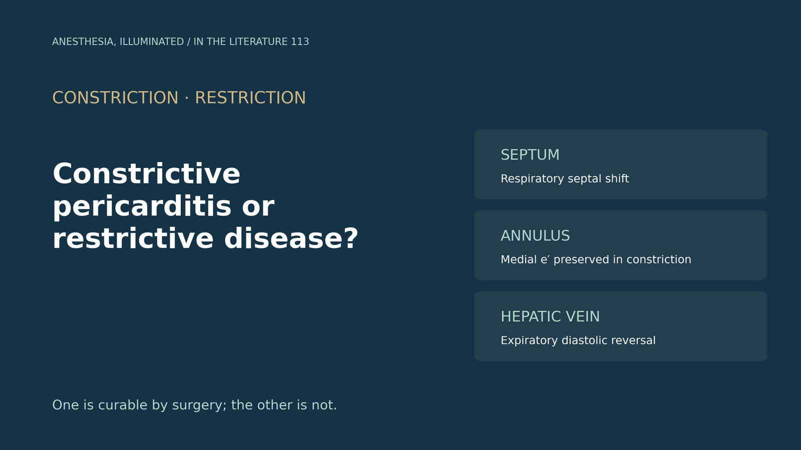
A patient with edema, ascites and a raised jugular venous pressure has a preserved ejection fraction on echocardiography and is referred for possible pericardiectomy. Is this constriction, which surgery could cure, or a restrictive cardiomyopathy, and what should the echocardiographer look for, including in the operating room?[1], [2]
Why the distinction matters
Constriction and restriction look alike clinically, but Geske and colleagues emphasize that constriction is a potentially curable cause of heart failure.[2] Lloyd and colleagues report that, in their practice, echocardiography obviates the need for invasive hemodynamics in most surgically proven cases.[1]
The echocardiographic criteria
Ventricular interdependence is the hallmark of constriction. In the criteria summarized by Geske and colleagues, respiratory septal shift had a sensitivity of 93% but a specificity of 69%; a mitral E-wave variation above 14.6% had 84% and 73%; a medial e′ above 9 cm/s had 83% and 81%; and an expiratory hepatic vein diastolic reversal ratio of 0.79 or more had 76% and 88%. Septal shift combined with either the medial e′ or the hepatic vein criterion gave 87% sensitivity and 91% specificity.[2]
In restriction the myocardium itself is abnormal, so medial e′ is low and may be as low as 2 to 3 cm/s, and longitudinal strain is lower than in constriction.[1] In constriction, tethering of the lateral annulus to the thickened pericardium explains why the lateral velocity can fall below the medial (annulus reversus).[3]
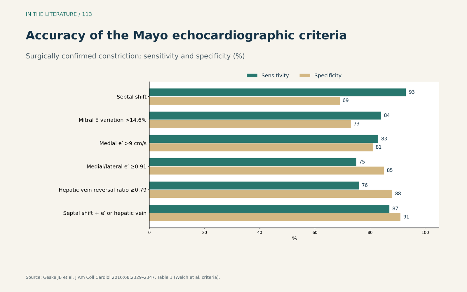
Figure 1. Accuracy of the Mayo echocardiographic criteria. Redrawn from Table 1 of Geske et al. (surgically confirmed constriction).[2] View larger.

Pitfalls of single findings
Mitral respiratory variation might be absent in up to one-third of cases, for example when filling pressures are very high. Septal shift is not specific: it occurred in 31% of patients with restrictive cardiomyopathy or severe tricuspid regurgitation. A normal-sized, collapsible inferior vena cava essentially excludes hemodynamically significant constriction.[1]
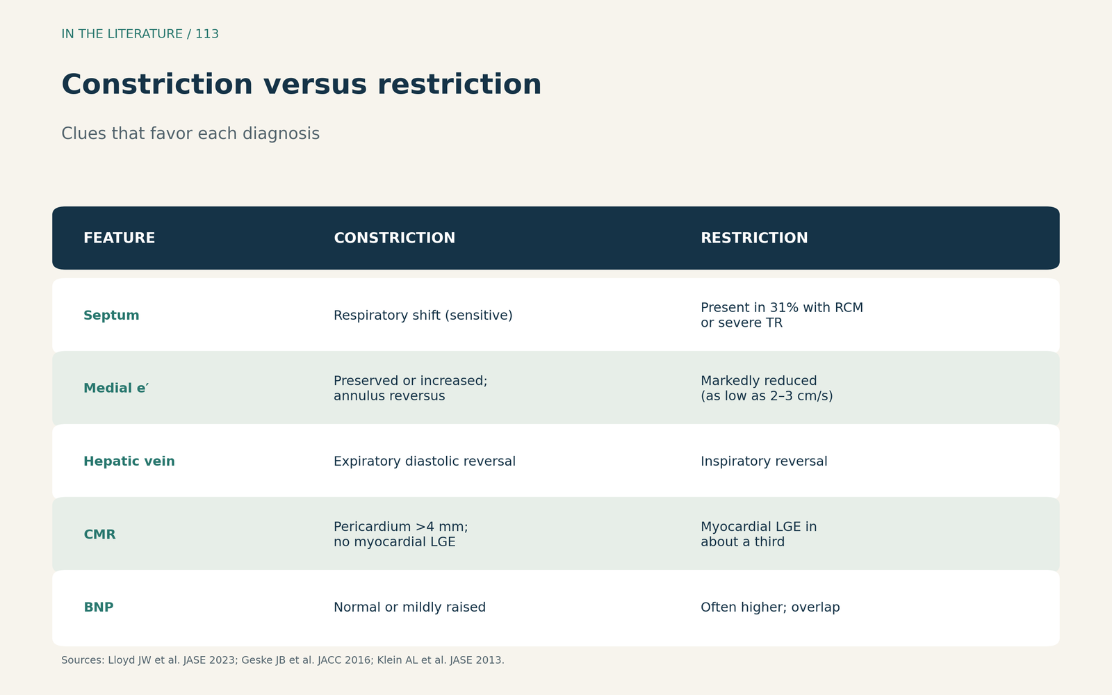
Figure 2. Constriction versus restriction. Synthesized from Lloyd et al., Geske et al. and the ASE recommendations; each feature shifts probability rather than proving a diagnosis.[1], [2], [3] View larger.

Cardiac MRI, CT and catheterization
A thick pericardium supports constriction, but its absence does not exclude it: the ESC guideline notes normal thickness in up to 20% of cases, and Lloyd and colleagues cite almost 28% in surgically confirmed disease.[4], [1] Cardiac MRI has an accuracy of 93% for pericardial thickening above 4 mm, and pericardial enhancement may predict reversibility.[3] The ESC notes that anti-inflammatory therapy may resolve transient constriction, which occurs in 10–20% of cases.[4] In restriction, myocardial enhancement is present in nearly a third of cases, and real-time cine imaging of septal motion can discriminate the two.[1]
When imaging is inconclusive, invasive hemodynamics help: a systolic area index above 1.1 was 97% sensitive and 100% specific for constriction. BNP tends to be higher in restriction than in constriction, with overlap.[2]
In the operating room
The echocardiographic criteria have been validated only in spontaneously breathing patients. Under positive-pressure ventilation the respiratory patterns change, and hepatic vein reversals may occur with inspiration, show no respiratory variation or disappear. Operating within 6 months of symptom onset was associated with lower mortality.[5] Helwani and colleagues released the left ventricular side before the right during pericardiectomy, and after release the hepatic vein pattern indicated improved, but not normalized, right atrial pressure.[6]
Outcomes depend on the cause. Geske and colleagues report perioperative mortality generally below 5%, but above 20% after prior radiation.[2] In a series cited by Liu and colleagues, 5-year survival was 79.6% after idiopathic constriction and 0% after radiation-induced constriction.[5]
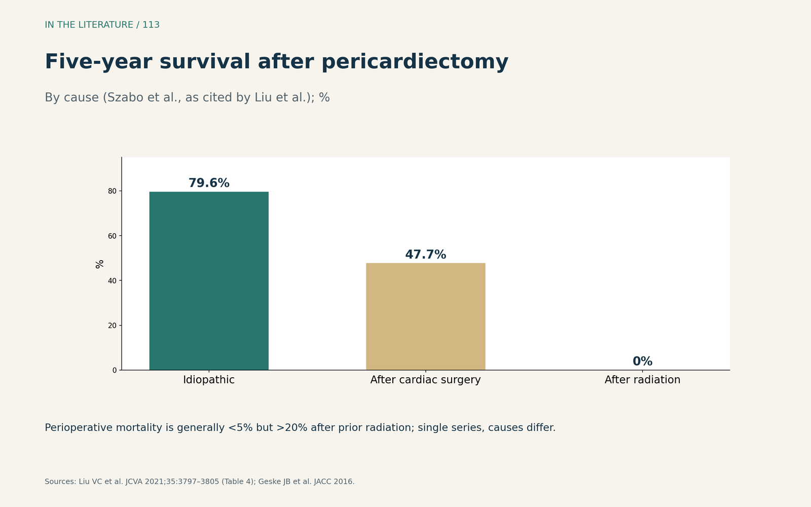
Figure 3. Five-year survival after pericardiectomy. Redrawn from the series of Szabo et al. as tabulated by Liu et al.; single series, causes not directly comparable.[5], [2] View larger.

Three takeaways
- Look for interdependence, the annulus and the hepatic vein together. Septal shift with a preserved medial e′ or expiratory hepatic vein reversal favors constriction.[1], [2]
- Do not trust one finding. Mitral variation can be absent, septal shift occurs in restriction, and the pericardium can be normal in thickness; use MRI or catheterization when in doubt.[1], [3], [4]
- Interpret carefully under ventilation and plan for risk. Criteria change with positive-pressure ventilation, and radiation-related constriction carries high surgical risk.[2], [5], [6]
References
- Lloyd JW, Anavekar NS, Oh JK, Miranda WR. Multimodality imaging in differentiating constrictive pericarditis from restrictive cardiomyopathy: a comprehensive overview for clinicians and imagers. J Am Soc Echocardiogr. 2023;36(12):1254–1265. doi:10.1016/j.echo.2023.08.016
- Geske JB, Anavekar NS, Nishimura RA, et al. Differentiation of constriction and restriction: complex cardiovascular hemodynamics. J Am Coll Cardiol. 2016;68(21):2329–2347. doi:10.1016/j.jacc.2016.08.050
- Klein AL, Abbara S, Agler DA, et al. American Society of Echocardiography clinical recommendations for multimodality cardiovascular imaging of patients with pericardial disease: endorsed by the Society for Cardiovascular Magnetic Resonance and Society of Cardiovascular Computed Tomography. J Am Soc Echocardiogr. 2013;26(9):965–1012.e15. doi:10.1016/j.echo.2013.06.023
- Adler Y, Charron P, Imazio M, et al. 2015 ESC guidelines for the diagnosis and management of pericardial diseases. Eur Heart J. 2015;36(42):2921–2964. doi:10.1093/eurheartj/ehv318
- Liu VC, Fritz AV, Burtoft MA, et al. Pericardiectomy for constrictive pericarditis: analysis of outcomes. J Cardiothorac Vasc Anesth. 2021;35(12):3797–3805. doi:10.1053/j.jvca.2021.02.020
- Helwani MA, Alber S, DeWet CJ, et al. Intraoperative assessment of hepatic vein tracings in constrictive pericarditis during surgical pericardiectomy. J Cardiothorac Vasc Anesth. 2020;34(4):1099–1102. doi:10.1053/j.jvca.2019.11.011