ITL-f74275a7bffd
Juan Li, MD · Updated November 17, 2026
Watch the video
Key points
- It can masquerade as mitral stenosis. Adults can develop gradual obstruction that mimics mitral stenosis, and the membrane inserts above the left atrial appendage.[1]
- Listen and look. There is no opening snap or loud S1, and a supravalvular ring lies below the appendage rather than above it.[2]
- Grade it like mitral stenosis. A Doppler velocity above 2 m/s indicates severe obstruction, and the opening matters: patients are usually asymptomatic when it exceeds 1 cm.[2]
- Use 3D and check the veins. 3D TEE can measure the fenestration, and pulmonary vein velocity over 125 cm/s with a monophasic waveform suggests venous obstruction.[3]
- Plan the anesthetic like mitral stenosis. Observation suits asymptomatic patients without obstruction, and a left superior vena cava accompanies 25% to 32% of cases.[4]
An adult with exertional dyspnea and pulmonary hypertension is thought to have mitral stenosis, but intraoperative TEE shows a thin membrane across the left atrium and a normal mitral valve. Is this cor triatriatum, and how is it distinguished from mitral stenosis or a supramitral ring?[1], [2]
Why it looks like mitral stenosis
Preston and Moreno-Duarte describe cor triatriatum in adults as gradual obstruction mimicking mitral stenosis; it is distinguished from a supravalvular mitral ring by insertion above the left atrial appendage, an atrial septal defect can decompress the proximal chamber, and echocardiography is indispensable for diagnosis.[1] Butt and colleagues note that only 170 adult cases had been reported, that patients are likely asymptomatic when the opening exceeds 1 cm, and that absence of an opening snap or loud S1 distinguishes it from mitral stenosis.[2]
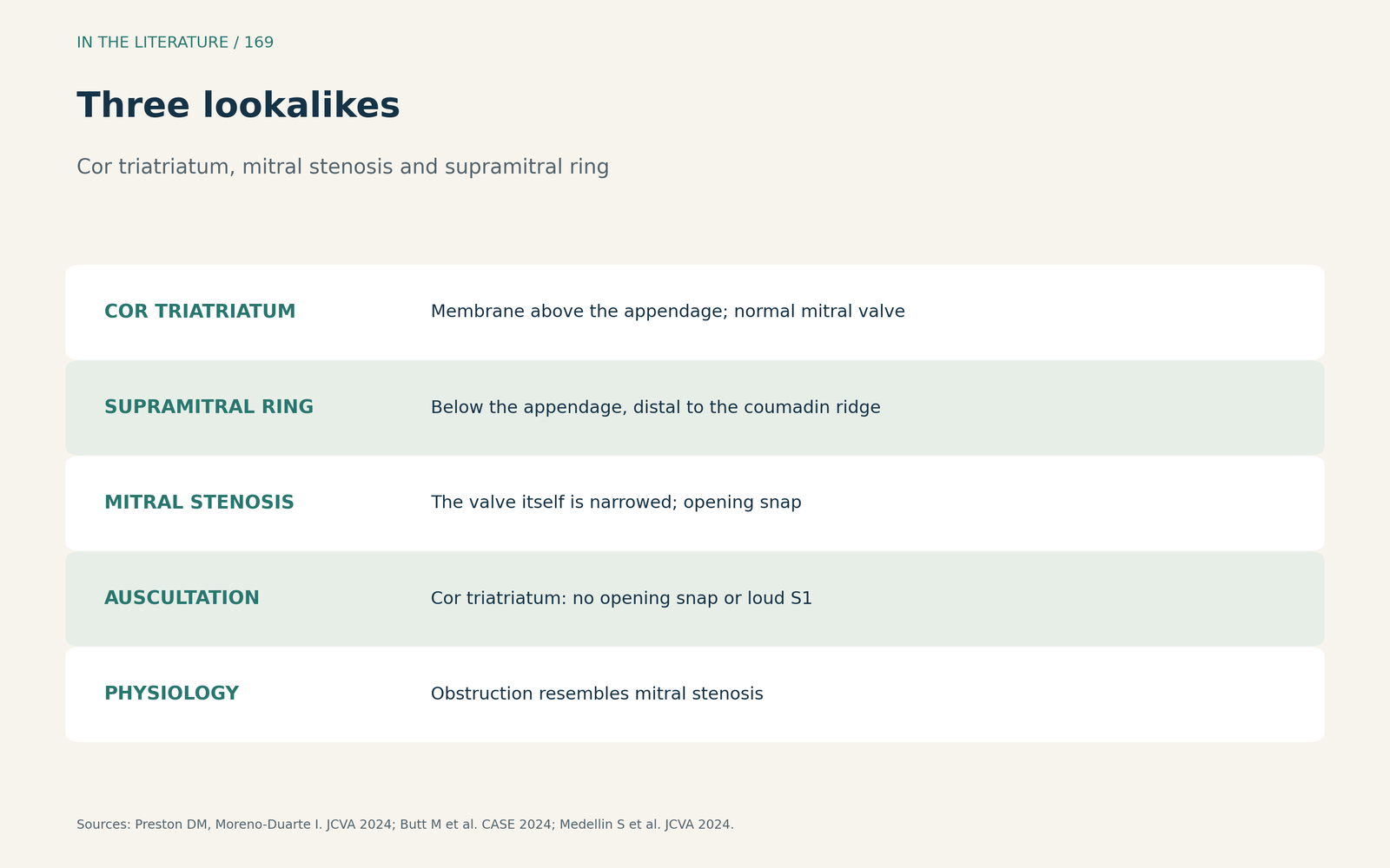
Figure 1. Three lookalikes. Synthesized from Preston and Moreno-Duarte, Butt et al. and Medellin et al.[1], [2], [3] View larger.

Echo and Doppler
In Butt's case the opening measured 0.8 cm2; severe obstruction is indicated by a Doppler velocity above 2 m/s, a supravalvular ring lies below the appendage, preoperative CT detected cor triatriatum 50% more often in a cited analysis, and surgery is indicated for symptoms from the obstruction.[2] Medellin and colleagues found that TTE showed the membrane but could not quantify its hemodynamics, whereas 3D TEE measured a fenestration of 1.14 cm2; a supramitral ring inserts distal to the coumadin ridge, the obstruction resembles mitral stenosis physiology, and pulmonary vein velocity over 125 cm/s with a monophasic waveform suggests venous obstruction.[3]
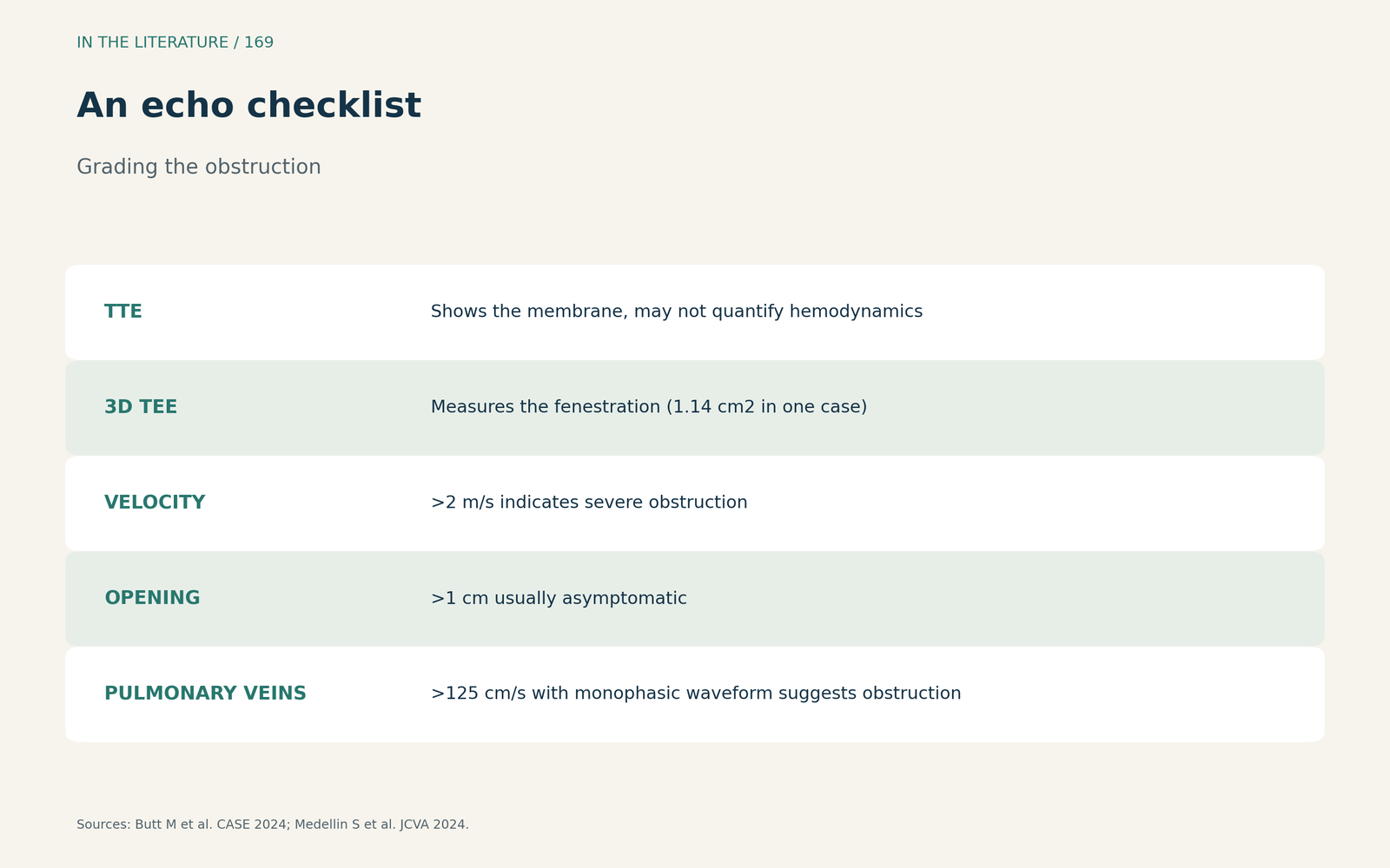
Figure 2. An echo checklist. Synthesized from Butt et al. and Medellin et al.[2], [3] View larger.

Natural history and anesthetic care
Fernando and colleagues describe a membrane attached to the warfarin ridge found incidentally before bypass surgery. Survival to adulthood requires an opening larger than 10 mm, a persistent left superior vena cava is present in 25% to 32%, observation is warranted without inlet obstruction and with normal pulmonary pressure, cor triatriatum can also be acquired, and anesthetic goals are similar to those of mitral stenosis.[4]
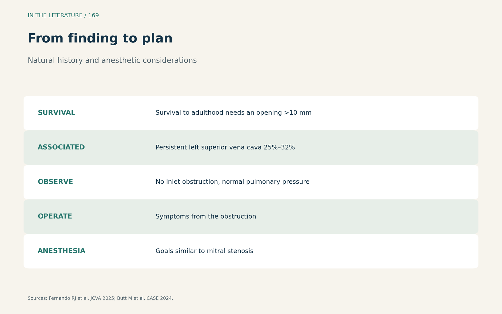
Figure 3. From finding to plan. Synthesized from Fernando et al. and Butt et al.[4], [2] View larger.

Three takeaways
- Check where the membrane inserts. Above the appendage suggests cor triatriatum; below it, a supramitral ring.[1], [2]
- Grade it like mitral stenosis. Velocity, gradient, opening size and the pulmonary veins.[2], [3]
- Anesthetize it like mitral stenosis. And look for a left superior vena cava.[4]
References
- Preston DM, Moreno-Duarte I. Decoding the enigma of cor triatriatum sinister manifesting in adulthood. J Cardiothorac Vasc Anesth. 2024;38(2):581. doi:10.1053/j.jvca.2023.11.004
- Butt M, Halim H, Qureshi A, et al. A tale of three chambers: cor triatriatum sinistrum. CASE (Phila). 2024;8(3):221–225. doi:10.1016/j.case.2023.12.019
- Medellin S, Burbano-Vera N, Alfirevic A. Obstructed supramitral inflow: cor triatriatum sinister presentation in adulthood. J Cardiothorac Vasc Anesth. 2024;38(2):576–580. doi:10.1053/j.jvca.2023.11.006
- Fernando RJ, Buck JK, Augoustides JG, et al. Incidental discovery of forme fruste cor triatriatum sinister in an adult presenting for emergent coronary artery bypass graft surgery. J Cardiothorac Vasc Anesth. 2025;39(5):1335–1344. doi:10.1053/j.jvca.2025.01.001