ITL-4cd99d54e789
Juan Li, MD · Updated November 17, 2026
Watch the video
Key points
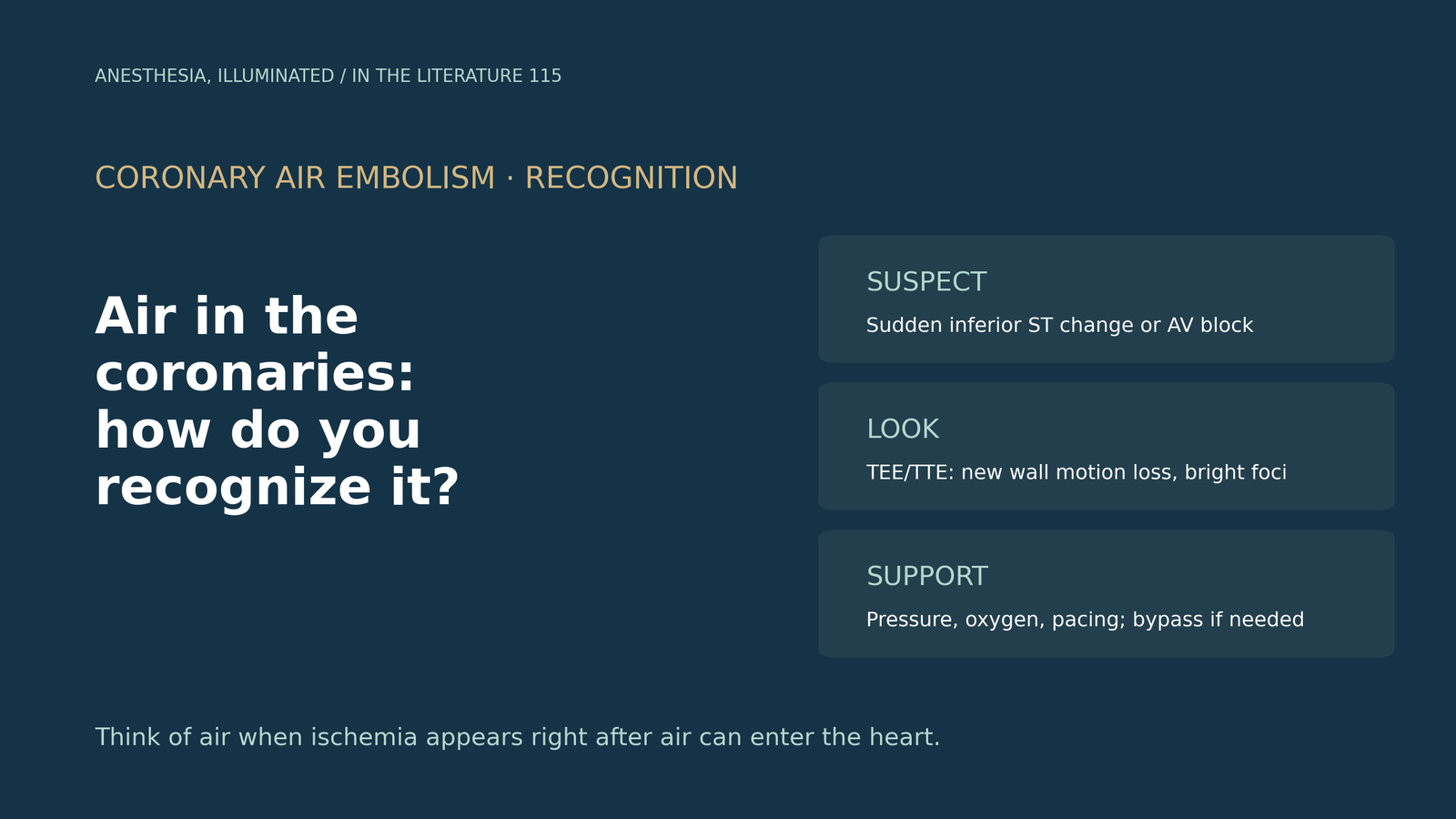
- Air can reach the coronary arteries whenever it enters the left heart or aortic root, during cardiac surgery, catheterization or ablation. A common presentation is acute inferior ischemia and/or heart block, because air tends to enter the right coronary artery, whose ostium sits uppermost in the supine patient, although the left anterior descending artery is most often reported after angiography.[1], [2]
- The electrocardiogram can be unremarkable. Keep a high index of suspicion, watch the ECG, and use echocardiography to look for new regional wall motion loss and bright foci within the myocardium, which suggest intramyocardial air.[3], [2]
- In cardiac surgery, microbubbles seen on transesophageal echocardiography (TEE) after unclamping were associated with perioperative arrhythmia in a retrospective myectomy cohort, and continuous carbon dioxide insufflation was associated with fewer arrhythmias (24.12% vs 36.59%).[4]
- Treatment is supportive: inspired oxygen fraction 1.0, a vasopressor to raise coronary perfusion pressure, pacing or cardiopulmonary resuscitation if AV block and hypotension persist, and extracorporeal support if other measures fail. Most cases resolve within minutes.[1], [2], [3]
- The evidence is thin: consensus statements, reviews, case reports and observational studies. No source gives a blood pressure target, and the hyperbaric oxygen data concern cerebral, not coronary, gas embolism.[5], [6]
Soon after separation from bypass, or during a catheter procedure, the patient suddenly develops inferior ST changes, bradycardia and hypotension. Could this be air in the coronary arteries, how can it be confirmed at the bedside, and what should the anesthesiologist do?[1], [3]
Why the right coronary artery
The 2017 Heart Rhythm Society (HRS) expert consensus on atrial fibrillation ablation states that the most common cause of air embolism during ablation is air introduced through the transseptal sheath. A common presentation is acute inferior ischemia and/or heart block, reflecting preferential migration of air into the right coronary artery, which may reflect the superior position of its ostium in the supine patient.[1]
Welle and colleagues, reporting a case during coronary angiography, note that the right coronary artery has a more anterior ostial position and is anatomically more susceptible, although case reports after angiography most often implicate the left anterior descending artery. They cite a proposed incidence of 0.1% to 0.3% during angiography and note that as little as 0.5 to 1.0 mL of air can be fatal.[2] Across all hospital care, Marsh and colleagues cite an estimate of 2.65 iatrogenic air embolisms per 100,000 hospitalizations.[5]
What it looks like
Oesterling and colleagues describe the presentation as chest pain, arrhythmia, ST segment changes, bradycardia and hypotension, and urge anesthesiologists to keep a high degree of suspicion and watch the ECG closely.[3] The ECG is not always diagnostic: in the case reported by Welle and colleagues, the ECG showed sinus rhythm without significant ischemic changes, while transthoracic echocardiography showed new anterior and septal akinesis and a bright focus in the anteroseptal myocardium, a pattern characteristic of intramyocardial air.[2] Marsh and colleagues add that intraoperative TEE allows early detection of air in the heart.[5]
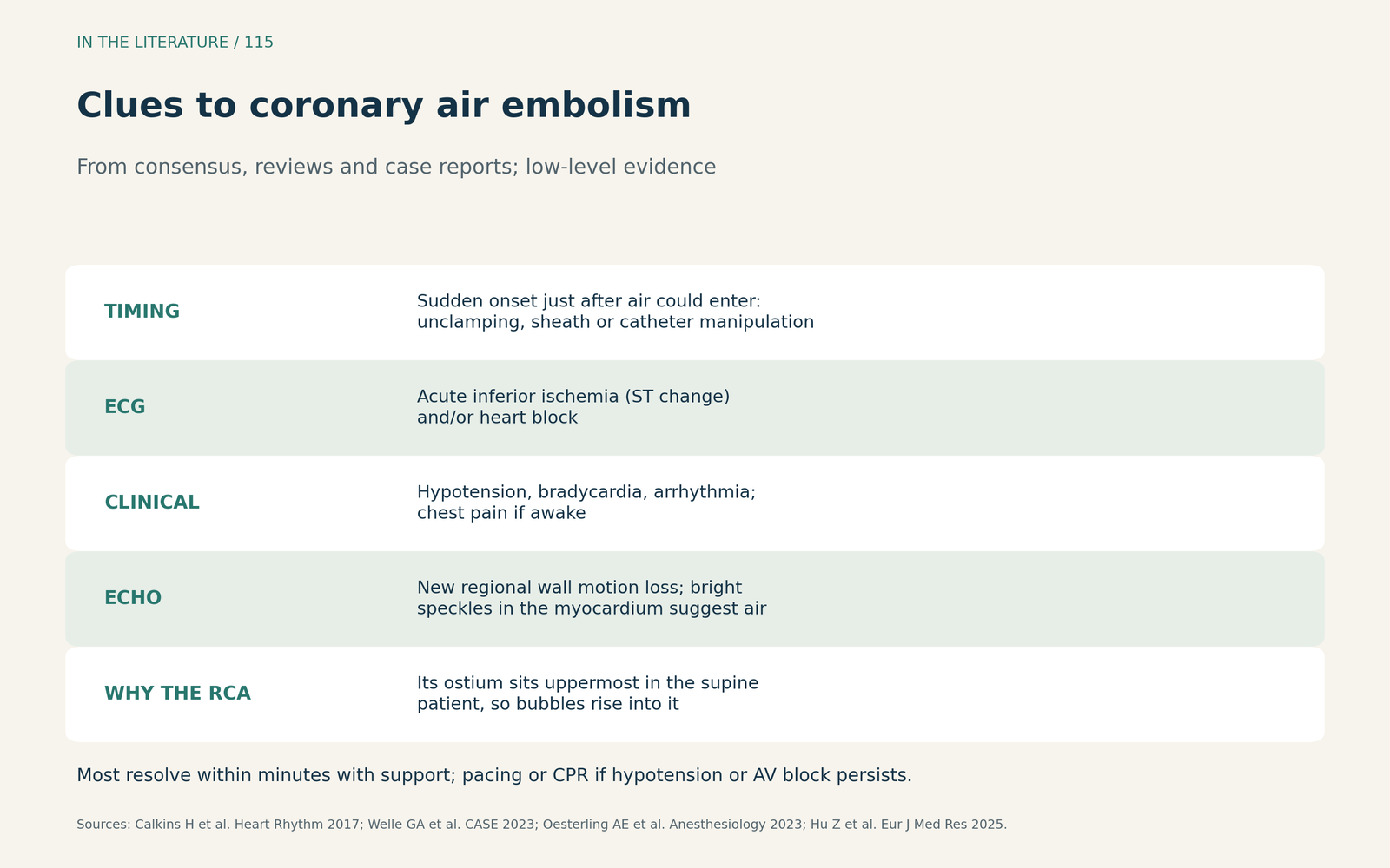
Figure 1. Clues to coronary air embolism. Synthesized from the HRS consensus, two case reports and a retrospective surgical cohort; low-level evidence.[1], [2], [3], [4] View larger.

In the cardiac operating room
No source in this review describes coronary air embolism after cardiac surgery as a clinical series, so the surgical evidence is indirect. Hu and colleagues graded TEE microbubbles in 351 patients after minimally invasive myectomy; the highest grade was microbubbles in the left atrium, left ventricle and aortic root at the same time. Their arrhythmia outcome included ischemic ECG changes (ST elevation or depression) within the first 10 minutes after cross-clamp removal. Patients with microbubbles had a higher risk of perioperative arrhythmia, which the authors attribute to coronary microemboli causing ischemia after reperfusion.[4]
Continuous carbon dioxide insufflation was associated with fewer microbubbles and fewer perioperative arrhythmias than insufflation limited to the period around aortotomy closure (24.12% vs 36.59%), and left ventricular venting was associated with a 93.3% reduction in the lowest-grade microbubbles. Carbon dioxide is about 25 times more soluble in blood than air. The study is retrospective, most arrhythmias were AV block, which is expected after myectomy, and the authors acknowledge selection bias.[4]
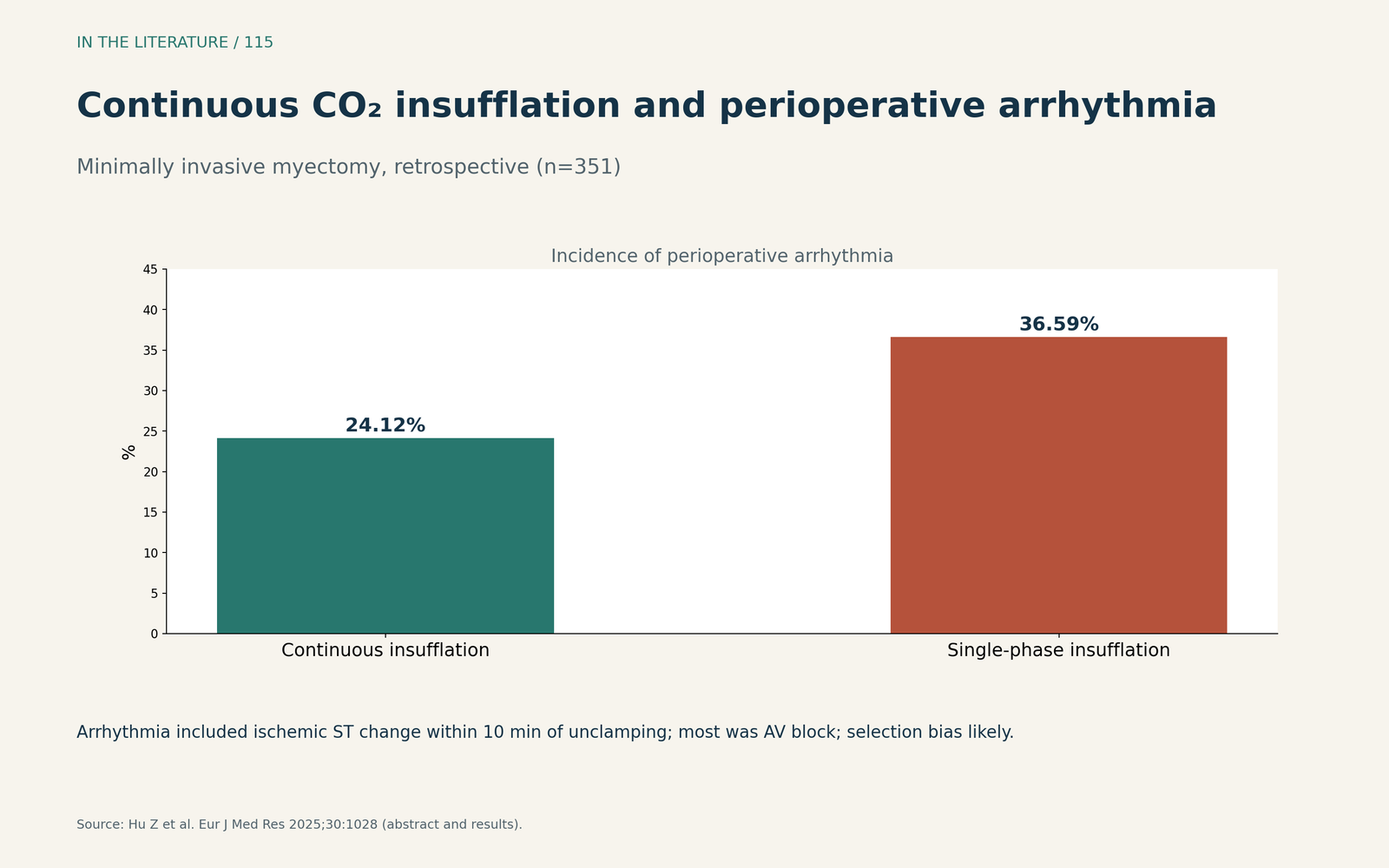
Figure 2. Carbon dioxide insufflation and perioperative arrhythmia. Redrawn from the results of Hu et al.; retrospective, single center.[4] View larger.

Management
The HRS consensus states that supportive care usually resolves symptoms and signs within minutes, but pacing and cardiopulmonary resuscitation may be needed if hypotension and AV block persist. In a series of massive air embolism during ablation, hemodynamic collapse and hypoxemia occurred in all 5 patients and lasted 10–35 minutes.[1] Welle and colleagues recommend maximizing coronary perfusion pressure by raising mean arterial pressure with a vasopressor, and 100% oxygen to speed diffusion of nitrogen out of the bubble.[2] Oesterling and colleagues list an inspired oxygen fraction of 1.0, central access and catecholamine support, with extracorporeal membrane oxygenation or cardiopulmonary bypass if other measures fail.[3]
Marsh and colleagues review animal work in which dogs given air into the left anterior descending artery had a 28% mortality without extracorporeal circulation and none on it. They also note that a higher blood pressure can improve washout of cerebral emboli, and that the left lateral head-down (Durant) position is reserved for venous air and should not be used for arterial air.[5] None of these sources gives a specific blood pressure target for coronary air.
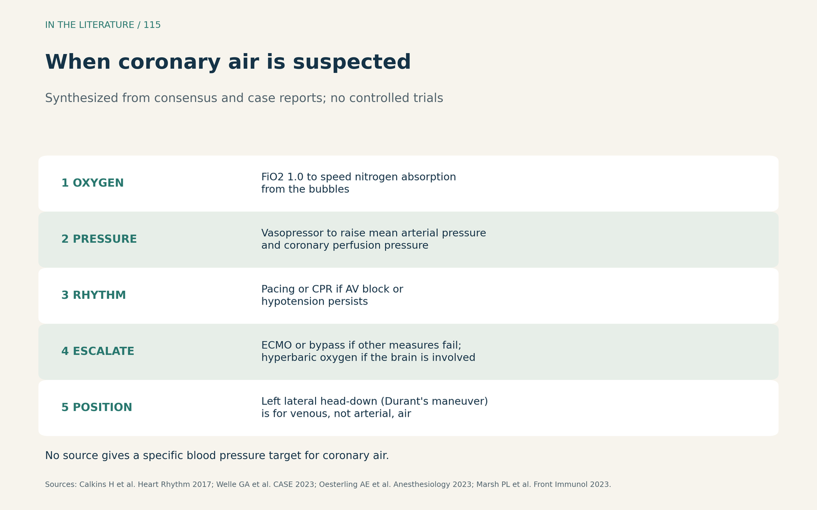
Figure 3. When coronary air is suspected. Synthesized from the HRS consensus, two case reports and a narrative review; no controlled trials.[1], [2], [3], [5] View larger.

When the brain is involved
Air that reaches the coronary arteries can also reach the brain. Marsh and colleagues advise that a symptomatic patient with arterial air embolism start hyperbaric oxygen as soon as cardiopulmonary stabilization has been achieved.[5] In an individual patient data meta-analysis of iatrogenic cerebral arterial gas embolism, whose causes included cardiac surgery, Fakkert and colleagues found that the probability of a favorable outcome fell from about 65% with immediate hyperbaric oxygen to 30% when it was delayed by 15 hours. The authors describe time to treatment as a continuum in which sooner is better, but the data are observational and an association does not prove causation.[6]
Three takeaways
- Think of air when ischemia follows air entry. Sudden inferior ischemia or heart block after unclamping, sheath or catheter work suggests coronary air, most often in the right coronary artery.[1], [3]
- Use echocardiography when the ECG is unclear. Look for new regional wall motion loss, bright intramyocardial foci and air in the left heart.[2], [4], [5]
- Support perfusion and escalate early. Give 100% oxygen and a vasopressor, pace if needed, consider extracorporeal support if this fails, and seek hyperbaric oxygen if the brain is involved.[1], [3], [6]
References
- Calkins H, Hindricks G, Cappato R, et al. 2017 HRS/EHRA/ECAS/APHRS/SOLAECE expert consensus statement on catheter and surgical ablation of atrial fibrillation. Heart Rhythm. 2017;14(10):e275–e444. doi:10.1016/j.hrthm.2017.05.012
- Welle GA, Chetram DK, Bryde RE, Bennett CE. Transthoracic echocardiography–assisted identification of coronary air embolism during coronary angiography. CASE (Phila). 2023;7(5):181–184. doi:10.1016/j.case.2022.12.013
- Oesterling AE, Kenlan DE, Cizman Z. Coronary artery air embolism in the non–operating room anesthesia setting. Anesthesiology. 2023;138(2):216–217. doi:10.1097/ALN.0000000000004413
- Hu Z, Mao W, Guo L, et al. Optimization of carbon dioxide insufflation strategies in minimally invasive cardiac surgery: the efficacy of continuous insufflation protocols with left ventricular venting. Eur J Med Res. 2025;30(1):1028. doi:10.1186/s40001-025-03184-0
- Marsh PL, Moore EE, Moore HB, et al. Iatrogenic air embolism: pathoanatomy, thromboinflammation, endotheliopathy, and therapies. Front Immunol. 2023;14:1230049. doi:10.3389/fimmu.2023.1230049
- Fakkert RA, Karlas N, Schober P, et al. Early hyperbaric oxygen therapy is associated with favorable outcome in patients with iatrogenic cerebral arterial gas embolism: systematic review and individual patient data meta-analysis of observational studies. Crit Care. 2023;27(1):282. doi:10.1186/s13054-023-04563-x