ITL-545ef377e679
Juan Li, MD · Updated November 17, 2026
Watch the video
Key points
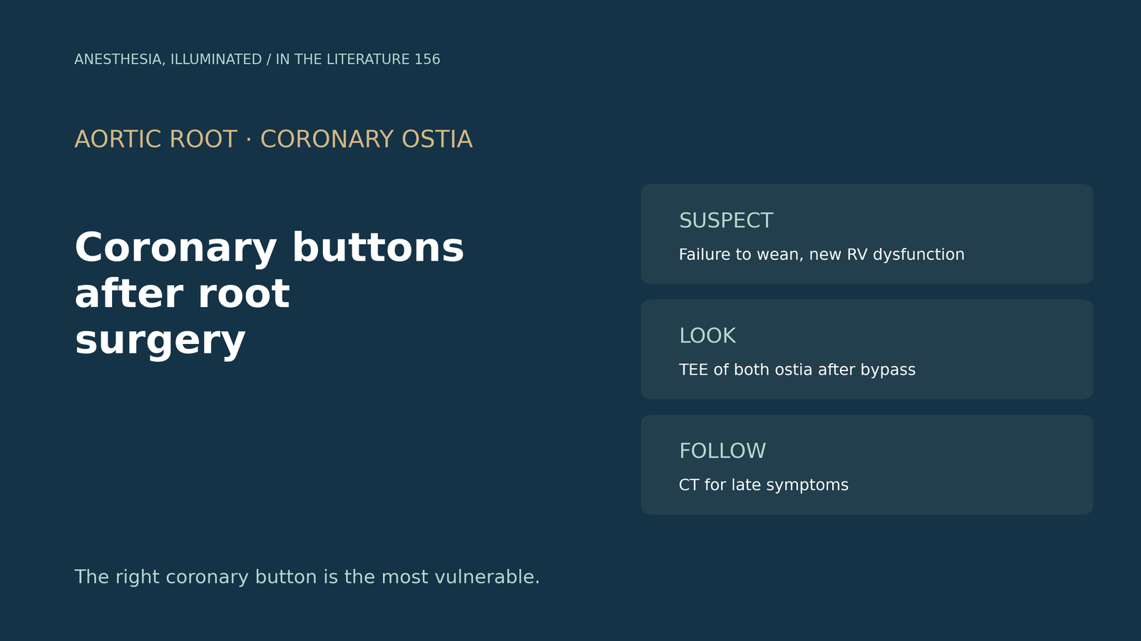
- It is not rare. After aortic root replacement, 4.3% of patients needed unplanned coronary bypass, most often a vein graft to the right coronary artery.[1]
- The right side is vulnerable. The right coronary button is more prone to malalignment, and kinking, coronary injury and poor right heart protection can all lead to rescue grafting.[1]
- TEE should look at both ostia. It images the ostia in nearly all patients, and the examination should begin as soon as bypass ends.[2]
- Not every bulge is a leak. A focal bulge at a coronary button can mimic an early pseudoaneurysm, while true pseudoaneurysms arise at suture lines or reimplantation sites; imaging quality control is recommended after every aortic procedure.[2], [3]
- Late stenosis occurs too. Coronary ostial stenosis after root replacement has an incidence of 1% to 3%, presents mostly within 6 months, and coronary CT detects it with high accuracy.[4]
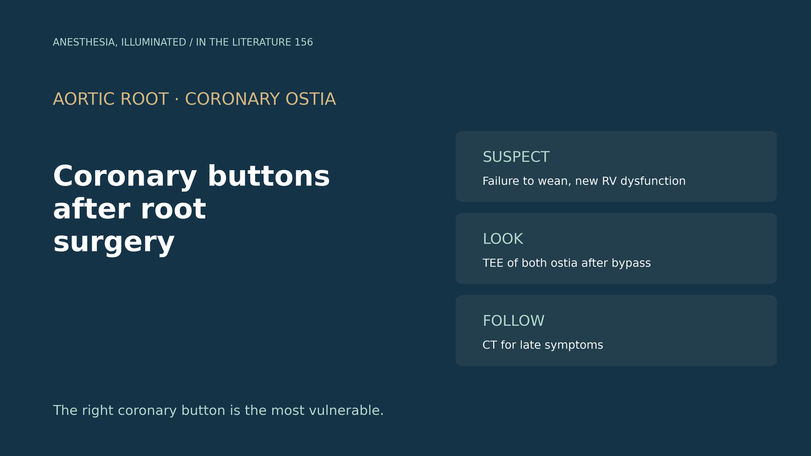
After a Bentall procedure with coronary button reimplantation, the patient cannot be weaned from bypass, and the right ventricle looks newly hypokinetic on TEE. Is a coronary button compromised, and how should the team find out?[1], [2]
How often and why
Ogami and colleagues reviewed aortic root replacements: 34 patients (4.3%) required unplanned coronary bypass, and a vein graft to the right coronary artery was the most frequent graft. Surgical causes included kinking of coronary buttons and coronary injury; the right coronary button has a higher chance of malalignment, and some unplanned grafting may reflect inadequate protection of the right heart. Renal failure was more common (20.6% vs 8.6%), a cited series reported 28.6% 30-day mortality after emergent grafting, and the authors advise that unexpected grafting should not be discouraged when needed.[1]
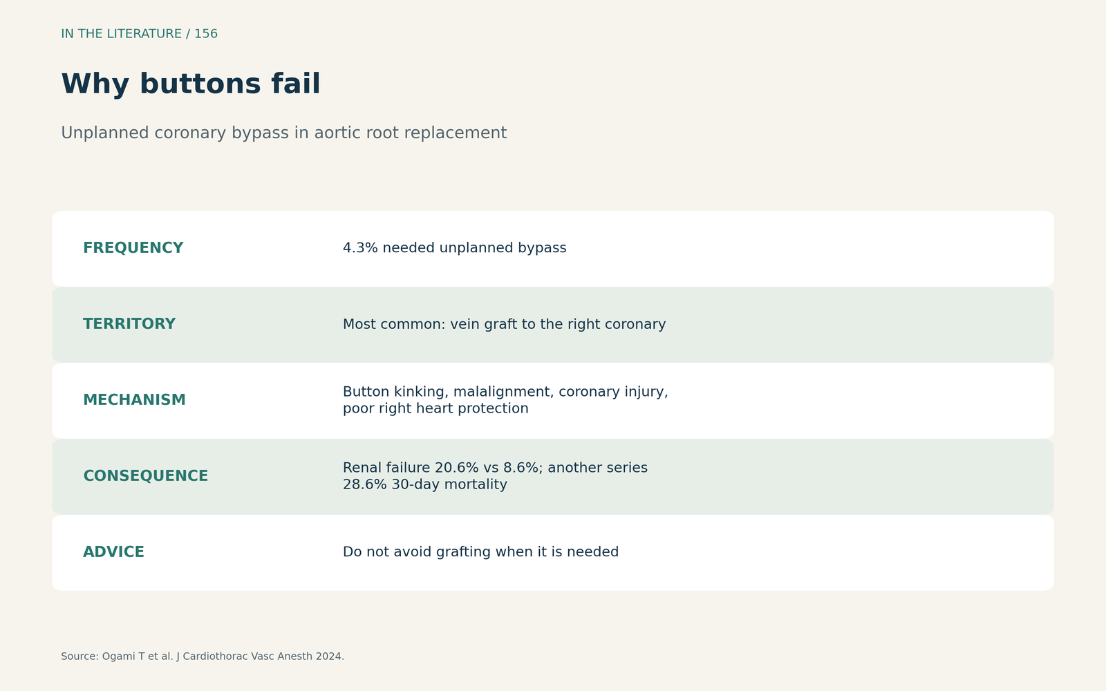
Figure 1. Why buttons fail. Redrawn from Ogami et al.[1] View larger.

What TEE and imaging should do
The American Society of Echocardiography and European imaging guideline states that TEE images the ostia and proximal coronary segments in nearly all patients; in dissection, both ostia should be examined for compromise, and the right coronary is affected more often than the left. Postoperative TEE should begin as soon as bypass ends. A focal bulge at a button can be misread as an early pseudoaneurysm; true pseudoaneurysms usually arise at suture lines or the coronary reimplantation site, and contrast CT is the optimal follow-up tool.[2] The 2024 EACTS/STS guideline notes that coronary malperfusion in acute type A dissection carries mortality of up to 40%, recommends transthoracic echo after any root surgery and imaging quality control after every aortic procedure.[3]
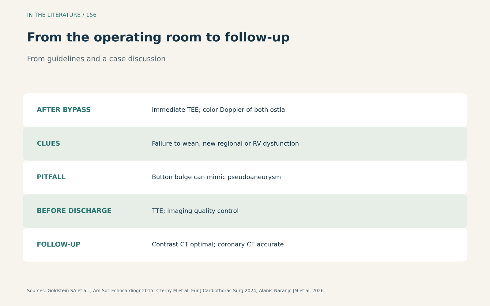
Figure 2. From the operating room to follow-up. Synthesized from the ASE/EACVI guideline, the EACTS/STS guideline and Alanís-Naranjo et al.[2], [3], [4] View larger.

Late ostial stenosis
Alanís-Naranjo and colleagues describe coronary ostial stenosis as a rare complication of root replacement (1% to 3%) that may cause unexplained postoperative deaths and presents most often within the first 6 months. Limited angiographic projections may underestimate it, whereas coronary CT has a sensitivity of 90% to 95% and specificity of 90% to 98% for postsurgical complications; percutaneous intervention is the most effective emergency salvage therapy.[4]
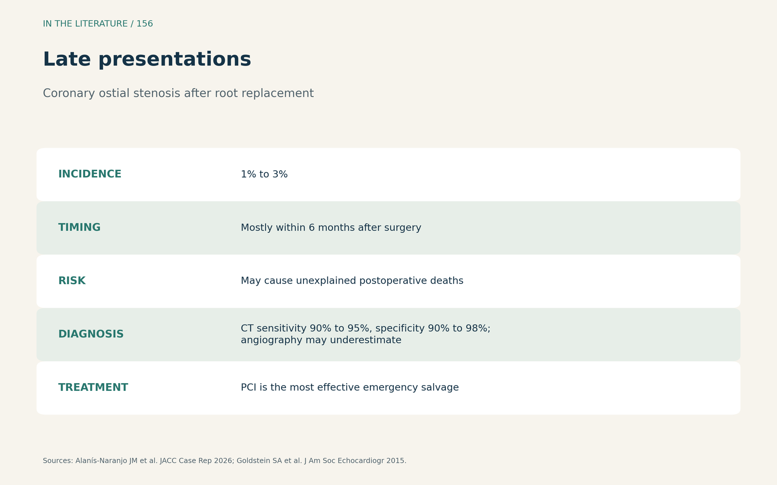
Figure 3. Late presentations. Synthesized from Alanís-Naranjo et al. and the ASE/EACVI guideline.[4], [2] View larger.

Three takeaways
- Think buttons when weaning fails. New ventricular dysfunction, especially on the right, after root reconstruction suggests ostial compromise.[1]
- Look at both ostia right after bypass. TEE sees them in nearly all patients; do not mistake a button bulge for a leak.[2]
- Keep watching after discharge. Late ostial stenosis appears mostly within 6 months; CT is accurate.[4], [3]
References
- Ogami T, Serna-Gallegos D, Yousef S, et al. The clinical significance of unplanned coronary artery bypass grafting in aortic root replacement. J Cardiothorac Vasc Anesth. 2024;38(4):918–923. doi:10.1053/j.jvca.2024.01.001
- Goldstein SA, Evangelista A, Abbara S, et al. Multimodality imaging of diseases of the thoracic aorta in adults: from the American Society of Echocardiography and the European Association of Cardiovascular Imaging: endorsed by the Society of Cardiovascular Computed Tomography and Society for Cardiovascular Magnetic Resonance. J Am Soc Echocardiogr. 2015;28(2):119–182. doi:10.1016/j.echo.2014.11.015
- Czerny M, Grabenwöger M, Berger T, et al. EACTS/STS guidelines for diagnosing and treating acute and chronic syndromes of the aortic organ. Eur J Cardiothorac Surg. 2024;65(2):ezad426. doi:10.1093/ejcts/ezad426
- Alanís-Naranjo JM, Campuzano-González D, Gamboa-Abundis OA, et al. Late coronary ostial stenosis after complicated Bentall-De Bono procedure: multimodal imaging assessment. JACC Case Rep. 2026;31(1):105946. doi:10.1016/j.jaccas.2025.105946