
ITL-677dab9143fe
Juan Li, MD · Updated November 17, 2026
Watch the video

Key points
- Reserve integrates two compartments. Flow passes through large epicardial conductance vessels and a network of small resistance vessels, and reserve is measured when microvascular resistance is minimized by vasodilators.[1]
- Read it with resistance. Microvascular dysfunction is diagnosed when reserve is below 2.0 and/or resistance is above 25; concordantly abnormal values carried the highest risk (major adverse events, odds ratio 3.23).[2]
- Without wall-motion abnormality or stenosis, a reduced reserve indicates microcirculatory dysfunction; a value below 2 separated 8-year cardiovascular mortality of 14.6% from 1.2%.[3]
- A better FFR does not mean better flow. After stenting, reserve fell in 25.1% of patients, and a lower post-procedure reserve was seen in patients with later adverse events.[4]
- In transplant vasculopathy, reserve was already lower in the earliest grade (3.1 vs 2.6), although it could not be measured in 11%.[5]
A patient with exertional angina and non-obstructive coronary arteries comes for noncardiac surgery; a stress echo report gives a left anterior descending flow reserve of 1.8. What does this number say about the microcirculation, and how should it be interpreted?[1], [2]
What flow reserve measures
Daubenspeck and Chaney describe the coronary circulation as large epicardial conductance vessels feeding an extensive network of small vessels. Reserve is assessed when microvascular resistance is minimized by potent vasodilators; systolic compression makes the subendocardium particularly vulnerable, and epicardial obstruction affects the microcirculation beyond the stenosis. Reserve, myocardial blood flow and FFR measure physiologically distinct things.[1] In transthoracic studies, flow velocity reserve is the ratio of hyperemic to resting diastolic velocity, usually in the left anterior descending artery, with a value below 2.0 considered abnormal.[5]
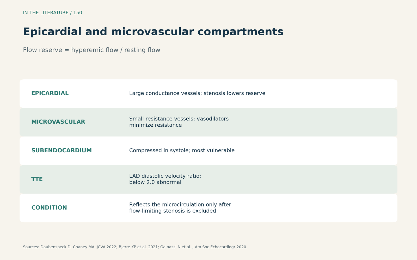
Figure 1. Epicardial and microvascular compartments. Synthesized from Daubenspeck and Chaney, Bjerre et al. and Gaibazzi et al.[1], [5], [3] View larger.

Reading reserve with microvascular resistance
Al-Gully and colleagues pooled four studies with 2310 participants in a network meta-analysis. Microvascular dysfunction was defined as reserve below 2.0 and/or an index of microcirculatory resistance above 25. Concordantly abnormal reserve and resistance, a structural form, had the highest risk of major adverse events (odds ratio 3.23) and death (odds ratio 2.27). Low reserve with normal resistance was described as a functional form, and risk was low with normal reserve but high resistance.[2]
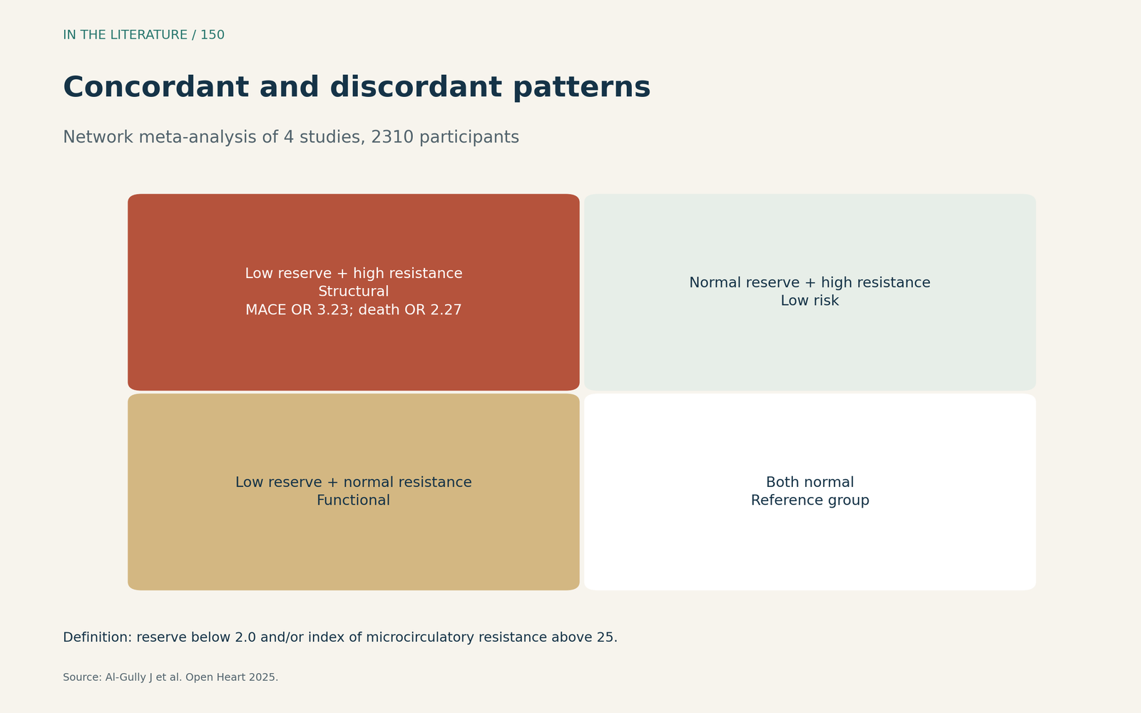
Figure 2. Concordant and discordant patterns. Redrawn from Al-Gully et al.[2] View larger.

What low reserve predicts
Gaibazzi and colleagues found that, in the absence of stress-induced wall-motion abnormality and flow-limiting stenosis, reduced flow velocity reserve was associated with microcirculatory dysfunction; a value below 2 separated 8-year cardiovascular mortality of 14.6% versus 1.2%, suggesting reserve may be a marker or mechanism preceding death.[3] Hanyu and colleagues measured reserve after stenting: although FFR improved in all, reserve fell in 25.1%; adverse events occurred in 21 patients (11.2%), who had lower post-procedure reserve, and FFR improvement did not necessarily mean increased flow.[4] In heart transplant recipients, Bjerre and colleagues found that vasculopathy also affects the microvasculature; reserve was already lower in grade 1 than grade 0, but could not be measured in 11%, and one vessel was assumed to represent global function.[5]
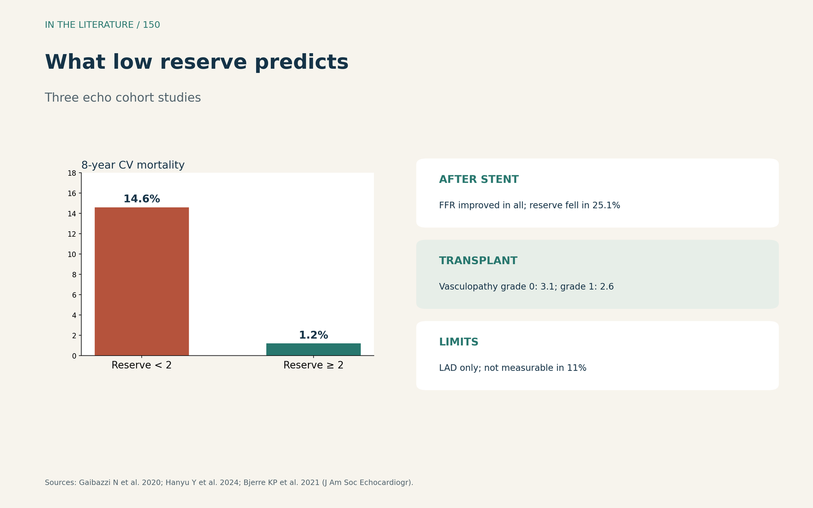
Figure 3. What low reserve predicts. Redrawn from Gaibazzi et al., Hanyu et al. and Bjerre et al.[3], [4], [5] View larger.

Three takeaways
- Exclude the epicardial lesion first. Only then does a low reserve speak for the microcirculation.[1], [3]
- Pair reserve with resistance. Concordantly abnormal values carry the most risk.[2]
- A normal FFR is not a normal microcirculation. Reserve can stay low after a successful stent.[4]
References
- Daubenspeck D, Chaney MA. Clinical importance of quantitative assessment of myocardial blood flow. J Cardiothorac Vasc Anesth. 2022;36(6):1511–1515. doi:10.1053/j.jvca.2022.01.030
- Al-Gully J, Oliveri F, Forouzanfar JP, et al. Prognostic role of con-/discordant coronary flow reserve and microvascular resistance in coronary microvascular disease: a systematic review and network meta-analysis. Open Heart. 2025;12(1):e003055. doi:10.1136/openhrt-2024-003055
- Gaibazzi N, Picano E, Suma S, et al. Coronary flow velocity reserve reduction is associated with cardiovascular, cancer, and noncancer, noncardiovascular mortality. J Am Soc Echocardiogr. 2020;33(5):594–603. doi:10.1016/j.echo.2020.01.007
- Hanyu Y, Hoshino M, Usui E, et al. Combined assessment of fractional flow reserve and coronary flow velocity reserve after drug-eluting stent implantation. J Am Soc Echocardiogr. 2024;37(4):428–438. doi:10.1016/j.echo.2023.12.006
- Bjerre KP, Clemmensen TS, Poulsen SH, et al. Coronary flow velocity reserve and myocardial deformation predict long-term outcomes in heart transplant recipients. J Am Soc Echocardiogr. 2021;34(12):1294–1302. doi:10.1016/j.echo.2021.07.012