ITL-6f05fd4c4c7b
Juan Li, MD · Updated November 17, 2026
Watch the video
Key points
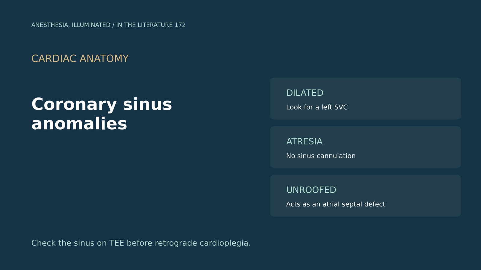
- Know the common variant. With a persistent left superior caval vein, the sinus and its orifice are usually dilated; the vein itself causes no hemodynamic problem.[1]
- Some variants block the route. An imperforate Thebesian valve produces sinus atresia and precludes catheterization, and an unroofed sinus acts as an interatrial communication.[1]
- Expect surprises on the table. A left superior vena cava occurs in 0.3% to 0.5% of people, TTE can miss sinus abnormalities, and knowing the alternative pathway matters for catheters and pacing leads.[2]
- TEE sees the sinus well. It was visible in the four-chamber view in 98%, TEE identifies variants that may preclude retrograde cardioplegia, and the modified bicaval view is preferred for cannulation.[3]
- Unroofing hides from bubbles. An unroofed sinus is almost always associated with a left superior vena cava, saline contrast can be negative, and TEE resolution was key.[4]
Before aortic valve surgery, TEE shows a markedly dilated coronary sinus, and the surgeon plans retrograde cardioplegia. What anomaly might be present, and how does it change cannulation, cardioplegia and later pacing?[1], [3]
The anatomy behind the problems
Anderson and colleagues explain that with a persistent left superior caval vein the sinus and its right atrial orifice are usually dilated; the vein itself causes no hemodynamic problem but can disconcert the catheterizer. An imperforate Thebesian valve produces atresia of the sinus and precludes catheterizing it, an unroofed sinus makes its orifice an interatrial communication, sinus diverticula account for about one-tenth of inferior paraseptal accessory pathways, the sinus is a conduit to the left heart for electrophysiologists, and right isomerism universally lacks a coronary sinus.[1]
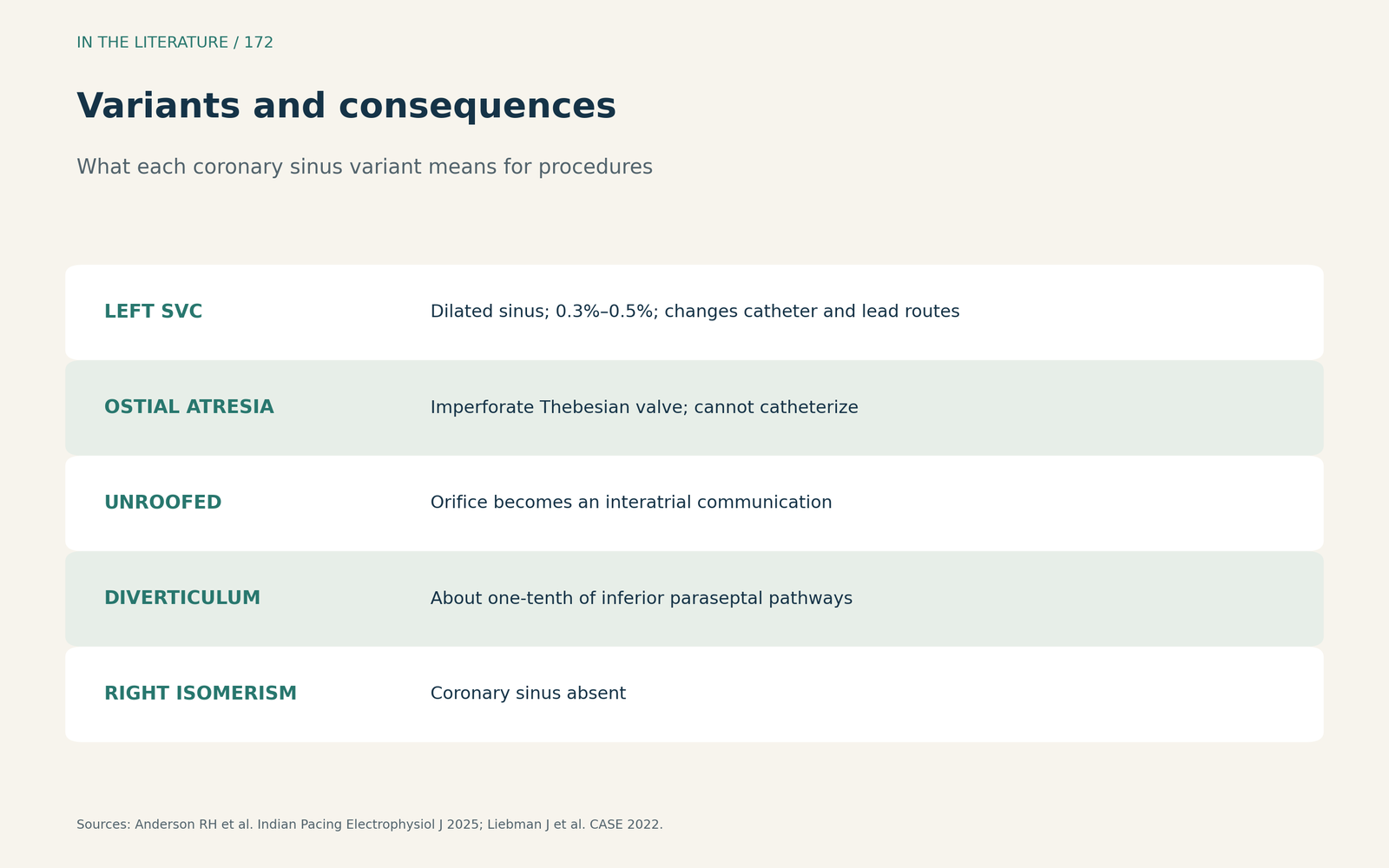
Figure 1. Variants and consequences. Synthesized from Anderson et al. and Liebman et al.[1], [2] View larger.

Imaging before cannulation
Liebman and colleagues report a left superior vena cava prevalence of 0.3% to 0.5%; in one case no sinus abnormality was visible on TTE, and they stress that knowing the alternative venous pathway matters when placing catheters or pacing leads.[2] Chen and colleagues found the sinus visible in the midesophageal four-chamber view in 98% (49/50) and in all 4 views in 45 patients. TEE identifies variants, such as a persistent left superior vena cava or a Thebesian valve, that may preclude retrograde cardioplegia, and the modified bicaval view is preferred for cannulation; their mostly normal cohort may not have captured variable anatomy.[3]
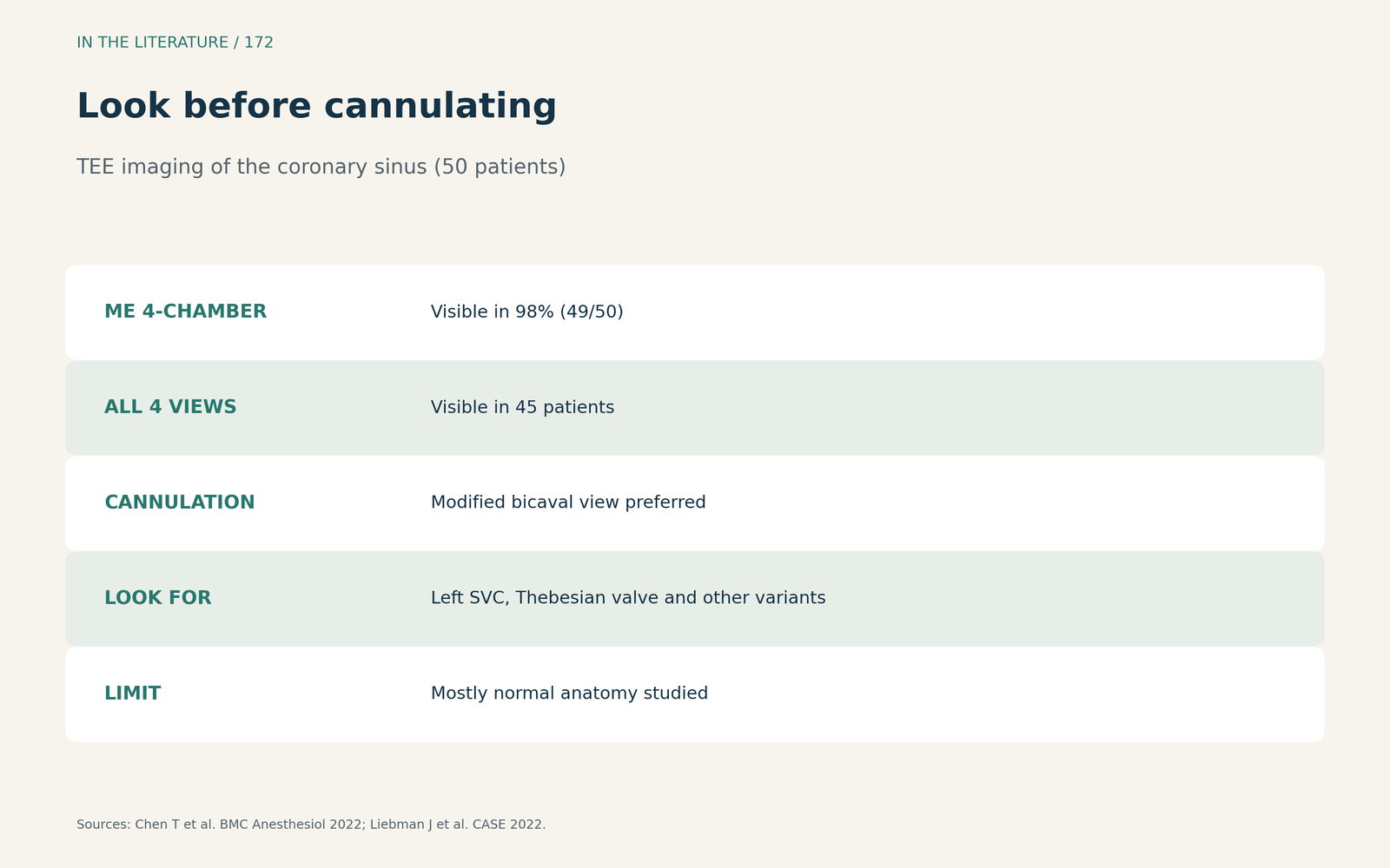
Figure 2. Look before cannulating. Redrawn from Chen et al. and Liebman et al.[3], [2] View larger.

The unroofed sinus
Osei and colleagues note that coronary sinus septal defects are under 1% of atrial septal defects and 0.1% of congenital defects, are almost always associated with a left superior vena cava, behave like a secundum defect, and can be missed by agitated saline; TEE resolution was key to their diagnosis.[4]
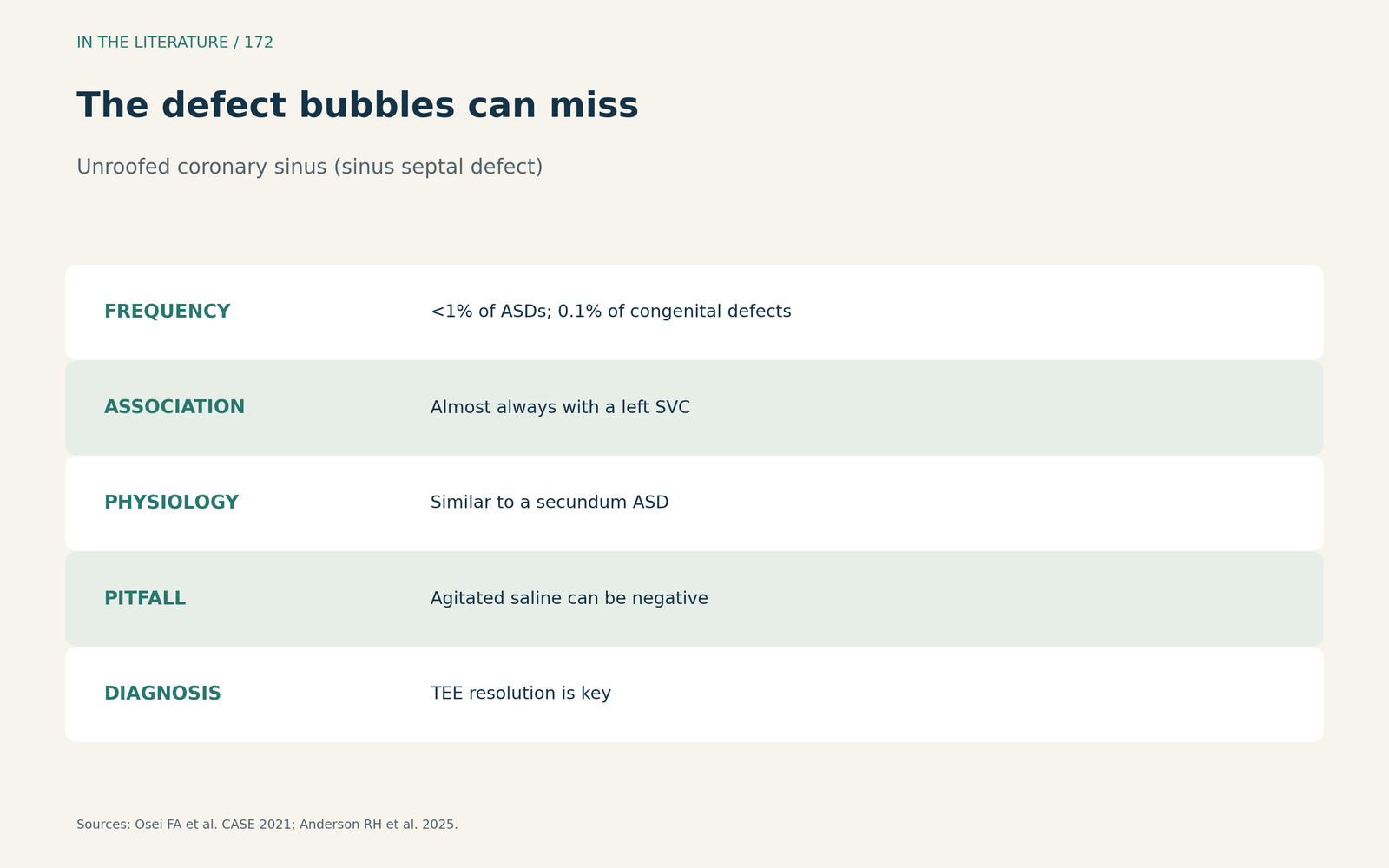
Figure 3. The defect bubbles can miss. Synthesized from Osei et al. and Anderson et al.[4], [1] View larger.

Three takeaways
- A dilated sinus means look for a left superior vena cava. It changes catheter and lead routes.[1], [2]
- Check the sinus on TEE before retrograde cardioplegia. Variants can preclude it.[3]
- Consider unroofing when an atrial-level shunt is unexplained. Bubbles can miss it; TEE finds it.[4]
References
- Anderson RH, Spicer DE, Sanchez-Quintana D. Developmental, normal, and abnormal anatomy of the coronary sinus. Indian Pacing Electrophysiol J. 2025;25(6):417–424. doi:10.1016/j.ipej.2025.12.013
- Liebman J, Bamira D, Ro R, et al. Multimodality imaging of caval and coronary sinus venous anomalies. CASE (Phila). 2022;6(8):366–376. doi:10.1016/j.case.2022.06.007
- Chen T, Hayward G, Apruzzese P, Maslow A. Transesophageal echocardiographic imaging of the coronary sinus: a retrospective analysis of mid-esophageal views and a novel transgastric view. BMC Anesthesiol. 2022;22(1):326. doi:10.1186/s12871-022-01873-5
- Osei FA, Hill S, Shakti D. A rare congenital cardiac defect: isolated coronary sinus septal defect and the challenges of diagnosis in a child. CASE (Phila). 2021;5(5):286–291. doi:10.1016/j.case.2021.07.005