
ITL-80c8e9b450ba
Watch the video

Figure 1. Conceptual illustration of spontaneous echo contrast; not an ultrasound image or a depiction of thrombus.
Key points
- Spontaneous echo contrast (SEC) is a dynamic, smoke-like blood-pool appearance; its presence alone does not diagnose thrombus.[1]
- Descending aortic SEC is associated with low local shear and atrial arrhythmia, but it is not a cardiac-output measurement.[2]
- Aortic and left atrial SEC may coexist, yet either can occur without the other.[3]
- Examine the aortic wall and lumen: SEC can also occur in a dissection's false lumen.[4]
- Management should follow the identified hemodynamic or structural problem and established treatment indications; these studies do not establish SEC alone as a trigger for anticoagulation.[1, 2, 3, 4]
During TEE, the descending aorta fills with slowly swirling echoes. The immediate temptation is to call the image “low flow” and assume that cardiac output has fallen. That may be a useful hypothesis, but it needs confirmation.
The clinically useful response is to establish what the image represents, assess circulation independently, and look for the setting that produced it. The distinction matters because local blood velocity, vessel diameter, rhythm, and underlying aortic disease can all influence the finding.[1, 2]
First establish that it is SEC
The classic description is an amorphous cloud of microechoes moving with blood flow and aortic pulsation. Review the moving image, optimize gain, and distinguish this appearance from excessive-gain noise, near-field artifacts, or bubbles. In the early aortic studies, observers specifically required a swirling pattern and adjusted gain to exclude noise.[1, 2]
Document the location and whether an associated mass, plaque, or flap is visible. The term SEC describes an appearance; it does not establish the composition of a separate lesion. deFilippi and colleagues found accompanying aortic or cardiac abnormalities in their small series, including plaque, aneurysm, thrombus, ventricular dysfunction, and severe mitral regurgitation.[1]
Low velocity is not the same measurement as low output
Sukernik and colleagues examined 102 patients referred for TEE; 19 had descending aortic SEC. Those with SEC had lower peak aortic velocity and lower estimated shear rate, as well as a larger aortic diameter. Measured descending aortic volumetric flow did not differ significantly between groups: 2.0 ± 0.9 versus 2.2 ± 1.1 L/min.[2]
In the multivariable analysis, atrial arrhythmia and shear rate remained associated with SEC. This small observational study used simplifying assumptions about the velocity profile, and descending aortic flow is not total cardiac output. Its practical lesson is that the image should prompt assessment of circulation, rather than substitute for it.[2]
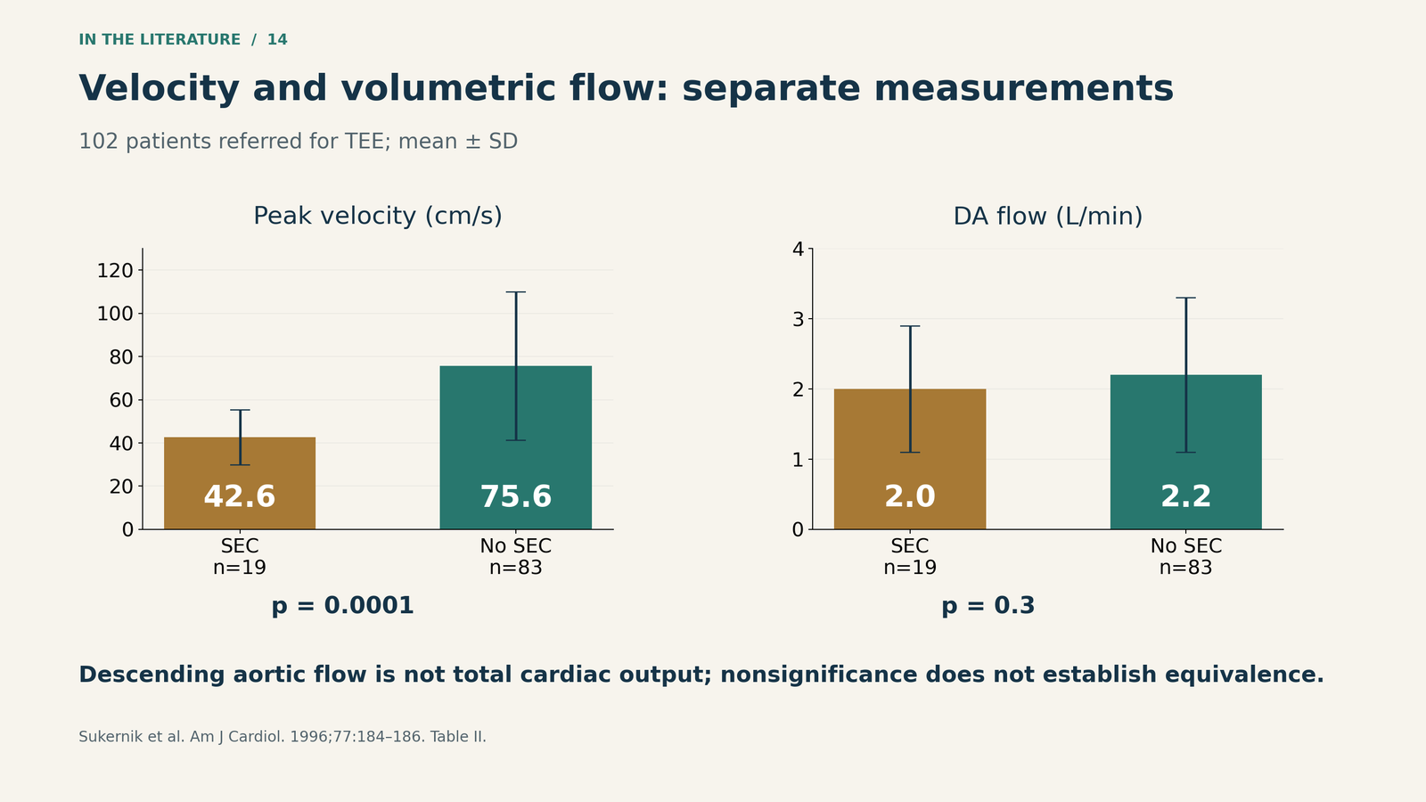
Figure 2. Mean ± SD in patients with SEC (n=19) and without SEC (n=83). These are study-group comparisons, not diagnostic cutoffs. Source: Sukernik et al., Table II.[2]

Look beyond the aorta without assuming the source
A 2024 retrospective study provides a useful reminder. Among 176 patients with nonvalvular AF undergoing TEE before ablation and/or cardioversion, 41 had descending aortic SEC. Of these, 23 also had left atrial SEC, while 18 had aortic SEC alone. Another 28 patients had left atrial SEC without aortic SEC.[3]
Left atrial SEC and EF <50% were independently associated with aortic SEC. The authors proposed that some aortic SEC might originate from the left atrium, but the study did not directly track that movement. It also did not evaluate future embolic outcomes. An association between the two locations is not proof of origin or a validated stroke prediction rule. Most patients were already receiving anticoagulation.[3]
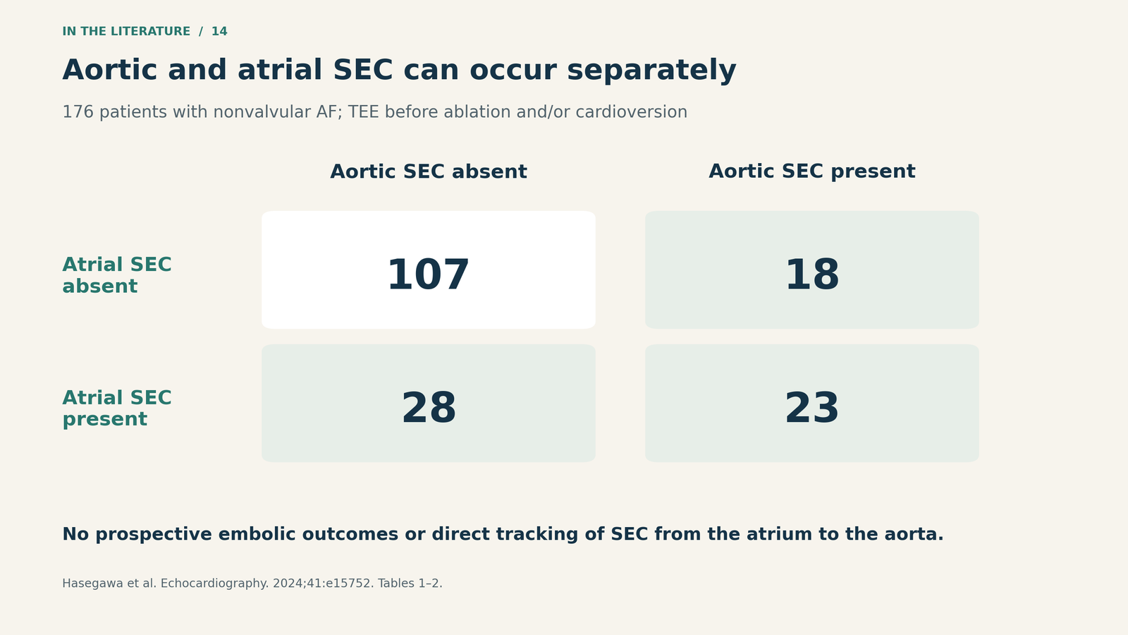
Figure 3. Aortic and atrial SEC overlap but are not interchangeable findings. Counts from Hasegawa et al., Tables 1–2.[3]

Separate the image from the treatment decision
A practical sequence is to confirm the appearance, assess ventricular function and perfusion, then inspect the aortic wall and the rest of the left heart. This is a clinical synthesis, not a validated SEC algorithm.[1, 2, 3, 4]
The structural examination is particularly important around aortic instrumentation. The ASE/SCA/STS intraoperative TEE guidance emphasizes identifying a dissection flap, distinguishing true from false lumen, and assessing complications. SEC may appear within the false lumen; conversely, SEC without a visible flap does not itself diagnose dissection.[4]
If circulation is inadequate, address the demonstrated cause. If there is an actual thrombus, aortic dissection, important plaque, or a separate indication for anticoagulation, that diagnosis drives the next decision. The available aortic SEC studies do not show that giving anticoagulation solely to clear the smoke improves clinical outcomes. Likewise, disappearance of SEC should not be treated as proof that embolic risk has resolved.[1, 2, 3]
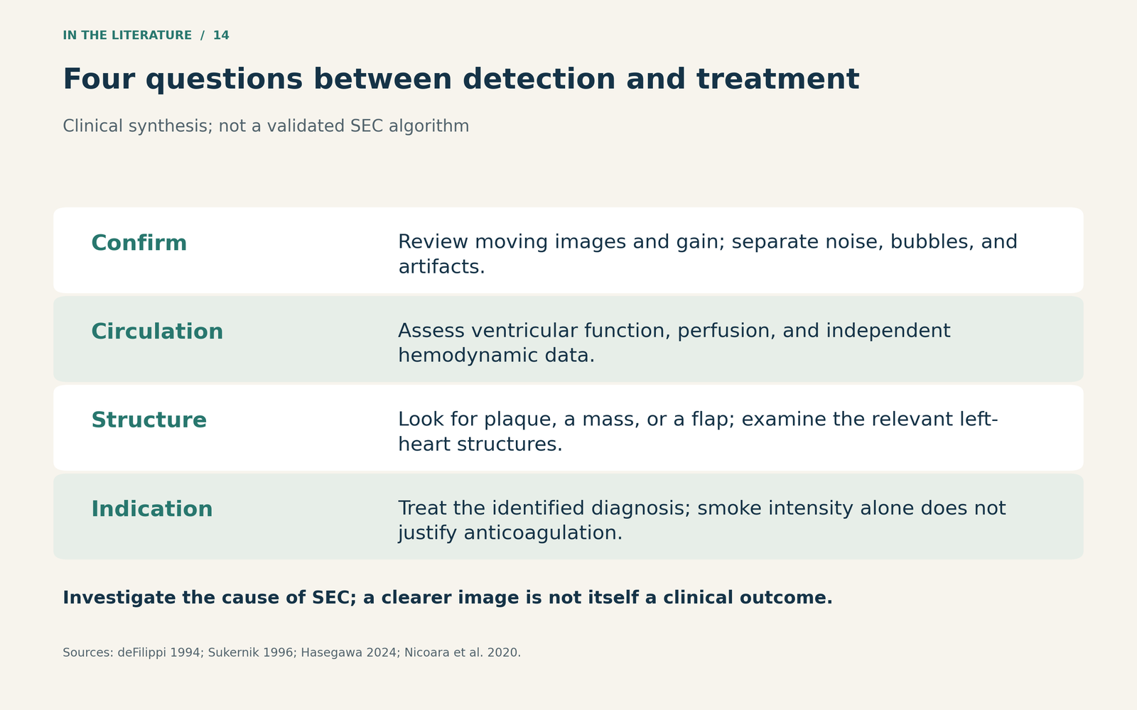
Figure 4. A source-based clinical assessment framework. Each step resolves a different uncertainty; SEC intensity alone is not a treatment target.[1, 2, 3, 4]

Three takeaways
- Confirm the phenomenon. Use the moving image and appropriate gain; describe any associated structural finding separately.
- Assess flow independently. SEC can reflect low local shear without demonstrating low total cardiac output.
- Treat the identified problem. Use hemodynamics, anatomy, and established indications to guide care; avoid deriving an anticoagulation order from smoke alone.
References
- deFilippi CR, Lacker M, Grayburn PA, Brickner ME. Spontaneous echo contrast in the descending aorta detected by transesophageal echocardiography. Am J Cardiol. 1994;74(4):410–411. doi:10.1016/0002-9149(94)90418-9.
- Sukernik MR, West O, Lawal O, Chittivelu B, Henderson R, Sherzoy AA, et al. Hemodynamic correlates of spontaneous echo contrast in the descending aorta. Am J Cardiol. 1996;77(2):184–186. doi:10.1016/S0002-9149(96)90593-7.
- Hasegawa H, Ito T, Akamatsu K, Hoshiga M. Clinical significance of aortic spontaneous echo contrast in patients with nonvalvular atrial fibrillation. Echocardiography. 2024;41(1):e15752. doi:10.1111/echo.15752.
- Nicoara A, Skubas N, Ad N, Finley A, Hahn RT, Mahmood F, et al. Guidelines for the use of transesophageal echocardiography to assist with surgical decision-making in the operating room: a surgery-based approach. J Am Soc Echocardiogr. 2020;33(6):692–734. doi:10.1016/j.echo.2020.03.002.