
ITL-e3a54f4a61d2
Juan Li, MD · Updated November 18, 2026
Key points
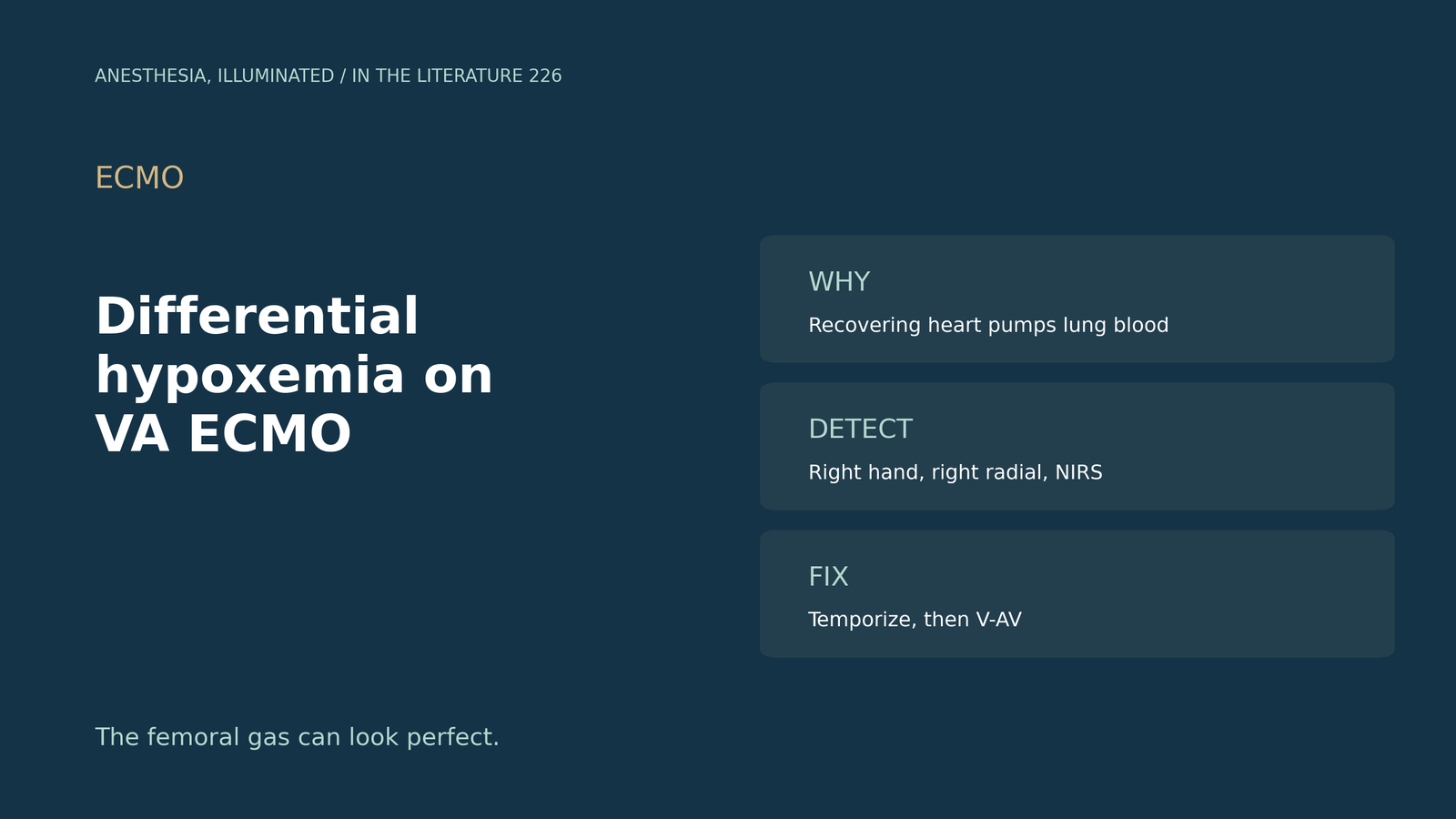
- Common but missed. Dual circulation is a common but underrecognized occurrence in peripheral VA ECMO.[1]
- Recovery moves the mixing zone. As native cardiac output improves, the watershed zone migrates caudally, toward the legs.[2]
- Monitor the right side. Right upper extremity saturation, by pulse oximetry or arterial gas, tracks upper-body oxygenation.[1]
- No perfect locator. No standardized clinical modality localizes the mixing zone in real time, and pulse pressure failed to do so.[3], [4]
- Temporize, then reconfigure. Raising flow and adjusting ventilation are temporizing; persistent hypoxemia warrants reassessment, and V-AV conversion may be preferable to repeated flow escalation.[3], [4]
On day 3 of femoral VA ECMO, the patient's heart is recovering. The femoral arterial gas looks excellent, but the right-hand saturation is 82%. Whose blood is reaching the brain?[1], [2]
Why it happens
Asija et al. (Crit Care 2023) describe dual circulation as common but underrecognized; higher ECMO flow moves the mixing point more proximally in the aorta.[1] Kumar et al. (J Cardiothorac Vasc Anesth 2023) note that as native cardiac output improves, the watershed zone migrates caudally.[2] Keebler et al. (JACC Heart Fail 2018) describe upper extremities with saturations below 90%, producing upper body cyanosis.[5]
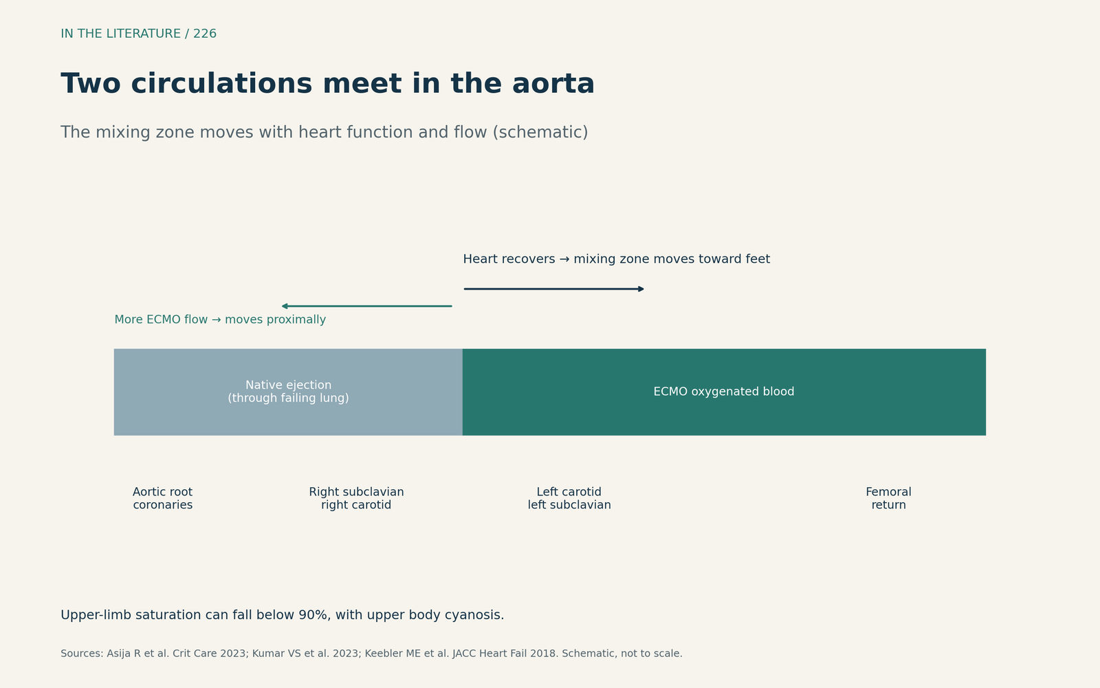
Figure 1. Two circulations meet in the aorta. Synthesized from Asija et al., Kumar et al. and Keebler et al.[1], [2], [5] View larger.

Recognizing it
Torre and Pirri (Rev Cardiovasc Med 2025) define differential hypoxemia as a significant upper-versus-lower body difference in oxygen saturation; continuous pulse oximetry is preferentially applied to the right upper limb, early detection of cerebral desaturation below 50% is emphasized, and no standardized modality localizes the mixing zone in real time.[3] Right-arm blood gases better reflect cerebral oxygen content.[5] Partial forms are easy to miss unless right upper limb oxygenation is monitored.[2] In a pilot study, cerebral oximetry desaturations occurred in 11 of 18 VA ECMO patients (61%), and NIRS may detect hypoxia continuously at the bedside.[6] Hasheminia et al. (Rev Cardiovasc Med 2026) report frequencies from 1.5% to 13.3% depending on definitions, separate the watershed itself from clinically significant hypoxemia, and found that pulse pressure failed to localize the mixing zone.[4]
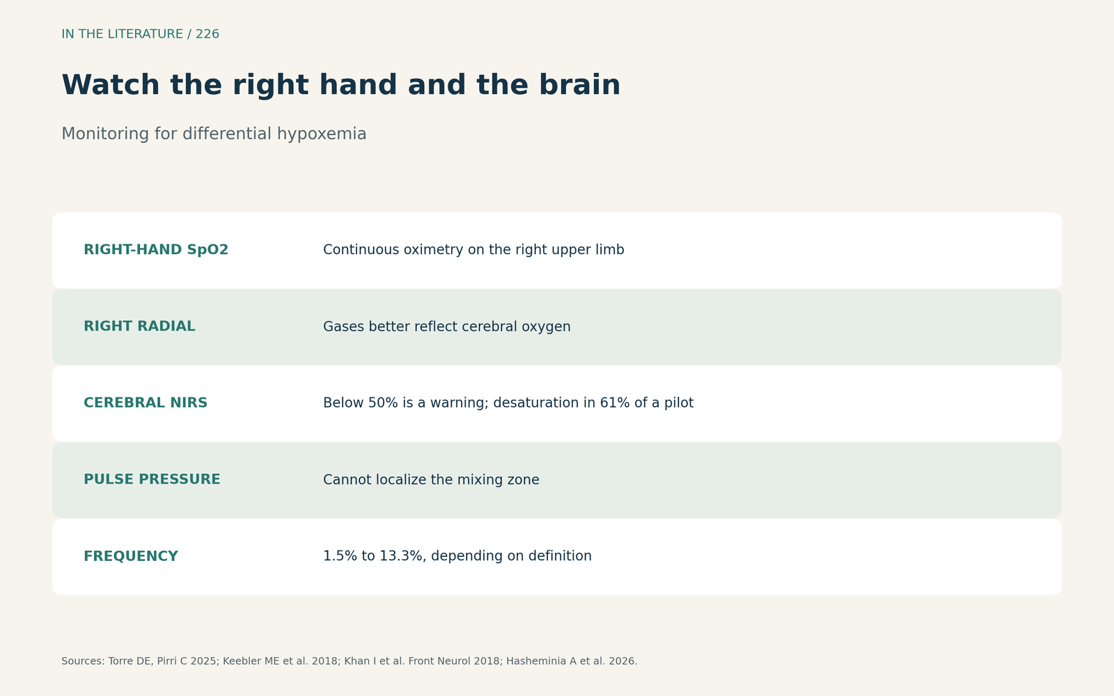
Figure 2. Watch the right hand and the brain. Synthesized from Torre and Pirri, Keebler et al., Kumar et al., Khan et al. and Hasheminia et al.[3], [5], [2], [6], [4] View larger.

Managing it
Temporizing steps include raising ECMO flow above 4 L/min and optimizing oxygenation, but these are temporizing because prolonged use may impair myocardial recovery.[3] Persistent hypoxemia warrants mechanism-based reassessment rather than reflexive flow escalation, and conversion to V-AV ECMO may be preferable to repeated flow escalation.[4] Options also include adjusting ventilation, and central cannulation mitigates the risk.[5] The evidence is reviews, case reports and small studies.
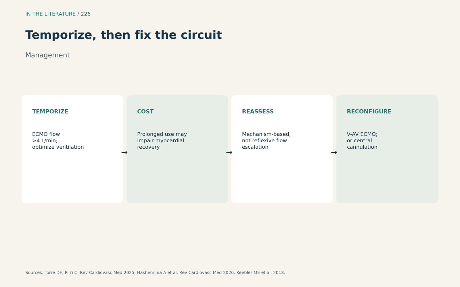
Figure 3. Temporize, then fix the circuit. Synthesized from Torre and Pirri, Hasheminia et al. and Keebler et al.[3], [4], [5] View larger.

Three takeaways
- Expect the mixing zone to move toward the legs as the heart recovers on peripheral VA ECMO.[1], [2]
- Monitor right-hand saturation, right radial gases and cerebral oximetry, not the femoral line.[1], [3], [5]
- Use flow and ventilator changes to buy time, then reconfigure, often to V-AV ECMO.[3], [4]
References
- Asija R, Fried JA, Siddall EC, et al. How I manage differential gas exchange in peripheral venoarterial extracorporeal membrane oxygenation. Crit Care. 2023;27(1):408. doi:10.1186/s13054-023-04703-3
- Kumar VS, Patangi SO, Warkari TA, Kothari RJ. "Partial" Harlequin syndrome in central veno-arterial extracorporeal membrane oxygenation. J Cardiothorac Vasc Anesth. 2023;37(11):2310–2313. doi:10.1053/j.jvca.2023.07.012
- Torre DE, Pirri C. Harlequin syndrome in venoarterial ECMO and ECPELLA: when ECMO and native or Impella circulations collide—a comprehensive review. Rev Cardiovasc Med. 2025;26(8):39992. doi:10.31083/RCM39992
- Hasheminia A, Kotb S, Elsherbini A, et al. The aortic watershed phenomenon and differential hypoxemia during peripheral venoarterial ECMO: a scoping review of clinical and experimental evidence. Rev Cardiovasc Med. 2026;27(10):52934. doi:10.31083/RCM52934
- Keebler ME, Haddad EV, Choi CW, et al. Venoarterial extracorporeal membrane oxygenation in cardiogenic shock. JACC Heart Fail. 2018;6(6):503–516. doi:10.1016/j.jchf.2017.11.017
- Khan I, Rehan M, Parikh G, et al. Regional cerebral oximetry as an indicator of acute brain injury in adults undergoing veno-arterial extracorporeal membrane oxygenation—a prospective pilot study. Front Neurol. 2018;9:993. doi:10.3389/fneur.2018.00993