ITL-a5d3fd5cd123
Watch the video
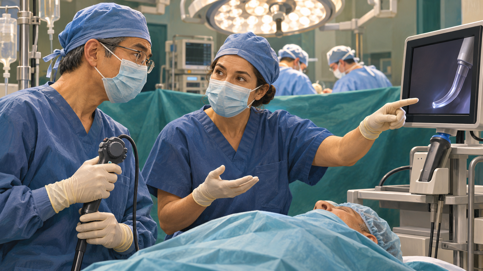
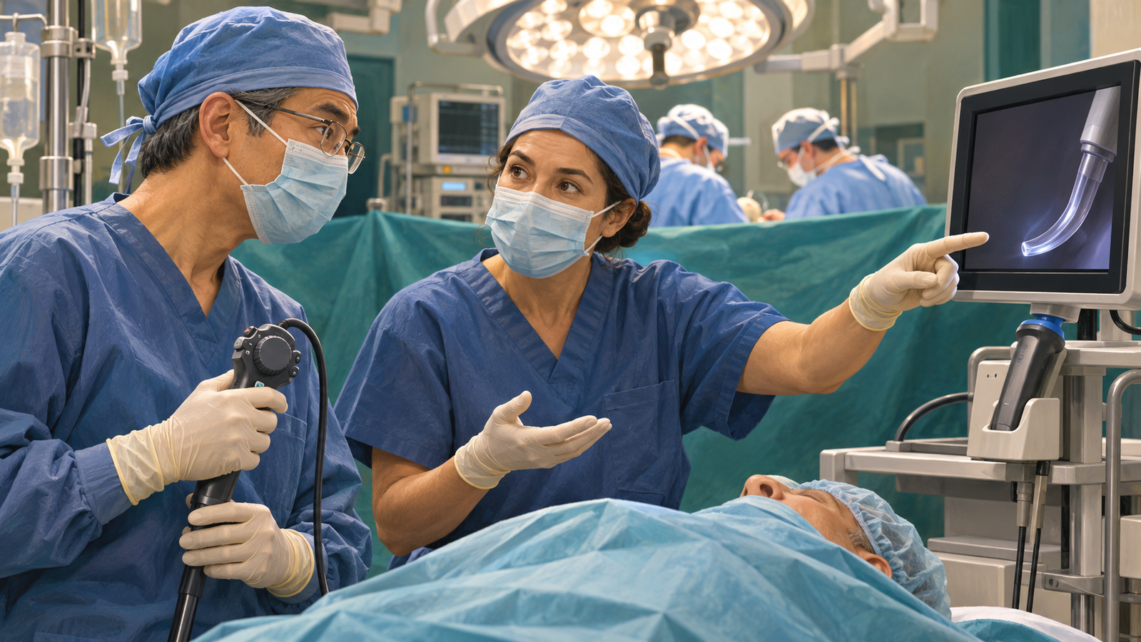
Figure 1. A difficult passage is a reason to reassess the plan. Conceptual illustration, not a demonstration of insertion technique.
Key points
- The closed-claims signal is injury. Ten of 13 TEE-related settled claims involved esophageal injury; those counts do not estimate the risk of injury per examination. [1]
- Reconsider whether and how TEE can be performed safely. Relevant esophageal disease, dysphagia and restricted neck mobility affect the plan; relative contraindications require an individual benefit–risk assessment. [2]
- Visualization has randomized evidence. In a multicenter trial, video-assisted insertion reduced the study's oropharyngeal-injury endpoint from 14.7% to 7.5%. It did not establish prevention of esophageal perforation. [3]
- Position is another modifiable factor. A small trial published in October 2026 favored the sniffing position over a defined neutral position, but did not compare it with video guidance or establish safety in difficult-airway patients. [4]
The patient is anesthetized, the tracheal tube is secure, and the TEE probe does not advance smoothly. The important question is what will make the next attempt safer. Repeating the same maneuver with more force does not answer it. The evidence supports reassessing the indication and anatomy, choosing an appropriate assisted approach, and recognizing when safe passage has not been established. [1–4]
What the malpractice paper actually tells us
McNeil and colleagues examined a sample of US anesthesia claims closed with settlement payments between 2000 and 2011. Of 2,564 claims in that dataset, 13 concerned TEE performed by an anesthesiologist: 10 involved esophageal injury, two an incorrect diagnosis, and one another cause. Six of the 10 patients with esophageal injury died; three injuries were diagnosed late. [1]
These are selected claims, not a denominator of TEE examinations. The study cannot tell a patient their probability of injury, estimate today's lawsuit rate, or establish that every injury reflected substandard care. Expert reviewers judged care appropriate in 10 of the 13 claims. Its useful clinical message is that probe-related harm and its recognition deserve attention alongside image interpretation. [1]
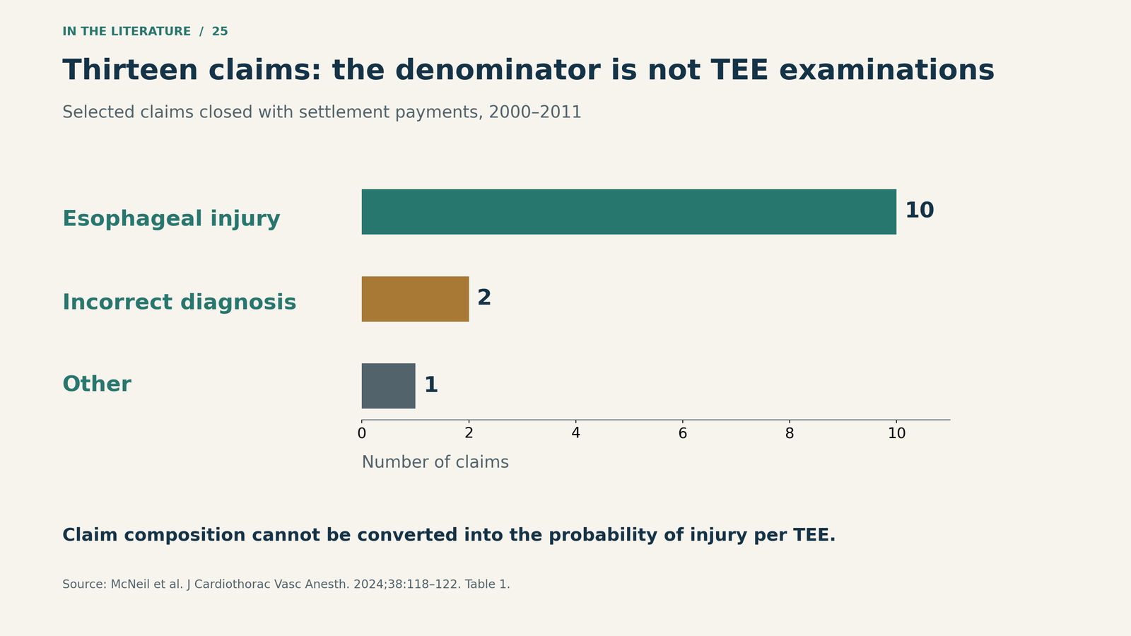
Figure 2. The composition of the 13 selected claims—not complication incidence among patients undergoing TEE. [1] Enlarge figure.

Before another attempt, revisit the risk assessment
The ASE/SCA comprehensive TEE guideline lists esophageal stricture, tumor, perforation or laceration, diverticulum, and active upper gastrointestinal bleeding among absolute contraindications. A history of dysphagia, prior gastrointestinal surgery, symptomatic hiatal hernia and restricted neck mobility are among the relative contraindications. The latter require an individualized assessment; uncertainty about gastrointestinal pathology warrants specialist input. [2]
That assessment matters again when passage proves difficult. Prior consent or an intended comprehensive examination does not resolve a new mechanical problem. Check the probe's condition and controls, ensure adequate anesthetic conditions, and protect the tracheal tube while reassessing access. The guideline supports laryngoscope-assisted placement and advises avoiding excessive probe flexion during advancement or withdrawal. [2]
A practical application is to pause advancement, identify what will change, and involve an experienced operator. Depending on the suspected obstacle, this may mean an appropriate positional adjustment or visualization of the esophageal inlet. If safe passage remains uncertain, reconsider the imaging plan with the surgical team. This is a clinical synthesis, not a validated rule allowing a fixed number of attempts. [1–4]
What video guidance has—and has not—proved
Borde and colleagues studied 363 adults undergoing elective cardiac surgery at five centers. Patients received conventional insertion or insertion under video-laryngoscope guidance. The primary endpoint combined blood on the probe tip at the end of surgery and/or injury identified on the subsequent video-laryngoscope examination. [3]
Injury occurred in 26/177 patients (14.7%) with conventional insertion and 14/186 (7.5%) with video guidance, an observed difference of approximately 7.2 percentage points. First-attempt success was 154/177 (87.0%) versus 176/186 (94.6%). These findings support visualization as a useful insertion strategy. [3]
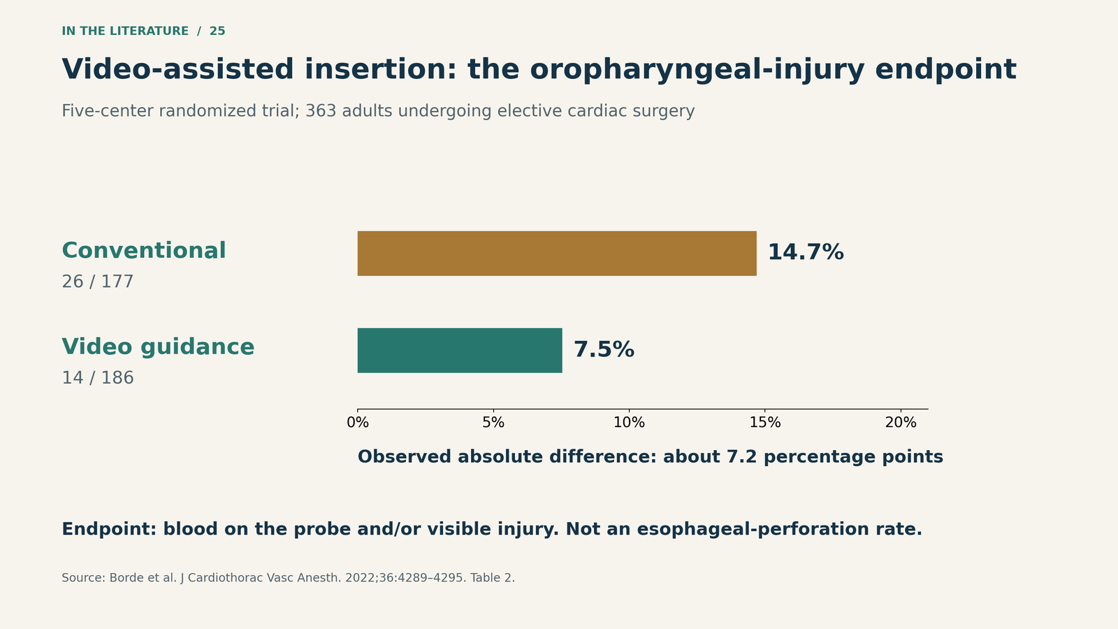
Figure 3. The trial's injury endpoint included blood on the probe tip and/or visible injury after the examination. It is not an esophageal-perforation rate. Percentages and denominators are from the original trial. [3] Enlarge figure.

The trial excluded patients with anticipated or unexpected difficult tracheal intubation, TEE contraindications and specified coagulation abnormalities. Experienced cardiac anesthesiologists performed the insertions. No insertion-related injury required further intervention, and the study was not powered to assess rare major perforation. It also did not evaluate thermal injury or harm from repeated manipulation during the examination. Better inlet visualization therefore does not remove the need for patient selection and careful subsequent handling. [3]
A recent positioning trial adds an option, not a universal maneuver
Bae and colleagues' October 2026 article reports a single-center trial conducted in 2020. Forty adults were randomized to a neutral head position without a pillow or a standardized sniffing position. Fiberoptically detected oropharyngeal injury occurred in 1/20 (5%) versus 9/20 (45%), respectively; the risk difference was −0.40 (95% CI −0.61 to −0.13). [4]
The comparison is with that specific neutral position, not every institution's usual practice. The study excluded known or predicted difficult airways and relevant anatomical or esophageal problems. It used one experienced operator, did not inspect the esophagus endoscopically, and did not establish a reduction in serious complications. It also did not test sniffing against video-assisted insertion. Positioning is worth reassessing, but neck movement must remain appropriate to the patient's cervical condition. [4]
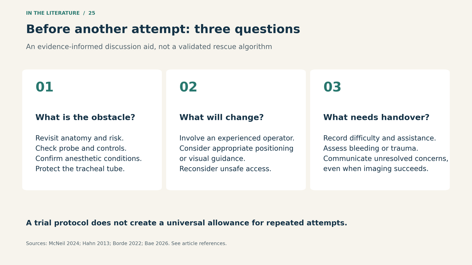
Figure 4. A practical pause before another attempt, derived from the guideline and clinical studies. This is a discussion aid, not a validated rescue algorithm. [1–4] Enlarge figure.

A successful image does not close the safety question
If passage was traumatic or bleeding occurred, communicate that explicitly and assess possible injury rather than treating successful imaging as reassurance. Record the difficulty, assistance used and relevant findings, and carry unresolved concerns into the postoperative handover. The closed-claims study's delayed diagnoses reinforce the importance of recognition; they do not quantify how much a particular monitoring strategy reduces harm. [1, 2]
Three takeaways
- Before repeating an attempt, change the assessment or approach. Revisit anatomy, contraindications, probe handling and the need for experienced assistance.
- Use visualization for what the trials support. It can reduce the measured oropharyngeal-injury endpoint; it does not guarantee freedom from perforation or later injury.
- Communicate a difficult passage even when TEE succeeds. Subsequent imaging quality does not establish that the passage was atraumatic. [1–4]
References
- McNeil JS, Singh KE, Gallo PD, Gehle B, Saunders MB, Mazzeffi MA. Medical malpractice claims related to performance of transesophageal echocardiography by anesthesiologists. J Cardiothorac Vasc Anesth. 2024;38(1):118–122. doi:10.1053/j.jvca.2023.10.002.
- Hahn RT, Abraham T, Adams MS, Bruce CJ, Glas KE, Lang RM, et al. Guidelines for performing a comprehensive transesophageal echocardiographic examination: recommendations from the American Society of Echocardiography and the Society of Cardiovascular Anesthesiologists. J Am Soc Echocardiogr. 2013;26(9):921–964. doi:10.1016/j.echo.2013.07.009.
- Borde D, Kumar C, Jasapara A, Shetty V, Juvekar N, Desurkar V, et al. Use of a video laryngoscope to reduce complications of transesophageal echocardiography probe insertion: a multicenter randomized study. J Cardiothorac Vasc Anesth. 2022;36(12):4289–4295. doi:10.1053/j.jvca.2022.07.017.
- Bae MI, Kim MH, Park SK, Go D, Lee S, Kim HC. Effect of sniffing position on transesophageal echocardiography probe insertion: a randomized controlled trial. BMC Anesthesiol. 2026 Oct 15. Article in press. doi:10.1186/s12871-026-04363-0.