ITL-452e2f36bb54
Juan Li, MD · Updated November 17, 2026
Watch the video
Key points
- Know the lesion. An anomalous muscle bundle divides the right ventricle into chambers, with intraventricular gradients above 20 mm Hg and often a membranous VSD.[1]
- Echo finds most cases, TEE shows more. Echocardiography confirmed 81% of adult diagnoses, TEE showed anatomy better than TTE, and CMR or catheterization fills the gaps.[1]
- Check the repair with TEE. A residual muscle ridge can be seen after bypass; target a residual gradient under 40 mmHg and an RV to LV pressure ratio under 0.7.[2]
- 3D helps find the level. 3D TEE showed the obstruction en face after the flow was first mistaken for a VSD.[3]
- The PA catheter can be the clue. A systolic RV pressure of 125 mmHg prompted TEE, which found a 100 mmHg gradient.[4]
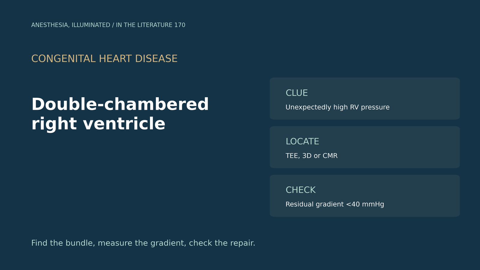
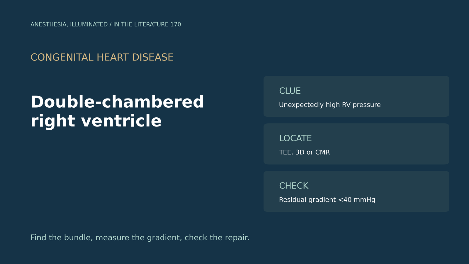
Before pulmonary valve surgery, the pulmonary artery catheter shows systemic pressure in the right ventricle, far above the pulmonary artery pressure. Where is the obstruction, and how should TEE find the muscle bundle?[1], [4]
What the lesion is and how it is imaged
Malone and colleagues describe double-chambered right ventricle as anomalous muscle bundles causing intraventricular gradients above 20 mm Hg, most often with a membranous VSD. In a cited study, echocardiography confirmed the diagnosis in 26 of 32 adults (81%), and TEE was more capable than TTE of showing the full anatomy; CMR visualizes the RVOT better, and catheterization helps if CMR is unavailable. Gradients in their cases ranged from 39 to 127 mm Hg.[1]
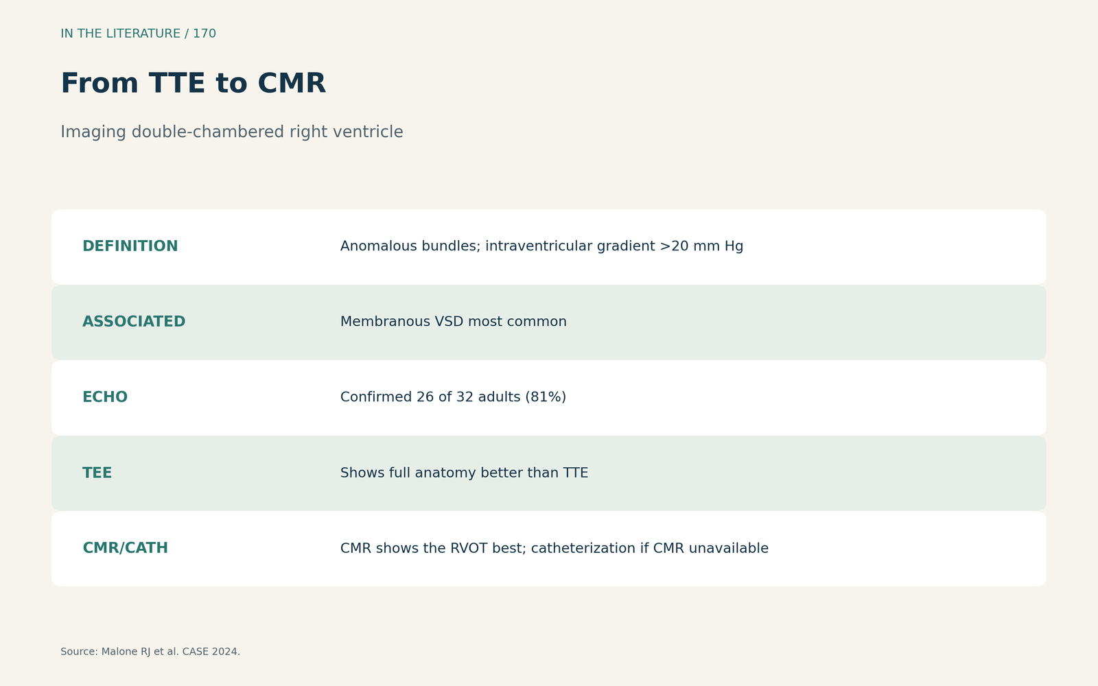
Figure 1. From TTE to CMR. Synthesized from Malone et al.[1] View larger.

TEE in the operating room
Narula and colleagues explain that the bundle divides the right ventricle into a proximal high-pressure and a distal low-pressure chamber; in type 1, an anomalous muscle bundle crosses the ventricle. After bypass, the midesophageal RV inflow-outflow view showed a residual muscle ridge, and continuous-wave Doppler should seek any high-velocity gradient. The authors target a residual peak gradient below 40 mmHg and a postrepair RV to LV pressure ratio below 0.7, and conclude that TEE is crucial at each step.[2] Ceremuga and colleagues describe a pulmonary artery catheter reading 125 mmHg in the right ventricle; only this systemic RV pressure triggered further evaluation, TEE showed a 100 mmHg gradient, and after repair TEE showed no RVOT gradient or residual VSD. They note that TEE or MRI is typically required for diagnosis.[4]
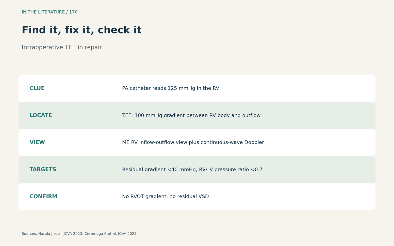
Figure 2. Find it, fix it, check it. Synthesized from Narula et al. and Ceremuga et al.[2], [4] View larger.

When the flow misleads
Sharma and colleagues describe flow first thought to be a VSD with left-to-right shunting; 3D TEE then showed double-chambered right ventricle with a presumably healed VSD. Echo can characterize the gradients and obviate invasive catheterization, and treatment is indicated for symptoms or asymptomatic obstruction.[3]
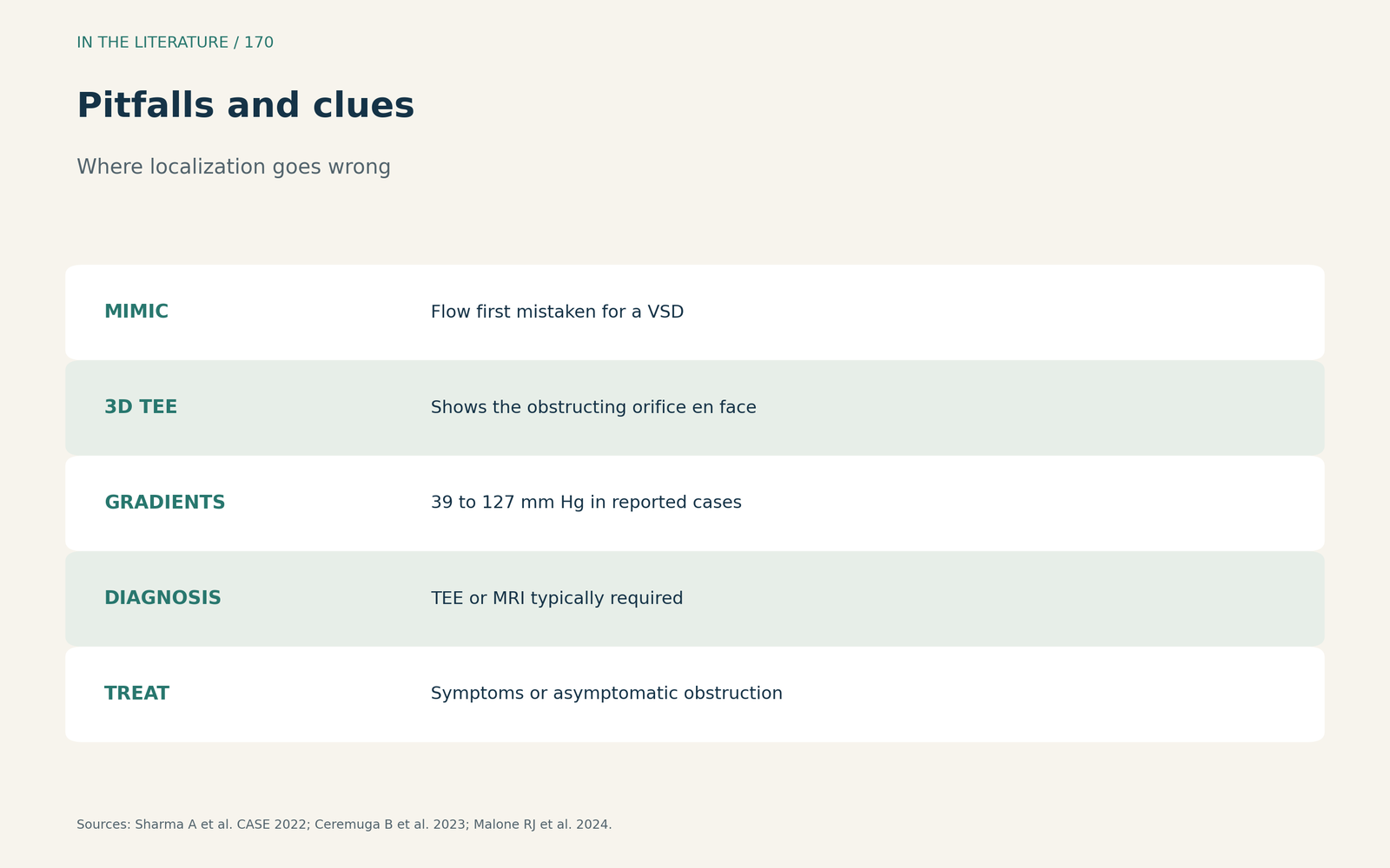
Figure 3. Pitfalls and clues. Synthesized from Sharma et al., Ceremuga et al. and Malone et al.[3], [4], [1] View larger.

Three takeaways
- Unexpectedly high RV pressure is a clue. Look for an intracavity gradient below the pulmonary valve.[4]
- Use the inflow-outflow view and Doppler. Add 3D TEE or CMR when the level is unclear.[2], [3], [1]
- Check the repair before leaving the room. Residual gradient under 40 mmHg, RV to LV ratio under 0.7.[2]
References
- Malone RJ, Henderson ER, Wilson ZR, et al. Double-chambered right ventricle in adulthood: a case series. CASE (Phila). 2024;8(3):202–209. doi:10.1016/j.case.2023.12.012
- Narula J, Bansal P, Rajput N. Indispensable role of transesophageal echocardiography in double-chamber right ventricle repair surgery. J Cardiothorac Vasc Anesth. 2023;37(7):1321–1323. doi:10.1053/j.jvca.2023.02.031
- Sharma A, Chera HH, Agarwal S, et al. Double chamber right ventricle with new-onset biventricular failure in an octogenarian. CASE (Phila). 2022;6(4):178–182. doi:10.1016/j.case.2022.01.012
- Ceremuga B, Kozik D, Sobczyk W, et al. Double-chambered right ventricle: an intraoperative surprise. J Cardiothorac Vasc Anesth. 2023;37(5):784–787. doi:10.1053/j.jvca.2023.01.031