
ITL-e60b88a4fb89
Juan Li, MD · Updated November 17, 2026
Watch the video

Key points
- A small wall-hugging colour jet can conceal important MR; jet area should not determine severity.1,2
- Eccentricity alone does not invalidate PISA. The question is whether flow convergence, radius measurement and Doppler alignment are adequate.1,2
- PISA can overestimate or underestimate regurgitation. Identify the failed assumption before assigning a direction to the error.1–3
- Use independent corroboration: valve anatomy, vena contracta, pulmonary venous flow and chamber response. Each has its own limitations.1,2
- If clinically important discordance persists, obtain better anatomical assessment with TEE and/or quantitative corroboration with CMR.1,2
The report gives an effective regurgitant orifice area (EROA), yet the eccentric mitral regurgitation (MR) jet seems small or another measurement suggests a different grade. Before deciding which number to believe, inspect how each was obtained. A technically unreliable measurement does not become reliable because the machine displays two decimal places.1,2
Separate the downstream jet from the upstream measurement
The colour jet in the left atrium and the proximal isovelocity surface area (PISA) describe different parts of the flow. A wall-hugging atrial jet can look small because of its interaction with the atrial wall—the Coandă effect. That appearance is not a dependable measure of regurgitant volume.1,2
PISA measures flow convergence upstream of the regurgitant orifice. Its conventional calculation assumes a hemispheric convergence surface. For an appropriate acquisition, flow rate is proportional to the square of the measured radius and to the aliasing velocity; EROA is then obtained by dividing flow rate by the corresponding MR velocity. Consequently, a small error in the radius is amplified, and poor continuous-wave (CW) Doppler alignment can underestimate velocity and overestimate EROA.1
The EACVI position paper explicitly allows PISA in eccentric jets when feasible. The useful question is therefore whether this particular acquisition satisfies the assumptions, rather than whether the jet carries an “eccentric” label.2
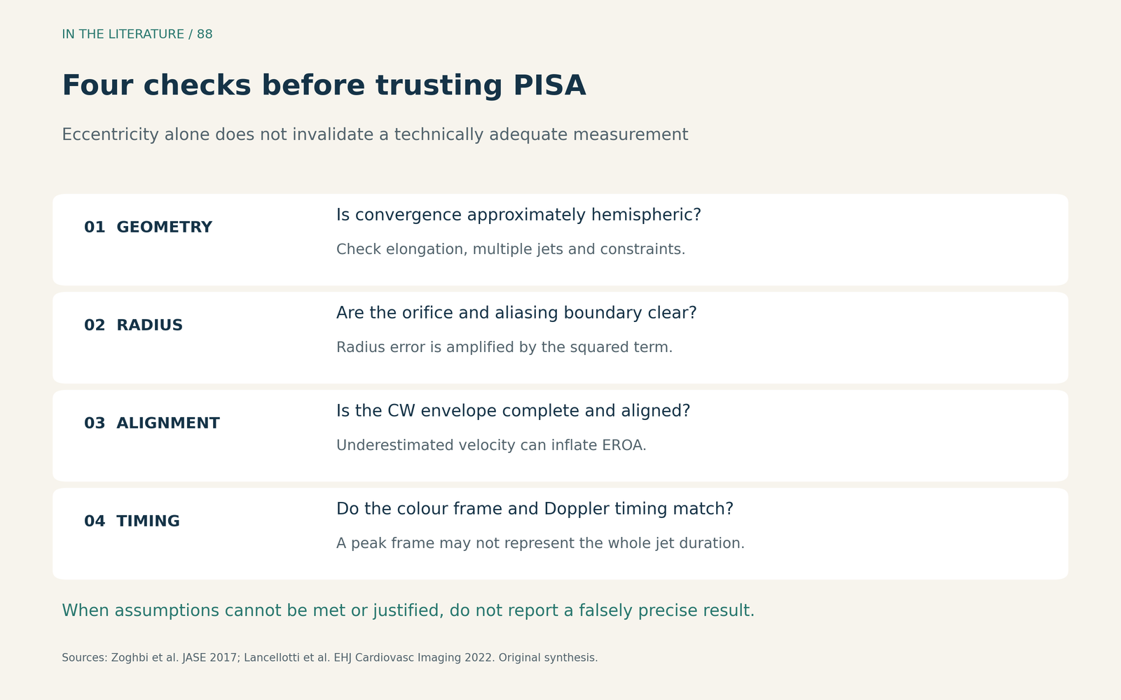
Figure 1. A measurement checklist based on ASE and EACVI guidance. These checks do not constitute a validated diagnostic score.1,2 Enlarge

Identify which assumption has failed
Geometry. Look at the convergence region in more than one view. A markedly elongated orifice can generate nonhemispheric convergence and cause conventional two-dimensional PISA to underestimate flow. Conversely, confinement by neighbouring structures can make an uncorrected hemispheric calculation inappropriate. Adjusting the acquisition may help; an angle correction is only defensible when the geometry can actually be characterized. If a usable hemisphere or justified correction cannot be obtained, do not report a falsely precise PISA result.1,2
Acquisition. Confirm the orifice location, the aliasing boundary and the radius between them. Obtain a complete, well-aligned CW envelope. A poorly aligned peak velocity is not evidence that the MR is mild; it also compromises the EROA calculation. Multiple jets require separate assessment rather than treating the most visible convergence zone as the whole lesion.1,2
Timing. Match the flow-convergence frame to the Doppler measurement. A peak single-frame EROA is not necessarily the average orifice area throughout systole. In late-systolic MR, treating that peak as representative of the entire regurgitant period can exaggerate the overall burden.1
The error is not predictably one-sided. In Altes and colleagues’ two-centre study of 188 patients with significant prolapse-related primary MR in sinus rhythm, PISA and CMR regurgitant volumes correlated weakly (r=0.29). The average PISA-minus-CMR difference was 8 mL, but the limits of agreement extended from −45 to 61 mL. PISA was not consistently higher in every patient. This selected cohort excluded atrial fibrillation and other important valve disease, and the examinations were not always performed on the same day.3
Corroborate with a method that answers a different question
Start with the valve lesion and its consequences. Does the anatomy explain the jet? Are the ventricular and atrial findings compatible with the suspected chronic burden? Normal chamber size cannot, by itself, exclude acute severe MR or important MR in a smaller patient.1
For vena contracta, use orthogonal views and, where adequate, three-dimensional assessment of the orifice. Do not add the widths of separate jets. Three-dimensional vena contracta area (VCA) can clarify complex geometry, but gain-related blooming, plane selection and systolic variation still affect it.1,2
For pulmonary venous flow, sample multiple veins, especially with TEE: an eccentric jet may preferentially affect one vein. Systolic blunting alone is nonspecific and can occur with atrial fibrillation or elevated left atrial pressure. Interpret the pattern alongside the other findings.2
A volumetric approach offers another estimate, but subtracting two measured stroke volumes can compound measurement errors. CMR is particularly useful when echo findings remain inconclusive or conflict with the clinical picture. Its preferred indirect calculation subtracts aortic forward stroke volume from total LV stroke volume and does not depend on the direction of the MR jet. Regurgitant fraction uses total LV stroke volume as the denominator. Technical quality and the chosen flow method still matter; discordance should be reconciled rather than resolved by automatically declaring either modality correct.1,2
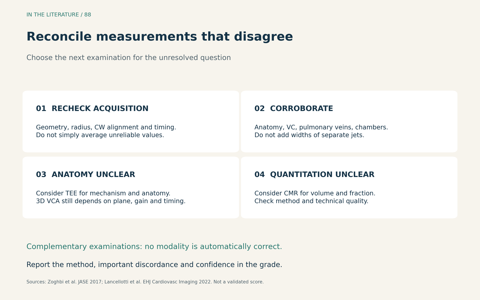
Figure 2. A practical synthesis: TEE can clarify mechanism and anatomy; CMR can corroborate regurgitant volume and fraction. The examinations are complementary and selected according to the unresolved question.1,2 Enlarge

What the latest comparison adds
Elgamal and colleagues’ 2026 retrospective study examined 85 patients undergoing elective mitral intervention under general anesthesia. PISA and volumetric severity classifications had limited concordance when used alone: κ=0.25 (95% CI 0.04–0.46). Incorporating their measurements into multiparametric grading increased concordance to κ=0.62 (95% CI 0.36–0.81).4
This supports documenting the method and integrating findings. It does not prove that PISA is superior. The study used 3D VCA as an echocardiographic comparator, without an independent CMR reference; shared measurements, timing effects and study-specific VCA thresholds limit the inference. Greater agreement is not proof of greater accuracy or better outcomes.4
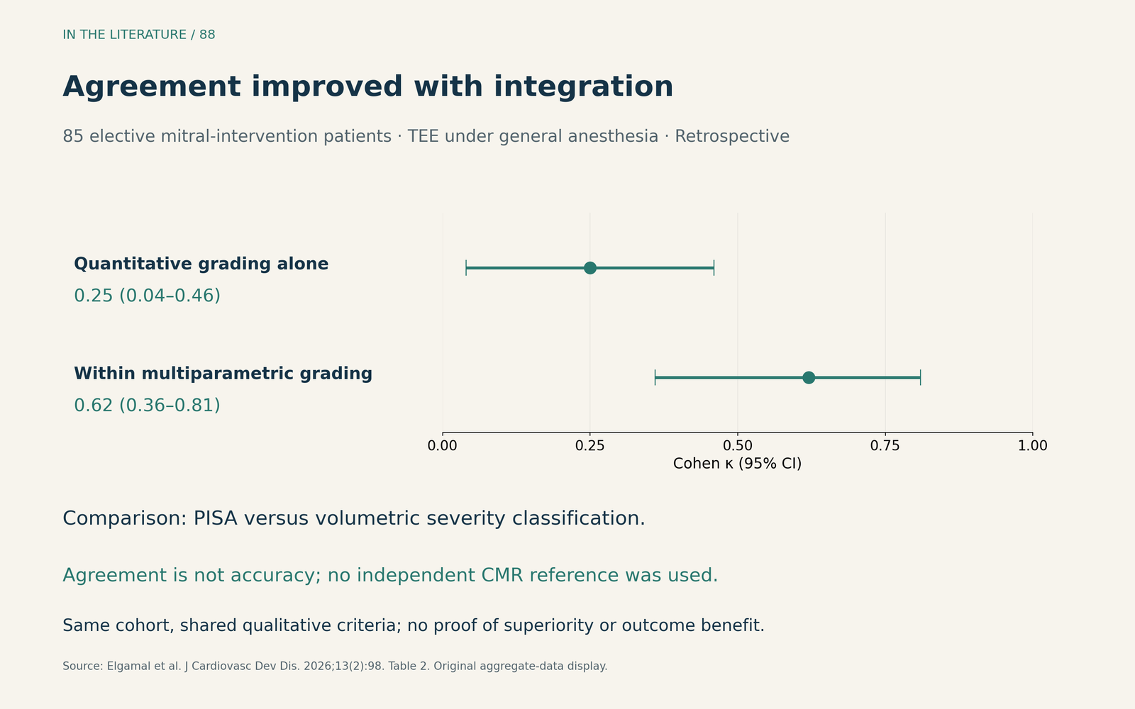
Figure 3. Original visualization based on Table 2 from Elgamal et al. The same selected intraoperative cohort contributed both comparisons; κ measures agreement beyond chance, not diagnostic accuracy.4 Enlarge

Three takeaways
- Check the assumptions before accepting the number. Eccentricity is a reason to scrutinize PISA, not an automatic reason to discard it.
- Explain discordance rather than averaging it away. Geometry, alignment and timing can produce errors in different directions; corroborate with independent findings.
- Make the next examination answer a specific question. Use TEE for unresolved anatomy or mechanism and CMR for persistent quantitative or clinical discordance; record the method and confidence in the final grade.
References
- Zoghbi WA, Adams D, Bonow RO, et al. Recommendations for Noninvasive Evaluation of Native Valvular Regurgitation: A Report from the American Society of Echocardiography Developed in Collaboration with the Society for Cardiovascular Magnetic Resonance. J Am Soc Echocardiogr. 2017;30(4):303–371. doi:10.1016/j.echo.2017.01.007
- Lancellotti P, Pibarot P, Chambers J, et al. Multi-modality imaging assessment of native valvular regurgitation: an EACVI and ESC council of valvular heart disease position paper. Eur Heart J Cardiovasc Imaging. 2022;23(5):e171–e232. doi:10.1093/ehjci/jeab253
- Altes A, Levy F, Iacuzio L, et al. Comparison of Mitral Regurgitant Volume Assessment between Proximal Flow Convergence and Volumetric Methods in Patients with Significant Primary Mitral Regurgitation: An Echocardiographic and Cardiac Magnetic Resonance Imaging Study. J Am Soc Echocardiogr. 2022;35(7):671–681. doi:10.1016/j.echo.2022.03.005
- Elgamal HR, Protsyk V, Meineri M, et al. Intraoperative Quantification of Severe Mitral Regurgitation: A Comparative Assessment of Two-Dimensional Flow Convergence, Three-Dimensional Volumetric, and Doppler-Based Methods. J Cardiovasc Dev Dis. 2026;13(2):98. doi:10.3390/jcdd13020098