ITL-0e1523f93e61
Juan Li, MD · Updated November 18, 2026
Key points
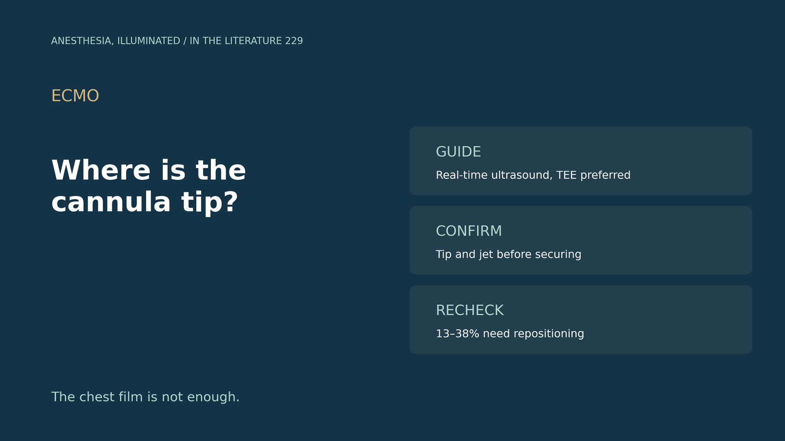
- Guide in real time. Real-time ultrasound guidance is recommended for cannulation to reduce complications and confirm position.[1]
- It works better than landmarks. In one cohort, optimal venous position was achieved in 91.30% with ultrasound vs 32.00% with landmarks.[2]
- TEE sees more. TEE shows wires traversing the SVC and IVC better than TTE, and transthoracic windows may be inadequate in about 60% of patients.[1], [3]
- Check the jet. A correctly placed dual-lumen cannula directs its outflow jet at the tricuspid valve.[4]
- Malposition happens later. During ECMO, 17 patients (38%) in one series required echo-guided repositioning.[5]
A patient with severe ARDS is being cannulated at the bedside, and the chest film looks acceptable. Is that enough, or should ultrasound decide where the cannula sits?[1], [6]
Guiding the insertion
Douflé et al. (ELSO state-of-the-art review, ASAIO J 2026) recommend real-time ultrasound guidance for cannulation; TEE shows wires traversing the SVC and IVC better than TTE, and cannula size in French can be guided by three times the vessel diameter in mm.[1] Sun et al. (Front Cardiovasc Med 2025) studied 48 VA ECMO patients: ultrasound guidance achieved optimal venous position in 91.30% vs 32.00% with landmarks, no IVC injury occurred with ultrasound vs two with landmarks, and set-up time was not longer.[2] Cha and Kostibas (J Clin Med 2024) note there is no recommended optimal imaging modality, but TEE-guided dual-lumen cannulation has success rates above 90%.[4]
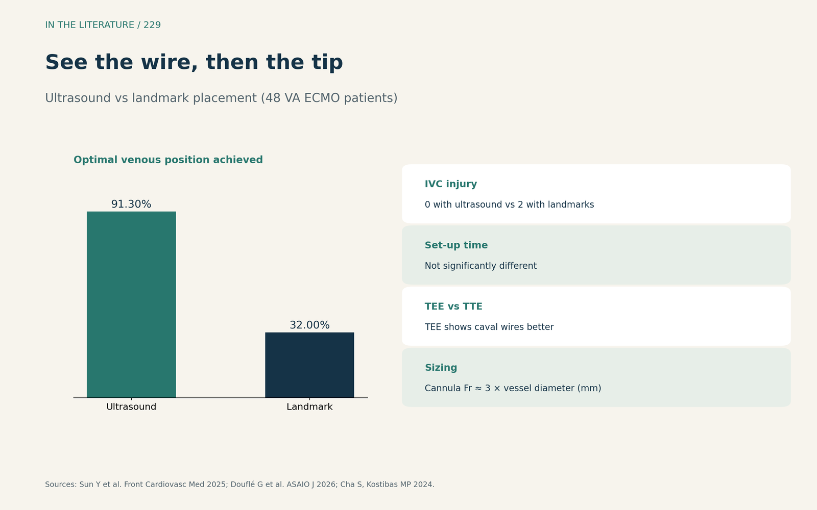
Figure 1. See the wire, then the tip. Synthesized from Douflé et al., Sun et al. and Cha and Kostibas.[1], [2], [4] View larger.

Confirming the tip
Obturators should be removed before confirming the cannula tip, and a post-cannulation echo is needed after ventilator changes and repositioning, before final securement; for bicaval dual-lumen cannulas, color Doppler identifies the reinfusion jet.[1] A correctly placed dual-lumen cannula directs its outflow jet at the tricuspid valve.[4] Sun et al. place the VA drainage tip in the mid-right atrium,[2] whereas one VV protocol placed the drainage cannula 2 to 3 cm above the IVC-RA junction.[3] Serial chest X-rays have limits for monitoring device position.[6]
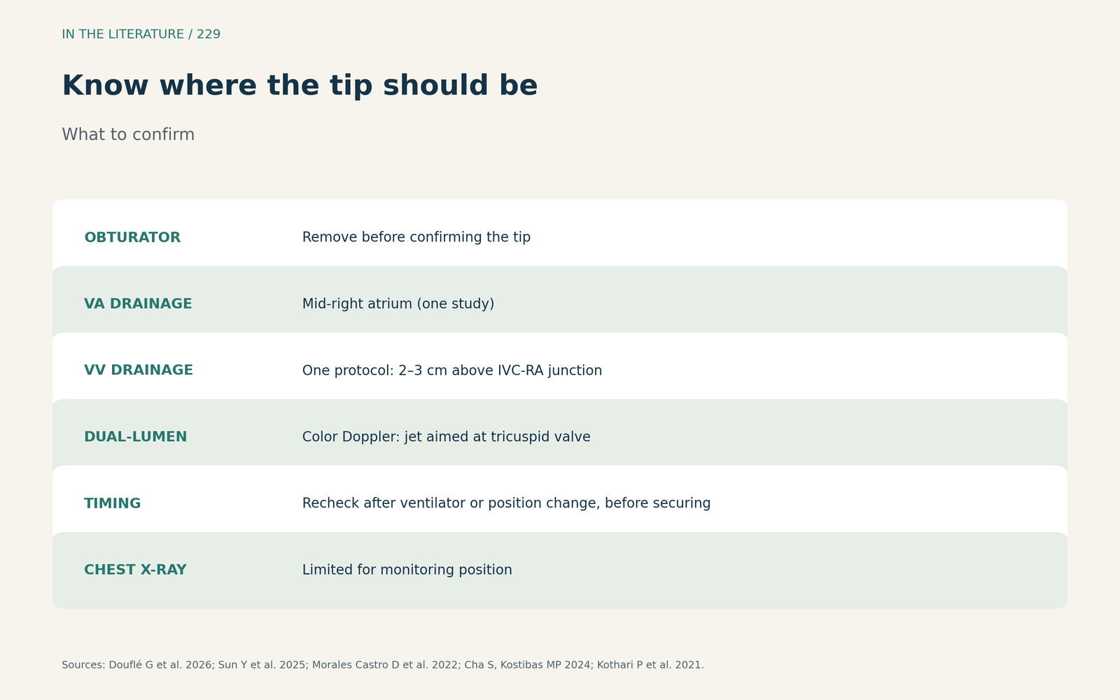
Figure 2. Know where the tip should be. Synthesized from Douflé et al., Cha and Kostibas, Sun et al., Morales Castro et al. and Kothari et al.[1], [4], [2], [3], [6] View larger.

Malposition and rechecks
In Griffee et al. (J Cardiothorac Vasc Anesth 2020), initial echo-guided dual-lumen cannulation succeeded in 87%, but 17 patients (38%) later required repositioning, and the cannula entered the RV in 2 cases (4%).[5] In Morales Castro et al. (J Cardiothorac Vasc Anesth 2022), TEE-guided cannulation succeeded in 99% of patients, yet 13 percent required repositioning.[3] After decannulation, ultrasound of the cannulation sites and venae cavae is recommended.[1] The evidence is observational and expert opinion.
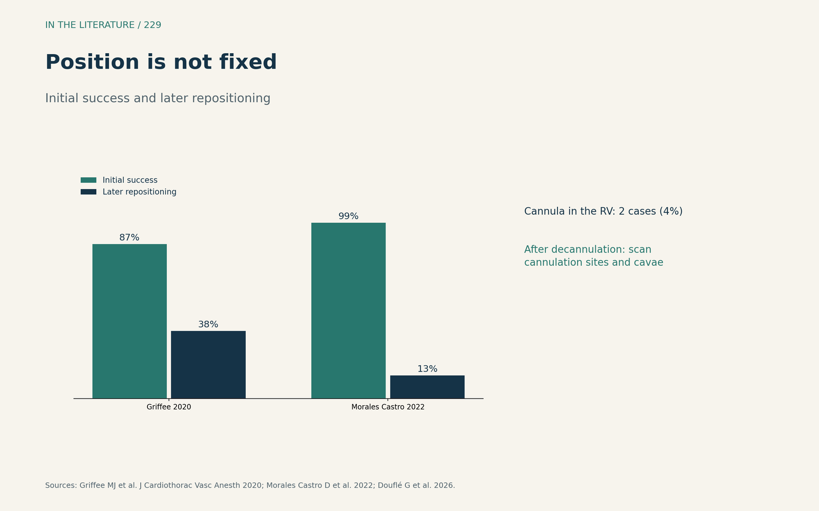
Figure 3. Position is not fixed. Redrawn from Griffee et al., Morales Castro et al. and Douflé et al.[5], [3], [1] View larger.

Three takeaways
- Use real-time ultrasound, preferably TEE, from wire to tip; do not rely on the chest film alone.[1], [2], [6]
- Remove obturators, confirm the tip and the reinfusion jet, then secure the cannula.[1], [4]
- Reimage whenever flow or oxygenation changes, and scan the vessels after decannulation.[1], [5], [3]
References
- Douflé G, O’Neil ER, Abreu S, et al. Ultrasound in extracorporeal membrane oxygenation: an ELSO state-of-the-art review. ASAIO J. 2026;72(2):85–98. doi:10.1097/MAT.0000000000002656
- Sun Y, Huang C, Ou W, et al. Efficacy of ultrasound guided venous cannulation positioning during venous-arterial extracorporeal membrane oxygenation. Front Cardiovasc Med. 2025;12:1656101. doi:10.3389/fcvm.2025.1656101
- Morales Castro D, Abdelnour-Berchtold E, Urner M, et al. Transesophageal echocardiography-guided extracorporeal membrane oxygenation cannulation in COVID-19 patients. J Cardiothorac Vasc Anesth. 2022;36(12):4296–4304. doi:10.1053/j.jvca.2022.07.020
- Cha S, Kostibas MP. Echocardiographic and point-of-care ultrasonography (POCUS) guidance in the management of the ECMO patient. J Clin Med. 2024;13(9):2630. doi:10.3390/jcm13092630
- Griffee MJ, Zimmerman JM, McKellar SH, Tonna JE. Echocardiography-guided dual-lumen venovenous extracorporeal membrane oxygenation cannula placement in the ICU—a retrospective review. J Cardiothorac Vasc Anesth. 2020;34(3):698–705. doi:10.1053/j.jvca.2019.10.024
- Kothari P, Ellis J, Pretorius VG, Khoche S. The risk of malposition: a tale of two devices. J Cardiothorac Vasc Anesth. 2021;35(3):963–966. doi:10.1053/j.jvca.2020.07.018