ITL-29e8096afbbd
Juan Li, MD · Updated November 17, 2026
Watch the video
Key points
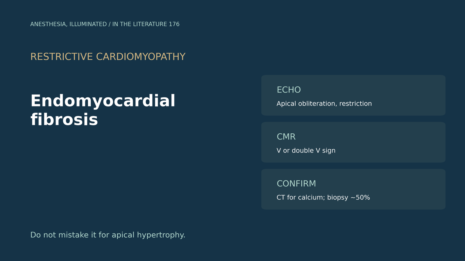
- Picture the apex. EMF is apical obliteration with fibrous tissue in one or both ventricles, and TTE is the first-line imaging test.[1]
- Use the echo criteria. EMF is diagnosed with two major criteria or one major plus minor criteria; a restrictive pattern is common, and both ventricles are involved in up to 55%.[2]
- Look for the V sign on CMR. Apical subendocardial enhancement over normal myocardium suggests EMF, whereas apical hypertrophic cardiomyopathy usually shows midwall enhancement.[3]
- Add strain and contrast. Contrast TTE is sensitive for thrombus, strain is a sensitive early tool, and biopsy remains the gold standard even without eosinophilia.[4]
- Know the limits. Biopsy yield is about 50%, and CT helps separate thrombus from calcium.[1]
A patient with heart failure is found to have an apex filled with echodense material and a restrictive mitral inflow pattern; apical hypertrophic cardiomyopathy is suggested. Is this endomyocardial fibrosis, and how should it be confirmed?[1], [2]
Echo recognition
Moscoso and colleagues describe EMF as apical obliteration with fibrous tissue of one or both ventricles, with TTE as the first-line test; pericardial effusion and an endocardial fibrous shelf are typical signs.[1] Velandia-Carrillo and Zuluaga state that echocardiography is necessary for the diagnosis, which requires two major criteria or one major plus minor criteria; their patient met three major and two minor criteria (score 13) with a restrictive pattern (E/A 3.4). Biventricular involvement occurs in up to 55%, and 10-year survival is about 37%.[2]
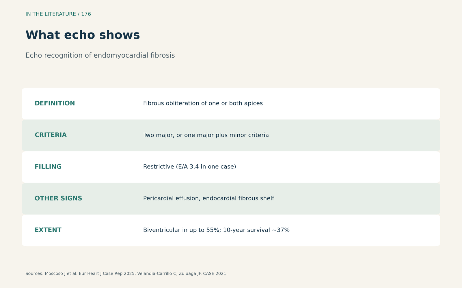
Figure 1. What echo shows. Synthesized from Moscoso et al. and Velandia-Carrillo and Zuluaga.[1], [2] View larger.

CMR, CT and the differential
The double V sign is a three-layered pattern of normal myocardium, thickened endocardium and overlying thrombus; without thrombus it is a single V sign, and CT distinguishes thrombus from calcification.[1] Sehly and colleagues describe apical subendocardial enhancement over normal myocardium (the V sign) as typical of EMF, whereas apical hypertrophic cardiomyopathy usually shows midwall enhancement and Loeffler endocarditis patchy or diffuse subendocardial enhancement; EMF was favored because eosinophilia was absent.[3]
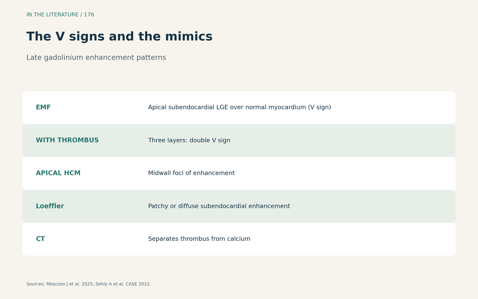
Figure 2. The V signs and the mimics. Synthesized from Moscoso et al. and Sehly et al.[1], [3] View larger.

Strain, thrombus and biopsy
Frederiksen and colleagues, describing eosinophilic endomyocardial disease, found large areas of subendocardial enhancement on CMR; contrast TTE is sensitive for intracavitary thrombus, myocardial strain is a highly sensitive tool on initial TTE, and reduced global strain is not typical of apical hypertrophic cardiomyopathy. Endomyocardial biopsy remains the gold standard and should be considered even without eosinophilia,[4] although its diagnostic yield is about 50%.[1]
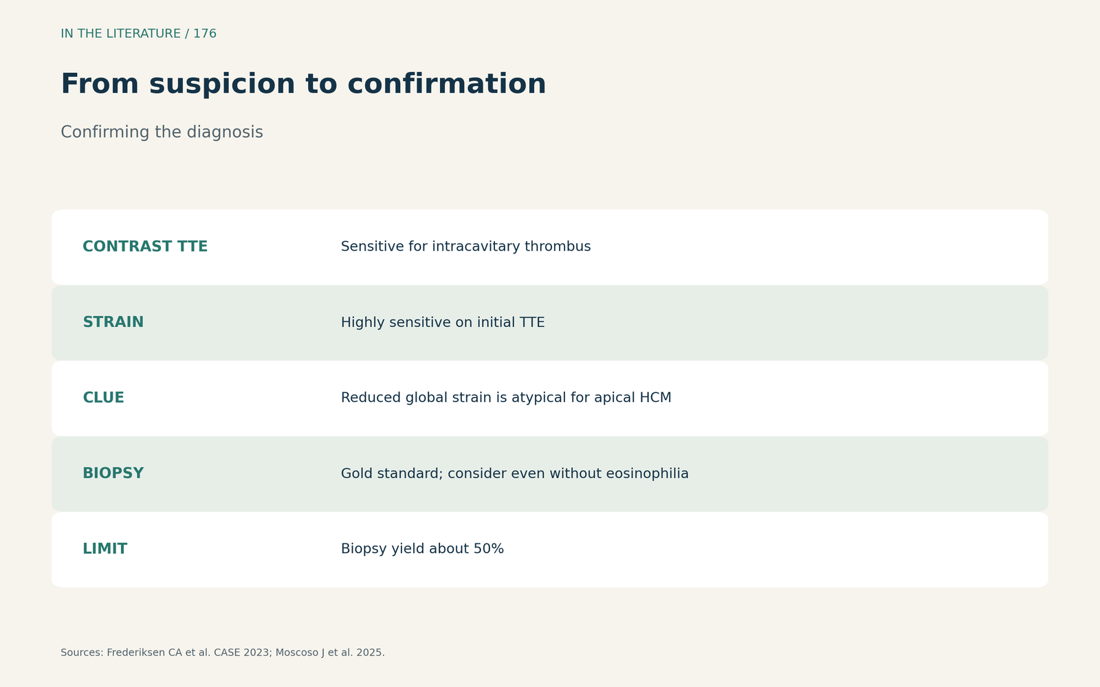
Figure 3. From suspicion to confirmation. Synthesized from Frederiksen et al. and Moscoso et al.[4], [1] View larger.

Three takeaways
- An obliterated apex with restrictive filling is the echo clue. Apply the major and minor criteria.[1], [2]
- CMR separates EMF from apical hypertrophy. Subendocardial V sign versus midwall enhancement.[3]
- Confirm thoughtfully. Contrast echo for thrombus, CT for calcium, biopsy when needed.[4], [1]
References
- Moscoso J, Velarde-Acosta K, Viñas A, et al. Multimodality imaging analysis of the spectrum of endomyocardial fibrosis involvement in a clinical case series. Eur Heart J Case Rep. 2025;9(2):ytaf039. doi:10.1093/ehjcr/ytaf039
- Velandia-Carrillo C, Zuluaga JF. Multimodality imaging in endomyocardial fibrosis: an unusual etiology of heart failure. CASE (Phila). 2021;5(5):301–304. doi:10.1016/j.case.2021.06.002
- Sehly A, Aleksova N, Chow BJ, Dwivedi G. Endomyocardial fibrosis, apical hypertrophy, or both? CASE (Phila). 2022;6(9):411–415. doi:10.1016/j.case.2022.08.001
- Frederiksen CA, Stilling C, Kim WY, Poulsen SH. Two different clinical presentations and stages of Loeffler endocarditis diagnosed by multimodality investigations. CASE (Phila). 2023;7(12):496–501. doi:10.1016/j.case.2023.09.003