
ITL-e287d3e19d43
Juan Li, MD · Updated November 18, 2026
Key points
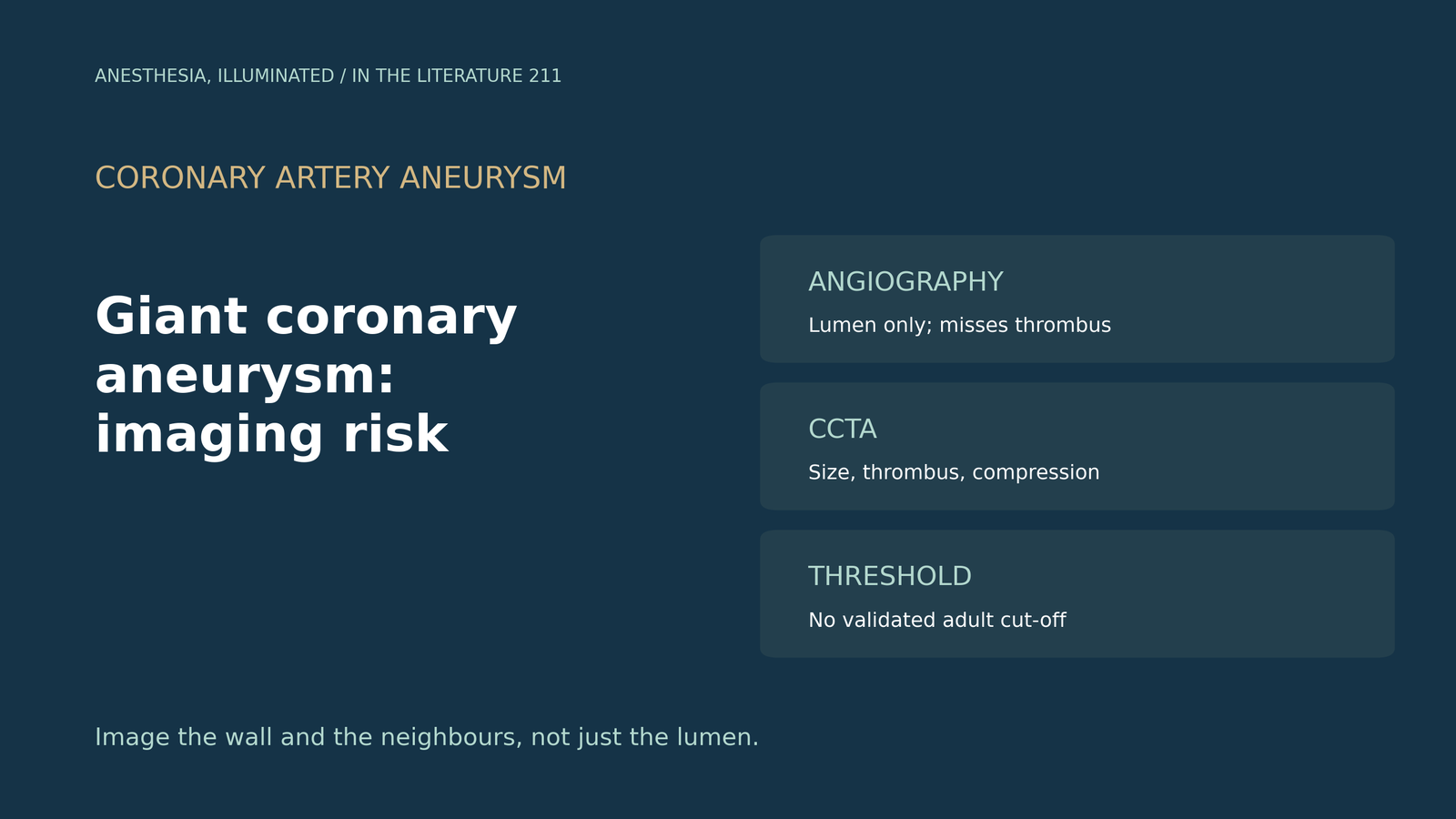
- "Giant" is a relative term. A dilation exceeding four times the reference diameter defines a giant aneurysm, and rupture is rare (below 1%) but catastrophic.[1]
- Angiography sees only the lumen. It can underestimate thrombus, whereas CCTA detects thrombi that angiography misses and is the modality of choice for follow-up.[2]
- Size predicts events. Longitudinal aneurysm diameter was linked to adverse events (OR 1.109), with MACCE in 13% during follow-up.[3]
- There is no rulebook. Unlike aortic aneurysms, no guidelines define size thresholds, and 85 to 97% of patients also have coronary stenosis.[4]
- Look beyond the coronary. Compression of the right heart and loss of preload with positive pressure ventilation can cause collapse.[4]
Before surgery for a giant right coronary artery aneurysm, angiography reports a modest lumen, but CT shows a much larger thrombosed sac pressing on the right atrium. Which images define the risk, and which findings should change the anesthetic plan?[2], [4]
Definition and natural history
Papoutsakis and colleagues define a giant aneurysm as a dilation exceeding four times the reference diameter; rupture is rare (below 1%) but catastrophic, a localized aneurysm was associated with 2.26-fold more MACE, and CABG with aneurysm exclusion is generally preferred for giant aneurysms.[1] Matta and colleagues found aneurysms in 100 of 26,962 angiograms; MACCE occurred in 13% during follow-up, and longitudinal aneurysm diameter was linked to MACCE (OR 1.109).[3] Scarpa and colleagues note that there is no consensus on investigating incidental aneurysms, that unlike aortic aneurysms no guidelines define size thresholds, and that 85 to 97% of patients also have coronary stenosis.[4]
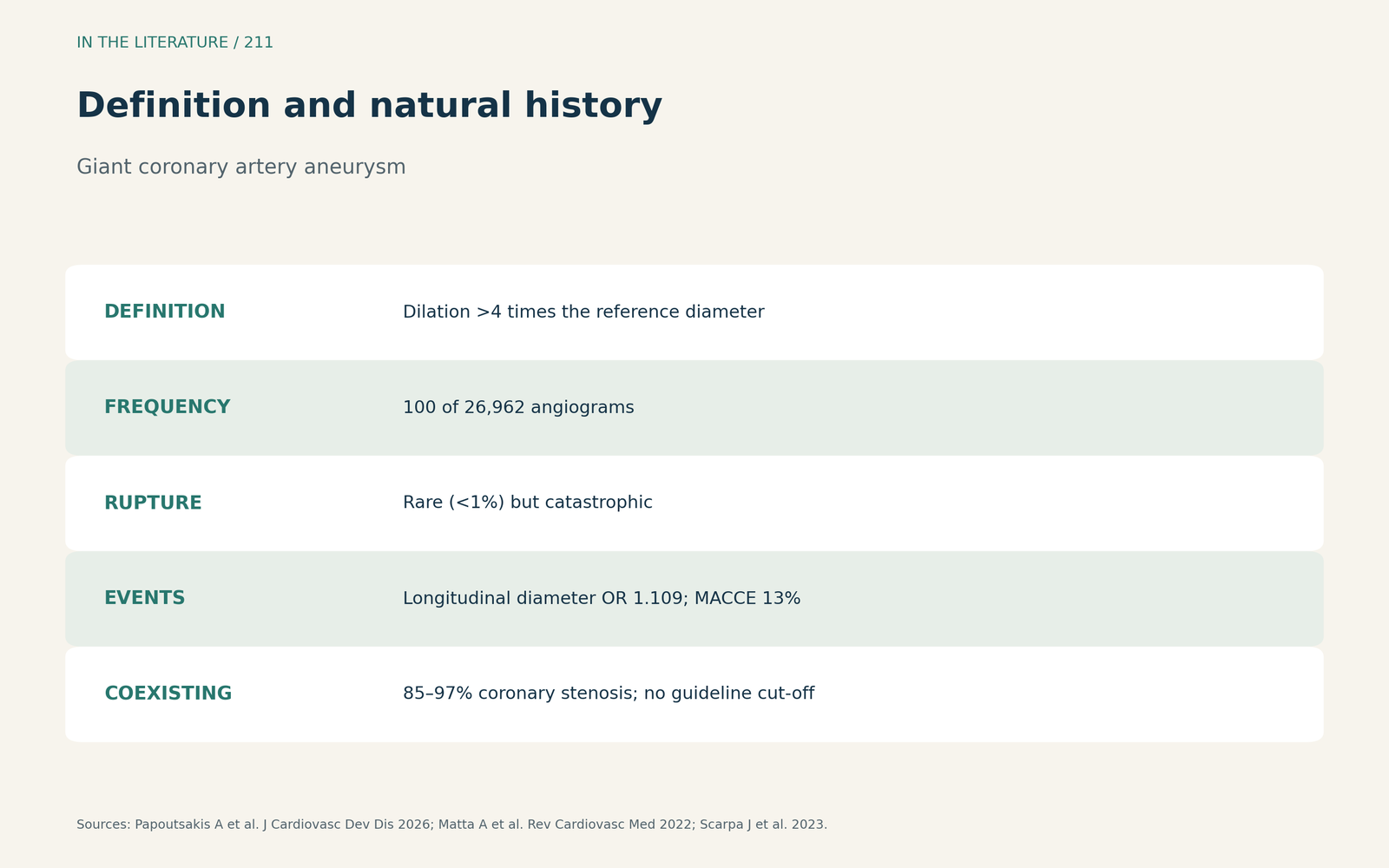
Figure 1. Relative size, rare rupture, common coexisting disease. Synthesized from Papoutsakis et al., Matta et al. and Scarpa et al.[1], [3], [4] View larger.

Which imaging shows what
Sorathia and colleagues explain that invasive angiography provides a luminogram and can underestimate thrombus, whereas CCTA detects thrombi that angiography misses and is the modality of choice for follow-up; MRA has lower spatial resolution and may miss calcification.[2] Papoutsakis and colleagues call CCTA the preferred initial modality because it delineates morphology, size, thrombus burden and wall calcification.[1] Meel and colleagues found that TTE could not define the origin and course of a giant aneurysm, that cardiac CT defines size, extent and relationships, and that differentials include tumors and coronary pseudoaneurysms.[5]
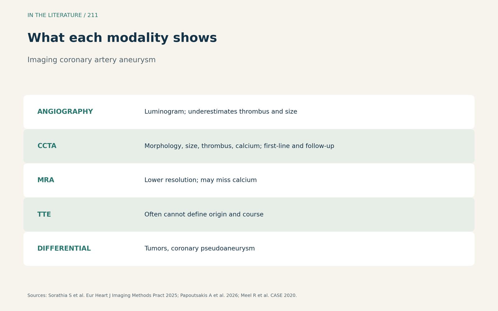
Figure 2. Lumen, wall, thrombus and neighbours. Synthesized from Sorathia et al., Papoutsakis et al. and Meel et al.[2], [1], [5] View larger.

Risk features that change management
Sorathia and colleagues list compression of adjacent structures causing compromise or a high rupture risk as reasons for intervention; in one conservatively treated cohort, 53% had MACE at 4 years. In Kawasaki disease, anticoagulation is advised for a Z score above 10, but this does not translate to an adult threshold.[2] Scarpa and colleagues describe imaging that showed right atrial and ventricular compression and warn that positive pressure ventilation can cause collapse from reduced preload.[4]
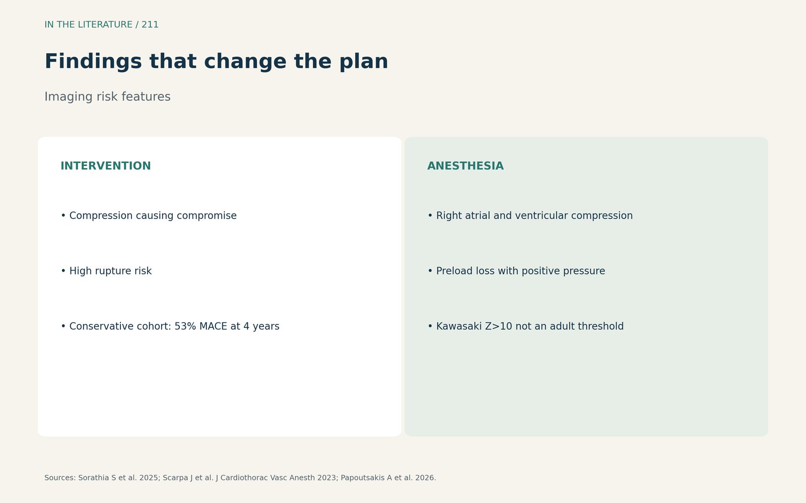
Figure 3. Compression, growth, symptoms and coexisting disease. Synthesized from Sorathia et al., Scarpa et al. and Papoutsakis et al.[2], [4], [1] View larger.

Three takeaways
- Do not size a giant coronary aneurysm by angiography alone; CCTA shows true size, thrombus and compression.[1], [2]
- No validated adult size threshold exists; weigh size, growth, symptoms and coexisting coronary disease.[3], [4]
- Look for compression of the right heart and plan induction for preload loss.[4]
References
- Papoutsakis A, Lempidakis D, Sideras-Marakas E, et al. Coronary artery ectasia and aneurysm: benign variant or high-risk substrate in need of tailored treatment? J Cardiovasc Dev Dis. 2026;13(7):336. doi:10.3390/jcdd13070336
- Sorathia S, Arockiam AD, Agrawal A, et al. Contemporary review of the clinical features, multi-modality imaging, and management of coronary artery aneurysms. Eur Heart J Imaging Methods Pract. 2025;3(1):qyaf054. doi:10.1093/ehjimp/qyaf054
- Matta A, Campelo-Parada F, Nader V, et al. Long-term outcomes of conservative versus invasive approach of coronary aneurysm. Rev Cardiovasc Med. 2022;23(8):281. doi:10.31083/j.rcm2308281
- Scarpa J, Zhu A, Morikawa NK, Chan JM. Perioperative management of giant coronary artery aneurysm. J Cardiothorac Vasc Anesth. 2023;37(10):2040–2045. doi:10.1053/j.jvca.2023.05.030
- Meel R, Lorentz L, Gonçalves R. Multimodality imaging of a giant circumflex coronary artery aneurysm in a septuagenarian. CASE (Phila). 2020;4(4):236–239. doi:10.1016/j.case.2020.02.004