ITL-375081e5c9e0
Juan Li, MD · Updated November 18, 2026
Watch the video
Key points
- Mismatch is overcalled. In a study of mechanism-proven cases, the most common overdiagnosis was PPM (22% to 29%), and overreliance on Doppler indexes was the culprit.[1]
- The catheter may disagree with Doppler. In one case, the Doppler mean gradient was 56 mmHg but the catheter gradient only 24 mmHg, a pressure recovery effect.[2]
- The LVOT is the weak link. Agreement is low for parameters requiring the LVOT diameter, and gradients are highly flow dependent.[3]
- Baseline matters. Obstruction is defined after excluding a high-output state, and stenosis is separated from mismatch by comparison with the early baseline.[4]
- Look at the leaflets. TEE best defines the cause of dysfunction, and CT can separate thrombus from pannus.[5]
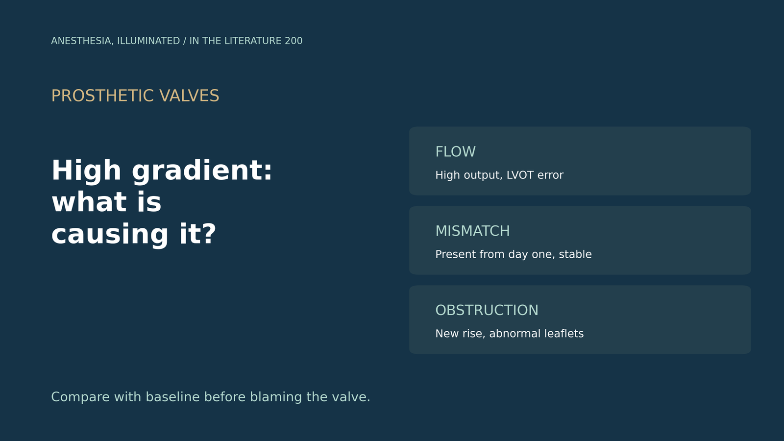
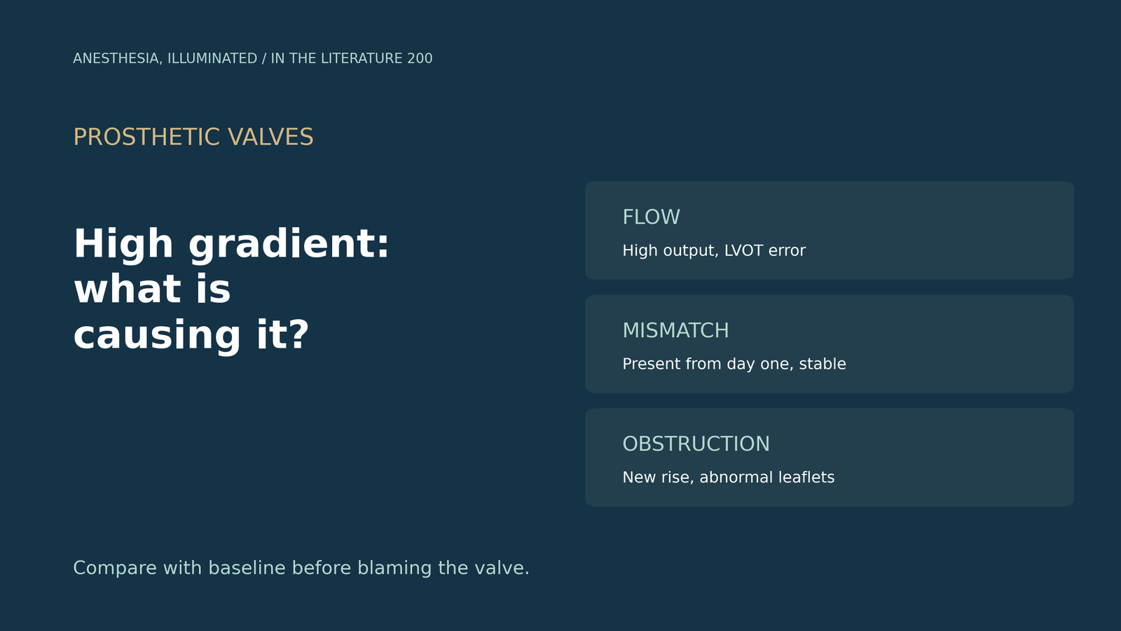
After cardiopulmonary bypass and a 21 mm aortic bioprosthesis, the TEE mean gradient is 32 mmHg with an inotrope running and a cardiac index of 3.8. Is the valve obstructed, too small for the patient, or simply being measured under high flow?[1], [4]
Why algorithms get it wrong
Roslan and colleagues tested echo algorithms in 266 mechanism-proven cases. AT, DVI and EOAi detected obstruction with AUCs of 0.84 to 0.94, yet the most common overdiagnosis was PPM (22% to 29%), pressure recovery or technical error was misdiagnosed in 23 patients (8%), and older algorithms recognized regurgitation in only 1 of 47. Neither prolonged AT nor reduced DVI is an absolute indicator of obstruction, and the authors blame overreliance on Doppler indexes. Their new algorithm raised accuracy from 57-62% to 83%, but it was tested on the data it was derived from.[1] Dietrich and Mankad describe a Doppler mean gradient of 56 mmHg with a catheter gradient of only 24 mmHg, note that pressure recovery matters more with small ascending aortas, and suggest that a DVI below 0.25 with an AT above 100 msec points to significant stenosis. PPM gradients, by contrast, are present immediately after implantation and stay constant.[2]
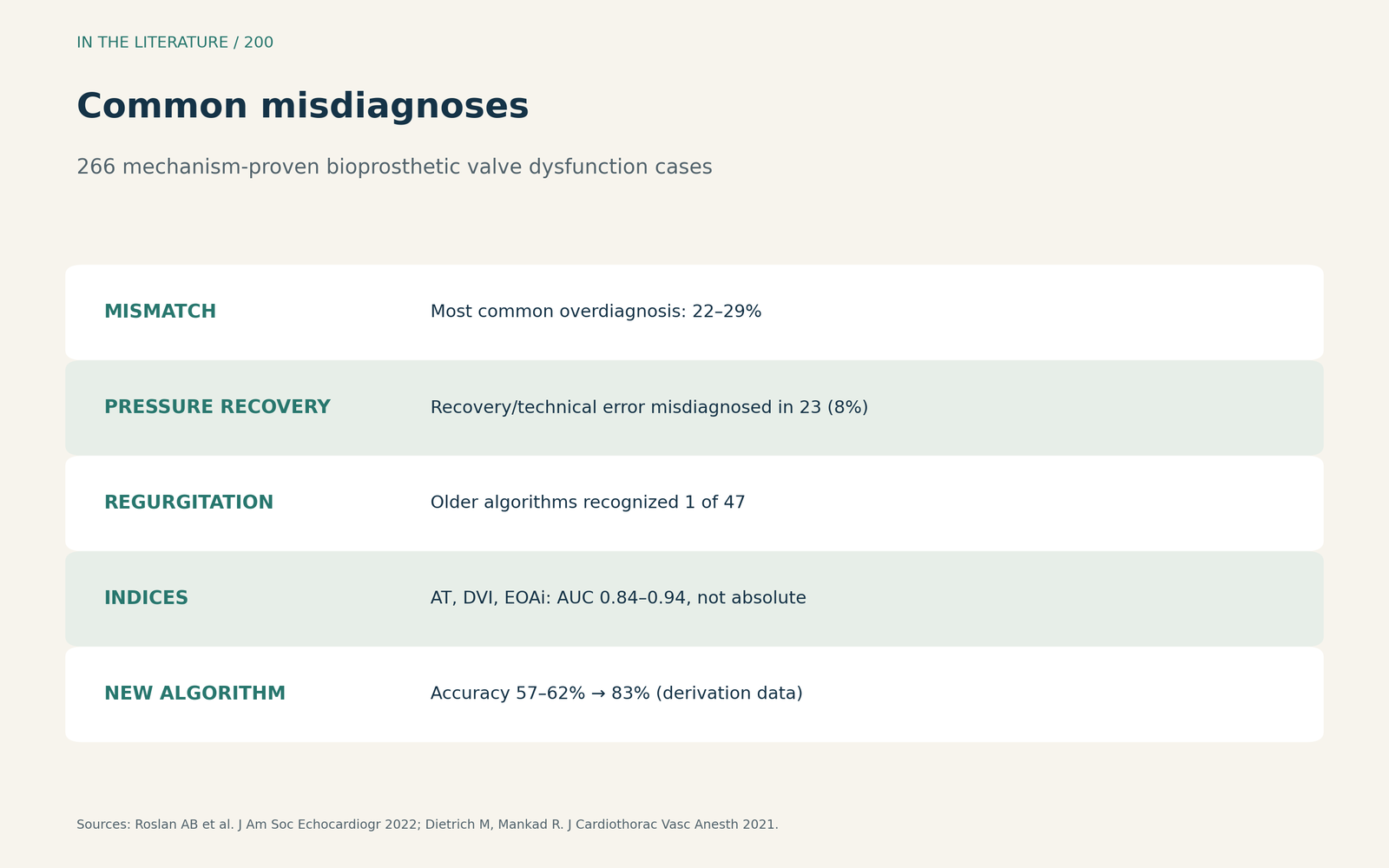
Figure 1. Mismatch is overcalled, pressure recovery underrecognized. Redrawn from Roslan et al. and Dietrich and Mankad.[1], [2] View larger.

Flow and measurement
In the PERIGON core-lab analysis, Velders and colleagues found low agreement for parameters requiring the LVOT diameter, noted that small errors greatly affect any parameter using LVOT area, and found that PPM classification was concordant in only 57%. Peak velocity and mean gradient are highly flow dependent.[3] The ACC/AHA guideline defines obstruction only after excluding causes such as a high-output state, distinguishes stenosis from mismatch by comparison with the early postoperative baseline, and notes that high gradients with normal leaflet motion and no thrombus suggest mismatch or pannus.[4]
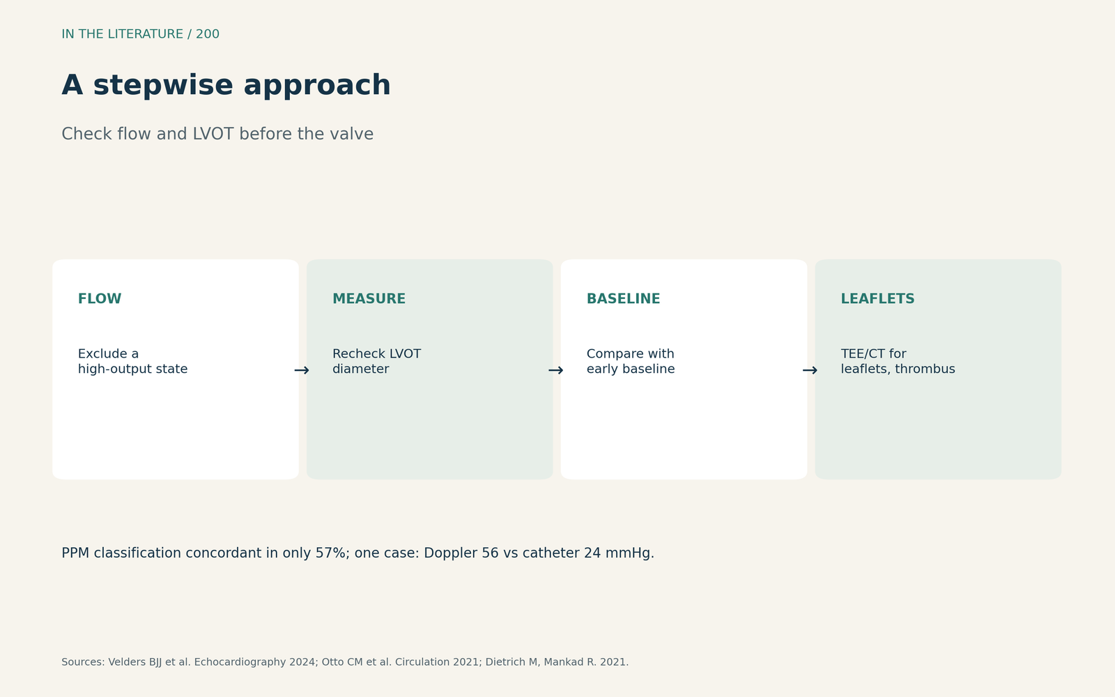
Figure 2. Check the flow and the LVOT before the valve. Synthesized from Velders et al., Otto et al. and Dietrich and Mankad.[3], [4], [2] View larger.

Mismatch or obstruction?
Ben Zekry and colleagues call PPM an important cause of high gradients across normally functioning prostheses and suggest that combining AT with AT/ET may be most helpful.[6] Talapaneni and colleagues stress that PPM reflects undersizing rather than leaflet pathology.[7] Muratori and colleagues state that TEE best defines the cause of prosthetic dysfunction, that up to 40% of pannus cases show normal opening angles, and that CT can differentiate thrombus from pannus by Hounsfield units.[5]
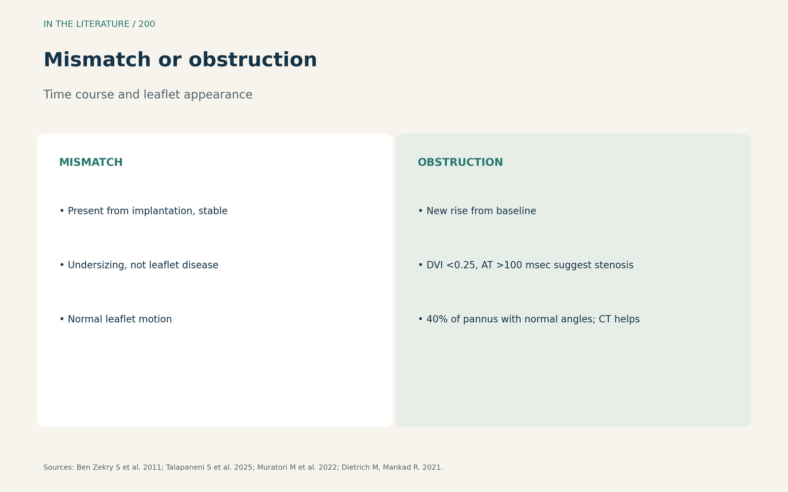
Figure 3. Constant from day one, or new and rising? Synthesized from Ben Zekry et al., Talapaneni et al., Muratori et al. and Dietrich and Mankad.[6], [7], [5], [2] View larger.

Three takeaways
- Before calling obstruction, check flow, LVOT measurement and the early postoperative baseline.[3], [4]
- High gradients present from implantation and stable suggest mismatch; pressure recovery can inflate Doppler gradients.[2], [1]
- A new rise with abnormal leaflets needs TEE or CT to separate thrombus from pannus.[5]
References
- Roslan AB, Naser JA, Nkomo VT, et al. Performance of echocardiographic algorithms for assessment of high aortic bioprosthetic valve gradients. J Am Soc Echocardiogr. 2022;35(7):682–691.e2. doi:10.1016/j.echo.2022.01.019
- Dietrich M, Mankad R. Elevated prosthetic valve gradients: what to consider when determining an etiology. J Cardiothorac Vasc Anesth. 2021;35(7):2223–2227. doi:10.1053/j.jvca.2021.02.021
- Velders BJJ, Vriesendorp MD, Weissman NJ, et al. Core laboratory versus center-reported echocardiographic assessment of the native and bioprosthetic aortic valve. Echocardiography. 2024;41(12):e70047. doi:10.1111/echo.70047
- Otto CM, Nishimura RA, Bonow RO, et al. 2020 ACC/AHA guideline for the management of patients with valvular heart disease: a report of the American College of Cardiology/American Heart Association Joint Committee on Clinical Practice Guidelines. Circulation. 2021;143(5):e72–e227. doi:10.1161/CIR.0000000000000923
- Muratori M, Fusini L, Mancini ME, et al. The role of multimodality imaging in left-sided prosthetic valve dysfunction. J Cardiovasc Dev Dis. 2022;9(1):12. doi:10.3390/jcdd9010012
- Ben Zekry S, Saad RM, Özkan M, et al. Flow acceleration time and ratio of acceleration time to ejection time for prosthetic aortic valve function. JACC Cardiovasc Imaging. 2011;4(11):1161–1170. doi:10.1016/j.jcmg.2011.08.012
- Talapaneni S, Ahmad D, Khandelwal M, et al. Prosthesis–patient mismatch following aortic valve replacement—a comprehensive review. J Clin Med. 2025;14(24):8868. doi:10.3390/jcm14248868