ITL-2c2895b32f10
Juan Li, MD · Updated November 17, 2026
Watch the video
Key points
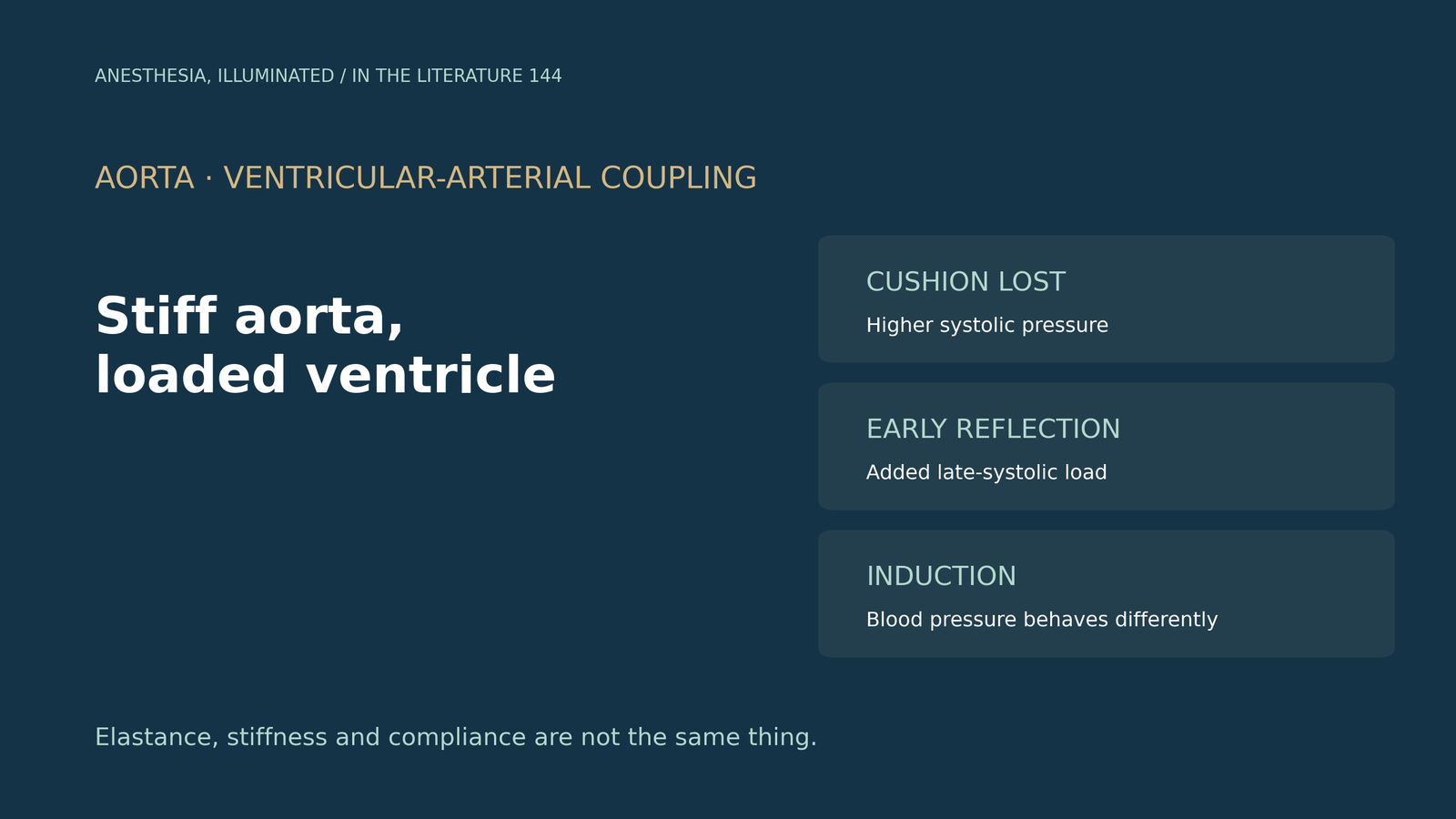
- The aorta is a cushion. In a stiffened aorta, systolic pressure and left ventricular afterload rise, leading to hypertrophy and higher myocardial oxygen demand.[1]
- Reflected waves add late-systolic load. When the aorta stiffens, pulse wave velocity rises and the reflected wave becomes an added burden on the ventricle during late systole.[2]
- The link appears early. In healthy normotensive adults, reduced aortic distensibility was associated with impaired atrio-ventricular-arterial coupling before hypertension or hypertrophy; in young adults with diabetes, stiffness preceded greater LV mass and worse diastolic indices, not systolic dysfunction.[2], [3]
- Terms are not interchangeable. Arterial elastance, stiffness and compliance describe different properties, and pulse pressure alone gives no real clue to mechanical properties.[4]
- Stiffness shapes induction hypotension both ways. In older patients, higher stiffness was associated with a 25% greater fall in systolic pressure at induction, whereas in patients taking ACE inhibitors, lower stiffness was associated with vasopressor-resistant hypotension.[5], [6]
An elderly patient with wide pulse pressure and concentric left ventricular hypertrophy is scheduled for surgery. How does the stiff aorta load the ventricle, and what does that mean around induction?[1], [5]
How the aorta loads the ventricle
Triposkiadis and colleagues describe the aorta as pivotal in optimizing the coupling between heart and circulation. With age, cross-linked collagen stiffens it; in the stiffened aorta, systolic pressure and left ventricular afterload rise, leading to hypertrophy and increased myocardial oxygen demand, and aging-related aortic stiffening is more pronounced in women.[1]
Kyhl and colleagues explain the timing: with distensible conductance arteries, pulse wave velocity is low and the reflected wave returns in diastole, but with stiff arteries it returns in late systole and becomes an added burden on the ventricle, whereas peripheral resistance mainly opposes outflow in early systole.[2]
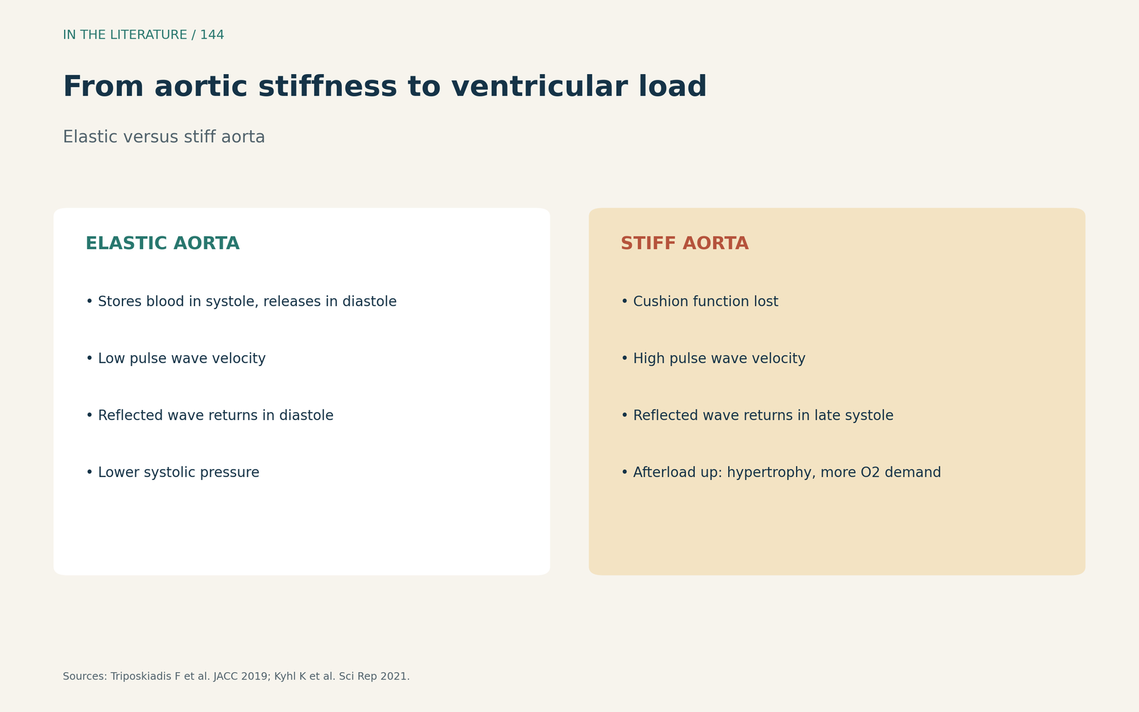
Figure 1. From aortic stiffness to ventricular load. Synthesized from Triposkiadis et al. and Kyhl et al.[1], [2] View larger.

Evidence that stiffness comes first
Kyhl and colleagues studied 52 healthy normotensive adults with magnetic resonance; lower aortic distensibility was associated with impaired left ventricular filling and atrial function before hypertension or hypertrophy, and they suggest distensibility as an early therapeutic target, although the study was cross-sectional.[2] In the TODAY study of 388 young adults with youth-onset type 2 diabetes, greater arterial stiffness predicted later higher left ventricular mass and worse diastolic indices, but stiffness was unrelated to systolic function; the authors suggest the path to heart failure may begin early in life.[3]
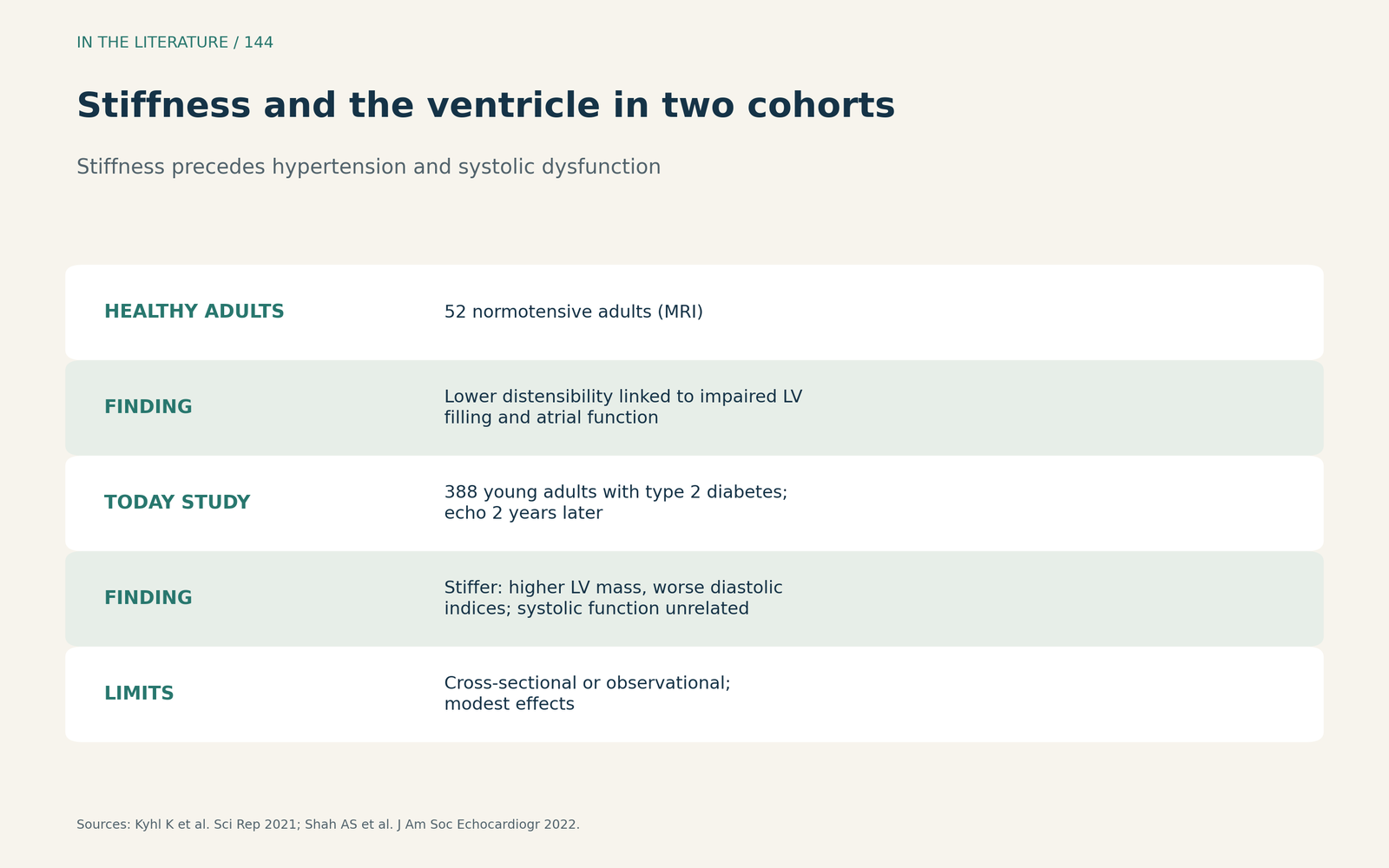
Figure 2. Stiffness and the ventricle in two cohorts. Synthesized from Kyhl et al. and the TODAY study.[2], [3] View larger.

Measuring stiffness and why terms matter
De Waal explains that carotid-femoral pulse wave velocity rises with arterial stiffness and is the method of first choice, that stiffness increases from proximal to distal arteries, and that arterial elastance, stiffness and compliance are not equivalent; pulse pressure alone does not provide a real clue about mechanical properties.[4]
What it means at induction
Alecu and colleagues found that older patients with higher arterial stiffness had a 25% more pronounced fall in systolic pressure at induction and concluded that stiffness is a useful predictor of instability, though not directly applicable to high-risk patients.[5] Ueda and colleagues studied 50 patients taking ACE inhibitors: those with vasopressor-resistant hypotension had lower carotid-femoral pulse wave velocity, and no patient with a value above the reference developed it, though generalizability is limited.[6]
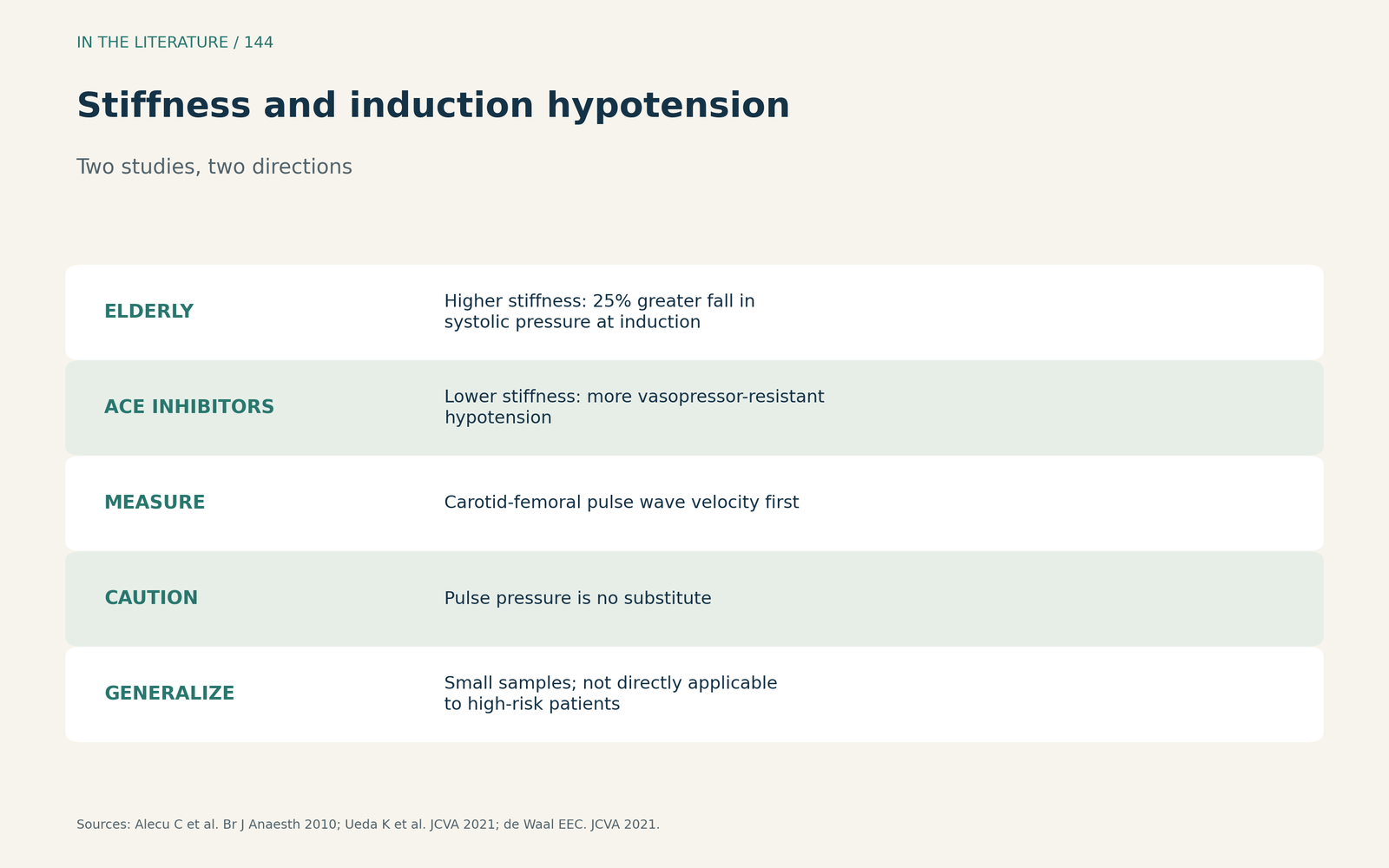
Figure 3. Stiffness and induction hypotension. Synthesized from Alecu et al., Ueda et al. and de Waal.[5], [6], [4] View larger.

Three takeaways
- A stiff aorta adds late-systolic load. Early wave reflection raises afterload and promotes hypertrophy.[1], [2]
- The ventricle feels it early. Stiffness is linked to diastolic changes before hypertension or systolic dysfunction.[2], [3]
- Expect blood pressure to behave differently at induction. Stiffness predicts larger falls in the elderly, but low stiffness was linked to vasopressor-resistant hypotension with ACE inhibitors.[5], [6]
References
- Triposkiadis F, Xanthopoulos A, Butler J. Cardiovascular aging and heart failure: JACC review topic of the week. J Am Coll Cardiol. 2019;74(6):804–813. doi:10.1016/j.jacc.2019.06.053
- Kyhl K, von Huth S, Bojer A, et al. Conductance artery stiffness impairs atrio-ventriculo-arterial coupling before manifestation of arterial hypertension or left ventricular hypertrophic remodelling. Sci Rep. 2021;11(1):14467. doi:10.1038/s41598-021-93614-w
- Shah AS, Gidding SS, El Ghormli L, et al. Relationship between arterial stiffness and subsequent cardiac structure and function in young adults with youth-onset type 2 diabetes: results from the TODAY study. J Am Soc Echocardiogr. 2022;35(6):620–628.e4. doi:10.1016/j.echo.2022.02.001
- de Waal EEC. Arterial stiffness, assessed with carotid-femoral pulse-wave velocity, and vasopressor response. J Cardiothorac Vasc Anesth. 2021;35(1):81–83. doi:10.1053/j.jvca.2020.09.124
- Alecu C, Cuignet-Royer E, Mertes PM, et al. Pre-existing arterial stiffness can predict hypotension during induction of anaesthesia in the elderly. Br J Anaesth. 2010;105(5):583–588. doi:10.1093/bja/aeq231
- Ueda K, Janiczek DM, Casey DP. Arterial stiffness predicts general anesthesia-induced vasopressor-resistant hypotension in patients taking angiotensin-converting enzyme inhibitors. J Cardiothorac Vasc Anesth. 2021;35(1):73–80. doi:10.1053/j.jvca.2020.08.040