ITL-739376ebb313
Juan Li, MD · Updated November 18, 2026
Key points
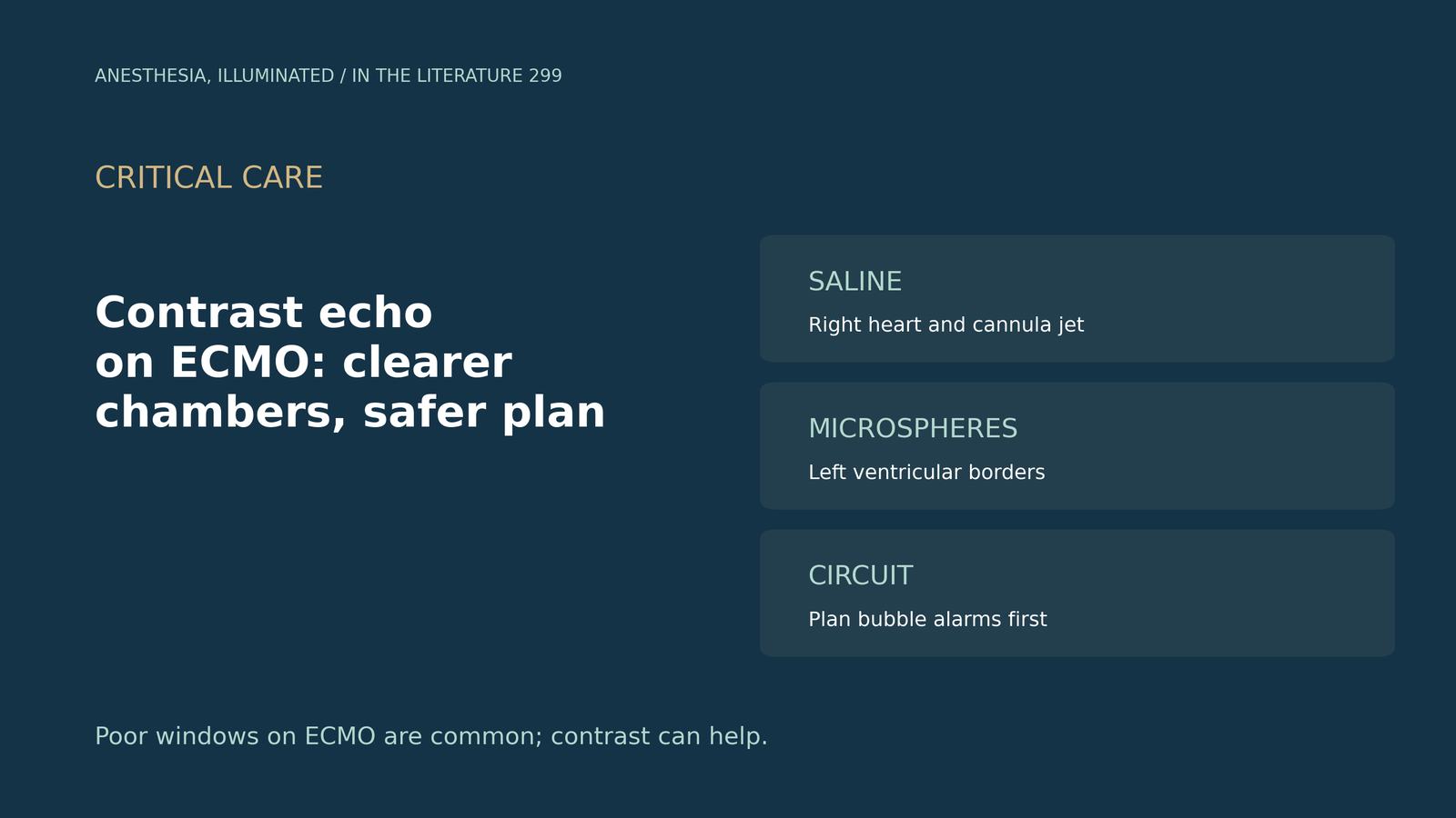
- Imaging is hard on ECMO. Poor acoustic windows are common, and suboptimal border definition is more frequent in critically ill patients.[1], [2]
- Contrast helps place cannulas. Saline contrast can show the return jet heading toward the tricuspid valve, where unenhanced imaging sometimes misses malposition.[1], [3]
- Contrast rescues chamber views. A VV-ECMO series obtained diagnostic images in every contrast study, and in COVID inpatients an agent changed the diagnosis in about a third.[4], [5]
- The circuit is the main hazard. Microspheres can trigger the bubble detector and stop flow, so the perfusionist must manage the circuit first.[1], [4]
- Serious reactions are rare. Mortality did not differ significantly with the agent, but most data come from non-ECMO patients.[2], [5]
A patient on VV-ECMO for severe ARDS has new hypotension, and the transthoracic views show almost nothing. Can an ultrasound-enhancing agent help, and is it safe with the circuit running?
Why imaging is hard, and which agent to use
Echocardiography aids ECMO implantation and monitoring, but poor acoustic windows often limit it; ventilated patients are hard to image because of positioning, oedema and hyperinflated or consolidated lungs (Lashin et al., J Cardiothorac Vasc Anesth 2022).[1] About 10% to 15% of routine echocardiograms have suboptimal border definition, rising to 25% to 30% in critically ill patients (Filippone et al., Front Cardiovasc Med 2023, a manufacturer-authored review).[2]
Agitated saline is commonly used to visualise the right heart, while microsphere agents are used for the left. Contrast imaging uses a low mechanical index of 0.4 to limit microsphere destruction, versus 1.4 for routine scans.[1]
Placing the cannula
When a dual-lumen cannula is correctly positioned, return flow should be directed toward the tricuspid valve (Mazzeffi et al., SCA consensus, J Cardiothorac Vasc Anesth 2021).[3] Unenhanced imaging can miss problems: in early reports, three of 11 patients had an inadequately positioned bicaval dual-lumen cannula despite non-enhanced echo guidance, and in a large series using unenhanced TEE to guide insertion, 13% had cannula malposition. In infants, saline-contrast TTE helped place the cannula tip 5 mm below the tricuspid valve, with better positioning and minimal recirculation. TEE carries higher risks, so contrast TTE may be used, reserving TEE for patients with poor TTE windows.[1]
Seeing the chambers
In four ECMO patients, contrast echo was diagnostic in all and influenced management in two.[1] Among 37 consecutive VV-ECMO patients, 10 (27%) needed an enhancing agent, and diagnostic images were obtained in all; boluses of 0.5 to 0.7 mL were given through the post-oxygenator limb, and all needed repeats, up to 2.5 mL in total (Bleakley et al., J Am Soc Echocardiogr 2020).[4]
Data outside ECMO point the same way. In COVID inpatients, standard TTE answered the question in 275 (75.8%), rising to 322 (88.7%) with an enhancing agent, and diagnosis changed in 22 of 61 patients (36%) and management in 13 of 61 (21.3%) (Bioh et al., Int J Cardiovasc Imaging 2024).[5]
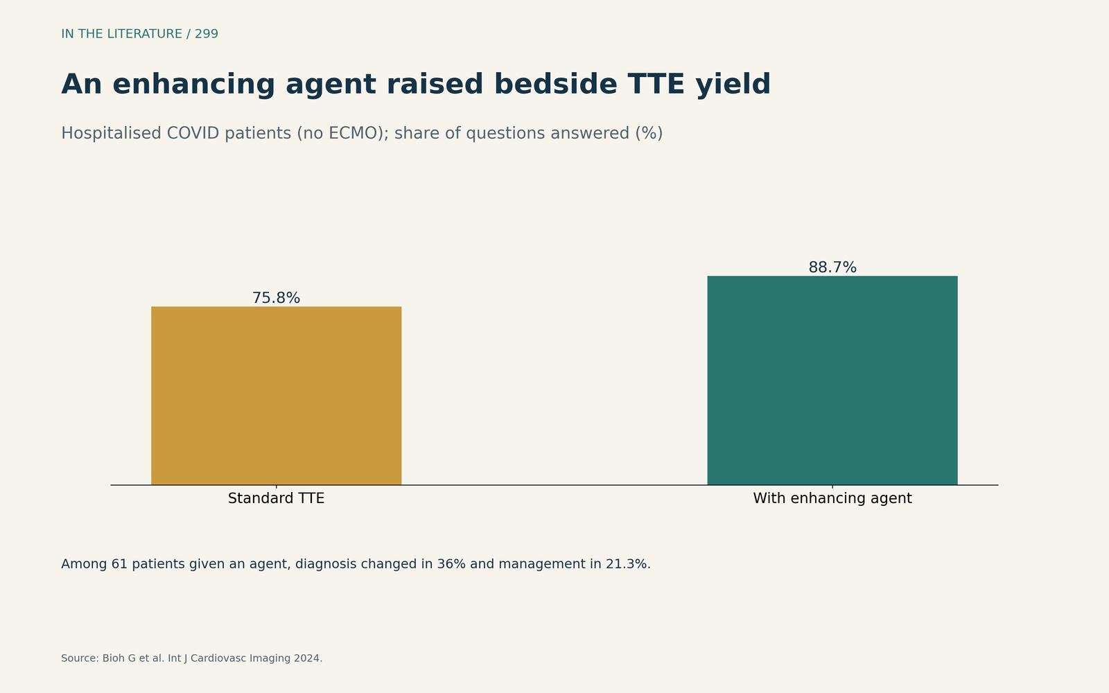
Figure 1. Adding an agent answered more clinical questions; this cohort had no ECMO patients. Data from Bioh et al.[5] View larger.

Contrast can also probe the circuit. In a two-patient pilot, raising ECMO flow above 5 L/min led to complete IVC opacification within 1,400 ms, indicating more recirculation.[1]
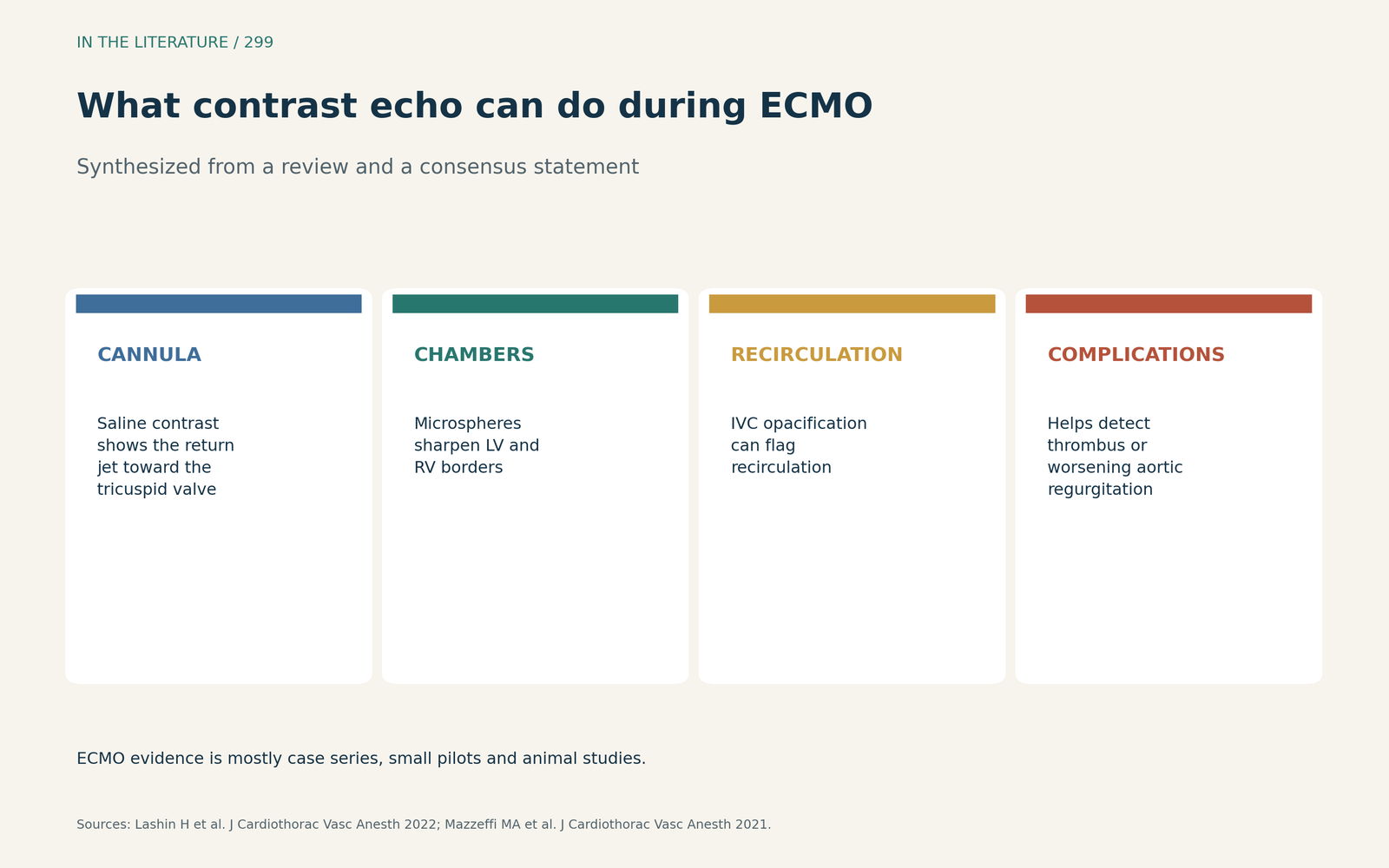
Figure 2. Cannula, chambers, recirculation and complications. Synthesized from Lashin et al. and Mazzeffi et al.[1], [3] View larger.

Safety
Cerebral ischaemic events were reported in 0.15% of agitated saline studies. In more than 5,000 patients, adverse events from microsphere agents occurred in 0.25%, and all were mild. In ten VA-ECMO patients given sulfur hexafluoride with the bubble detector disabled, pump function and flow were unaffected. The main safety concern is triggering of the circuit bubble detector, which can suspend flow.[1] In the VV-ECMO series, in every case the enhancing agent triggered the integrated air-bubble detector, so perfusionists disabled console interventions first.[4]
Serious hypersensitivity reactions were estimated at 0.007%, fewer than 1 in 10,000, and in propensity-matched critically ill patients, mortality did not differ significantly with or without the agent.[2] In the COVID cohort, mortality at 48 hours did not differ significantly, but events were few and power was limited.[5]
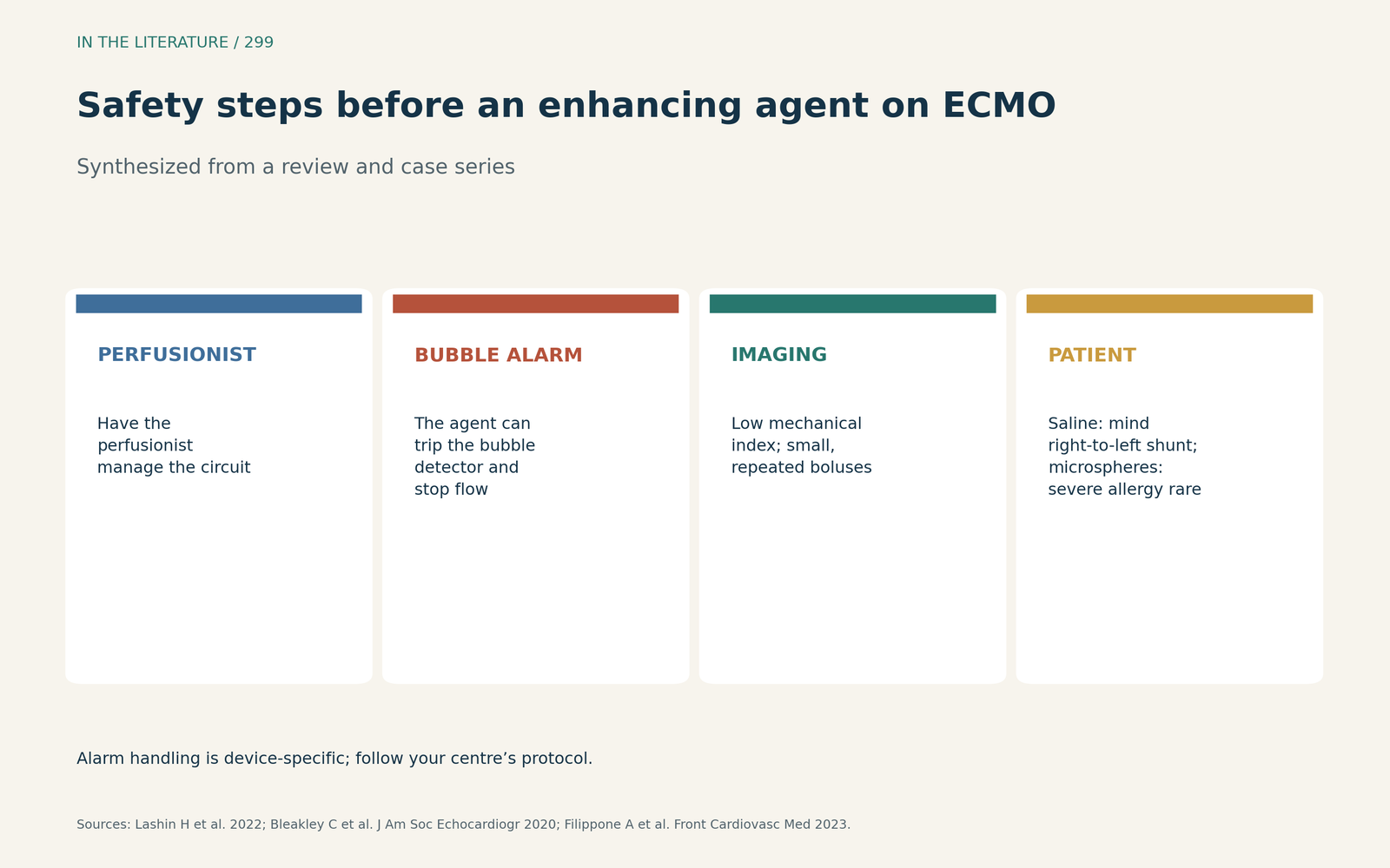
Figure 3. Plan the circuit before the injection. Synthesized from Lashin et al., Filippone et al. and Bleakley et al.[1], [2], [4] View larger.

Three takeaways
- When ECMO windows are poor, consider contrast: saline for the right heart and cannula jet, microspheres for the left ventricle.[1], [3]
- Expect better yield: one VV-ECMO series obtained diagnostic images in every contrast study, and an agent answered more clinical questions in COVID inpatients.[4], [5]
- Agree a protocol with the perfusionist for bubble-detector alarms before injecting; ECMO-specific safety data remain limited.[1], [2], [4]
References
- Lashin H, Shepherd S, Smith A. Contrast-enhanced echocardiography application in patients supported by extracorporeal membrane oxygenation (ECMO): a narrative review. J Cardiothorac Vasc Anesth. 2022;36:2080–2089. doi:10.1053/j.jvca.2021.04.031
- Filippone A, Kirchin MA, Monteith J, et al. Safety of Lumason® (SonoVue®) in special populations and critically ill patients. Front Cardiovasc Med. 2023;10:1225654. doi:10.3389/fcvm.2023.1225654
- Mazzeffi MA, Rao VK, Dodd-o J, et al. Intraoperative management of adult patients on extracorporeal membrane oxygenation: an expert consensus statement from the Society of Cardiovascular Anesthesiologists—Part I, technical aspects of extracorporeal membrane oxygenation. J Cardiothorac Vasc Anesth. 2021;35:3496–3512. doi:10.1053/j.jvca.2021.07.046
- Bleakley C, Smith R, Garfield B, et al. Contrast echocardiography in VV-ECMO-dependent patients with COVID-19. J Am Soc Echocardiogr. 2020;33:1419–1420. doi:10.1016/j.echo.2020.07.012
- Bioh G, Botrous C, Senior R. Efficacy and safety of use of ultrasound enhancing agent in patients hospitalized with COVID-19. Int J Cardiovasc Imaging. 2024;40(3):625–632. doi:10.1007/s10554-023-03032-4