
ITL-823f99bff66f
Juan Li, MD · Updated November 18, 2026
Key points
- A force imbalance. Atelectasis forms when pleural pressure and surface tension overcome the forces holding alveoli open.[1]
- Almost universal. Dependent, caudal atelectasis is found in 90% of patients under general anaesthesia.[1]
- Oxygen speeds it up. After preoxygenation with FiO2 1.0, atelectasis area was 20 times higher than with 0.3.[2]
- Collapse is not inert. Atelectatic sheep lung showed 2,363 differentially expressed genes.[3]
- PEEP can prevent it. At induction, atelectasis occurred with PEEP 0 and 5 but not 10 cmH2O.[4]
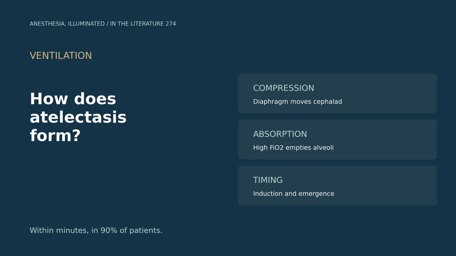
A healthy patient is preoxygenated, induced and ventilated for a laparoscopic operation. Where, how fast and why does the lung collapse?
The mechanisms
Atelectasis forms when collapsing forces from positive pleural pressure and surface tension overcome expanding forces; three mechanisms contribute: increased pleural pressure, low alveolar pressure and surfactant impairment. Dependent, caudal atelectasis is detected in 90% of patients under general anaesthesia; lungs ventilated without PEEP for hours progressively lose aeration in dorsal regions, lung units with greater gas outflow than inflow ultimately collapse, a high inspired oxygen fraction facilitates that outflow through gas absorption, and lung collapse causes local hypoxia, a potent inducer of inflammation (Zeng et al., Anesthesiology 2022).[1] General anaesthesia displaces the diaphragm cephalad almost instantly, atelectasis appears in the early minutes after loss of consciousness, and obese patients are at greater risk (Lagier et al., Anesthesiology 2022).[2] In sheep, atelectatic regions showed 2,363 differentially expressed genes, with impaired immune and barrier function (Zeng et al., Anesthesiology 2020).[3]
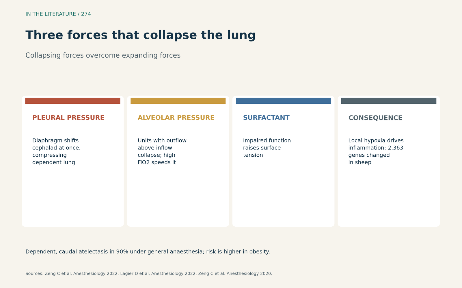
Figure 1. Compression, absorption and surfactant impairment combine in dependent lung. Synthesized from Zeng et al. and Lagier et al.[1], [2] View larger.

Induction and oxygen
Atelectasis area was 20 times higher after preoxygenation with FiO2 1.0 than with 0.3, and current recommendations limit recruitment manoeuvres to patients with respiratory dysfunction.[2] Induction causes atelectasis in 85–90% of patients within minutes; in a three-arm trial (Yang et al., BMC Anesthesiol 2025), facemask ventilation produced atelectasis with PEEP 0 and 5 but not with PEEP 10 cmH2O, and the mean closing pressure is estimated at 6 cmH2O.[4] A meta-analysis of 10 RCTs with 3471 participants (Wang et al., J Int Med Res 2025) found that high FiO2 increased postoperative atelectasis after abdominal surgery (RR 1.43, 95% CI 1.14–1.80), although definitions of atelectasis varied.[5]
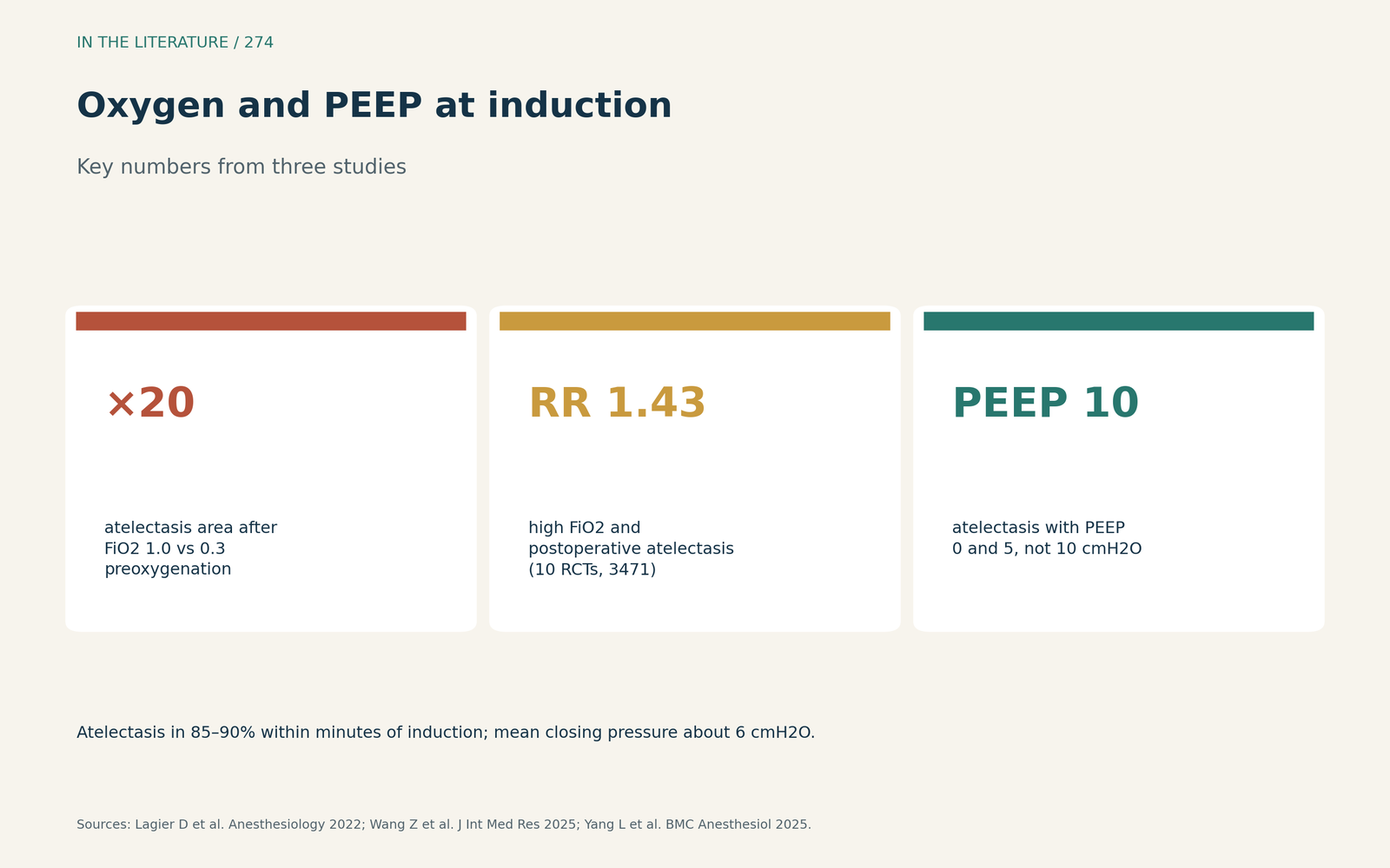
Figure 2. High FiO2 increases atelectasis; enough PEEP at induction prevents it. Data from Lagier et al., Yang et al. and Wang et al.[2], [4], [5] View larger.

Emergence
Emergence and extubation may hasten alveolar derecruitment.[2] The emergence period contributes about 39% of total postoperative atelectasis, and in a randomized trial (Jeong et al., Anesthesiology 2021), pressure support at emergence reduced atelectasis in the recovery room (33% vs 57%; RR 0.58).[6]
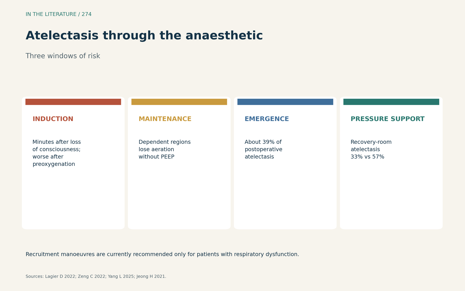
Figure 3. Where atelectasis arises during an anaesthetic. Synthesized from Lagier et al., Yang et al. and Jeong et al.[2], [4], [6] View larger.

Three takeaways
- Atelectasis is a predictable result of compression, gas absorption and surfactant changes, and it begins within minutes of induction.[1], [2]
- Limit FiO2 where safe and use adequate PEEP at induction.[4], [5]
- Treat emergence as a second risk window: pressure support reduced atelectasis compared with unsupported breathing.[6]
References
- Zeng C, Lagier D, Lee JW, Vidal Melo MF. Perioperative pulmonary atelectasis: part I. Biology and mechanisms. Anesthesiology. 2022;136(1):181–205. doi:10.1097/ALN.0000000000003943
- Lagier D, Zeng C, Fernandez-Bustamante A, Vidal Melo MF. Perioperative pulmonary atelectasis: part II. Clinical implications. Anesthesiology. 2022;136(1):206–236. doi:10.1097/ALN.0000000000004009
- Zeng C, Motta-Ribeiro GC, Hinoshita T, et al. Lung atelectasis promotes immune and barrier dysfunction as revealed by transcriptome sequencing in female sheep. Anesthesiology. 2020;133(5):1060–1076. doi:10.1097/ALN.0000000000003491
- Yang L, Wu L, Ye J, Zhao Z, Zhang J. Applying positive end-expiratory pressure to facemask ventilation improves post-induction lung atelectasis: a three-arm randomized controlled trial. BMC Anesthesiol. 2025;25:509. doi:10.1186/s12871-025-03352-z
- Wang Z, Jiang M, Huang C, et al. A low perioperative fraction of inspired oxygen reduces the incidence of postoperative atelectasis in patients undergoing abdominal surgery under general anesthesia: a systematic review and meta-analysis. J Int Med Res. 2025;53(11):3000605251392431. doi:10.1177/03000605251392431
- Jeong H, Tanatporn P, Ahn HJ, et al. Pressure support versus spontaneous ventilation during anesthetic emergence—effect on postoperative atelectasis: a randomized controlled trial. Anesthesiology. 2021;135(6):1004–1014. doi:10.1097/ALN.0000000000003997