
ITL-62ee30fd3c1a
Juan Li, MD · Updated November 18, 2026
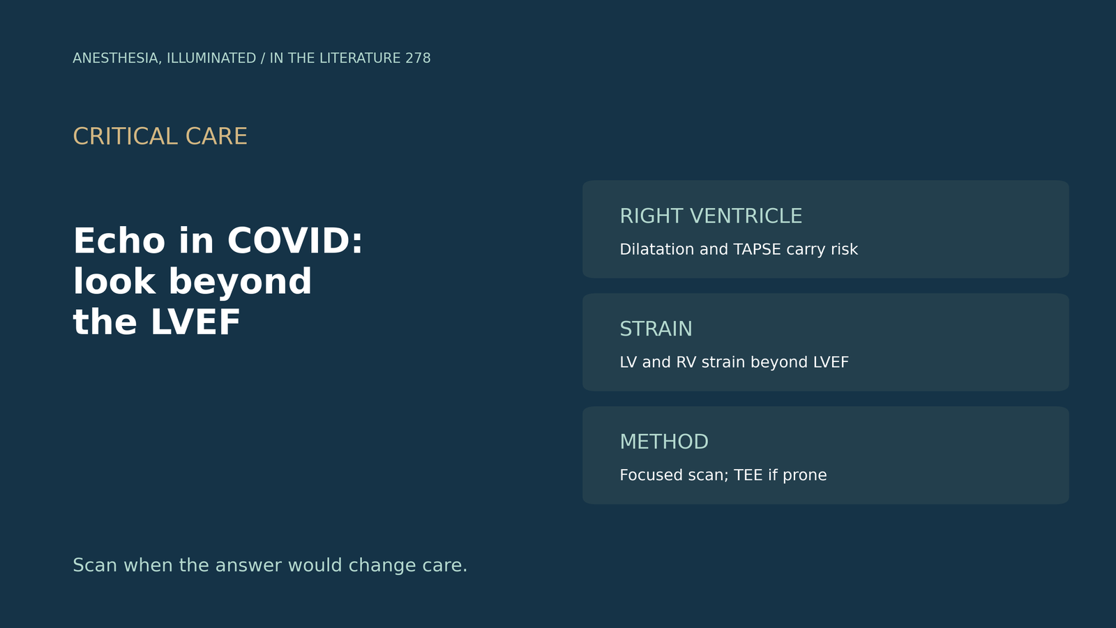
Key points
- Both ventricles are involved. In WASE-COVID, about 20% had LV and about 30% had RV dysfunction.[1]
- Troponin rise has several echo faces. With significant myocardial injury, 46% had regional wall motion abnormalities and only 8% diffuse hypokinesis.[2]
- RV and flow measures carry prognosis. Stroke volume index and TAPSE predicted outcome;[3] RV dilatation predicted death in the ICU[4] and, unlike in bacterial pneumonia, in COVID (adjusted relative risk 3.02).[5]
- Strain sees what LVEF misses. Abnormal LV strain was found in 67.9% and, unlike troponin, was linked to death.[6]
- Choose the scan for the setting. Post-COVID echo changes are small;[7] focused scans limit exposure,[8] and TEE helps when prone patients have poor windows.[9]
A ventilated patient with COVID pneumonia becomes hypotensive and the troponin is rising. What should the echo look for, and which numbers matter?
What echo finds in acute COVID
WASE-COVID (Karagodin et al., J Am Soc Echocardiogr 2021) studied 870 patients with acute COVID from 13 medical centres. LV dysfunction was seen in about 20% and RV dysfunction in about 30%; among patients with both strains measured, 19.7% had biventricular dysfunction and 51.1% had normal function of both ventricles. In-hospital mortality was 21.6%. LV longitudinal strain and RV free-wall strain were independently associated with in-hospital death, but LVEF was not.[1]
When troponin is high, the pattern matters. Among 24 patients with peak troponin I above 1 ng/mL (Sud et al., J Am Soc Echocardiogr 2020), 46% had regional wall motion abnormalities, only 8% had diffuse LV hypokinesis, 17% had isolated RV dysfunction, and 33% had more than trivial pericardial effusion.[2] The authors suggest that ischaemia or a prothrombotic state may be a common mechanism.
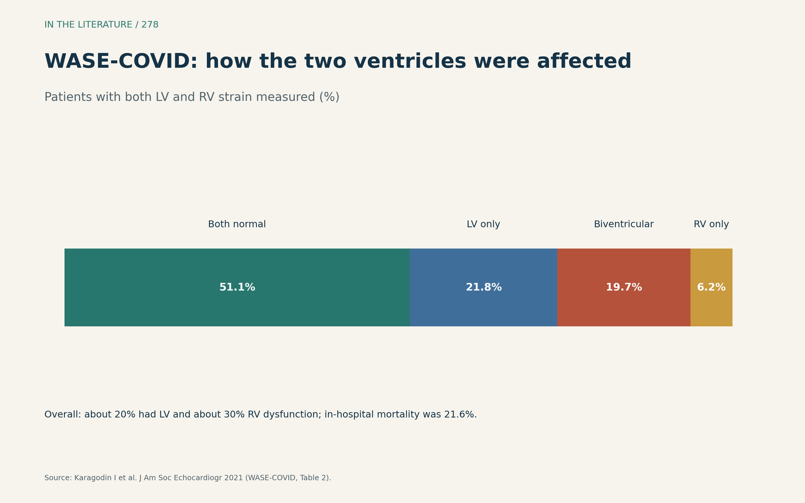
Figure 1. About half had normal function of both ventricles; the rest had LV, RV or biventricular dysfunction. Data from WASE-COVID.[1] View larger.

Which findings carry prognosis
In a prospective cohort scanned regardless of indication (Szekely et al., J Am Soc Echocardiogr 2021), stroke volume index, TAPSE and pulmonary acceleration time were the only independent echo predictors. Abnormal TAPSE, LVEF and SVI were found in 18%, 14% and 28%, and 63% had none of these. LVEF and TAPSE from the four-chamber view alone carried prognostic information, though a Doppler-based model performed better.[3]
In 153 ICU patients with COVID ARDS who had echo (Sprockel Diaz et al., Heart Lung 2022), 59.5% died within 60 days. RV dilatation (HR 3.33) and lower LVEF (HR 0.94 per point) were independently associated with death; the authors note selection bias, because echo was done in patients with an unfavourable course.[4]
RV dilation was equally common in COVID and bacterial pneumonia (22.2% vs 22.8%) in a San Francisco cohort (Sun et al., J Am Soc Echocardiogr 2023). Compared with bacterial pneumonia and a normal RV, COVID with RV dilation had an adjusted relative risk of death of 3.02, whereas bacterial pneumonia with RV dilation carried no clear excess risk (1.09).[5]
Strain adds information that LVEF misses. In 131 COVID inpatients (Hegde et al., Open Heart 2024), 67.9% had abnormal LV global longitudinal strain, and LV strain was associated with in-hospital death whereas troponin was not; the authors state that their small sample limits the strength of the conclusions.[6]
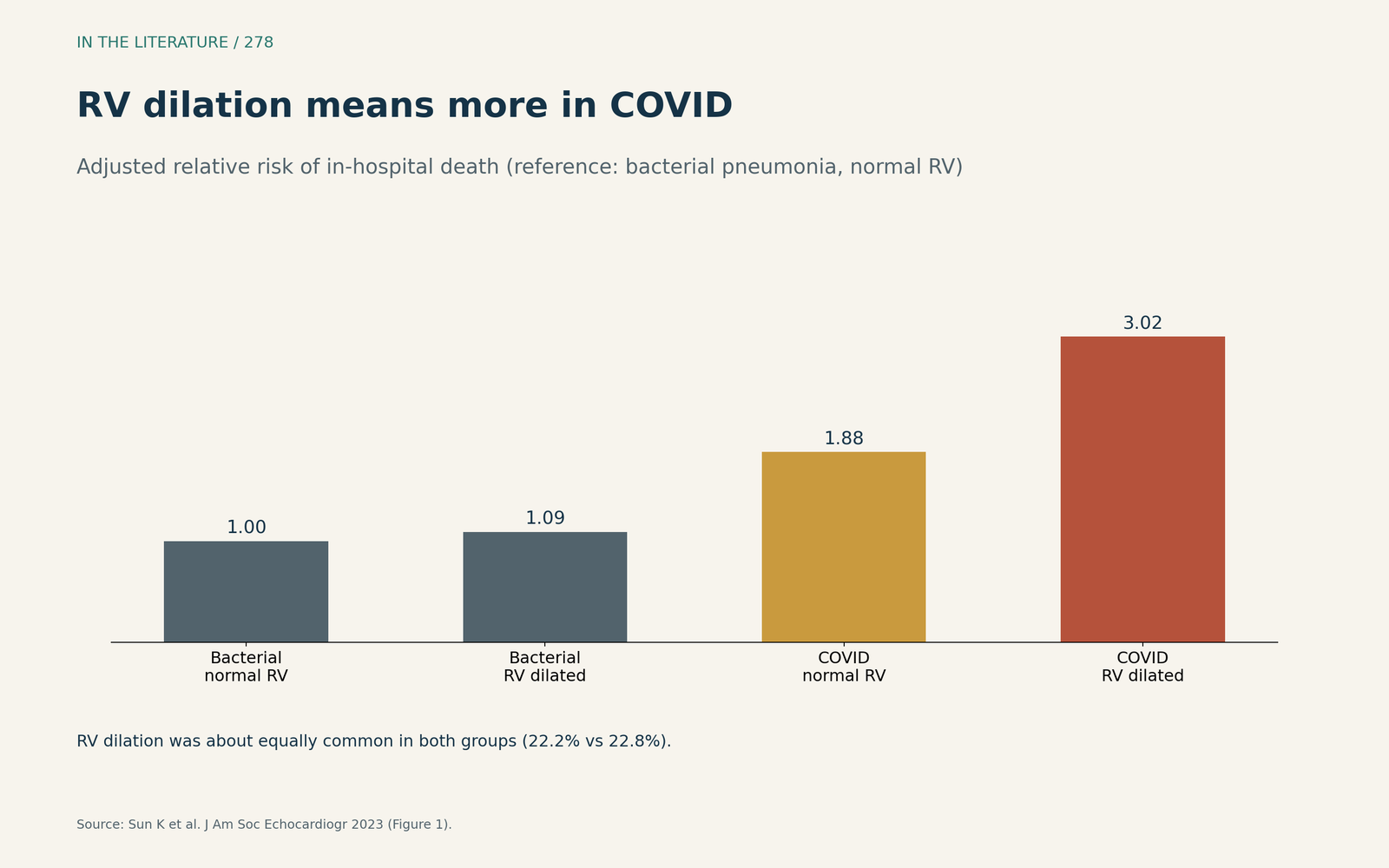
Figure 2. RV dilation carried excess risk in COVID but not in bacterial pneumonia. Data from Sun et al.[5] View larger.

After the acute illness
A meta-analysis of 16 studies compared echo in people with and without post-COVID condition (Khawandi et al., J Clin Med 2026). Across 11 studies (1764 patients), LVEF was about 1 point lower in post-COVID condition, a statistically lower but clinically trivial difference, and no study evaluated the diagnostic accuracy of echo; certainty was very low.[7]
How to scan
A Wuhan report (Zhang et al., J Am Soc Echocardiogr 2020) used binary point-of-care ultrasound for LV function, which can be done with limited training, and kept fuller studies for questions that needed them.[8] In prone ventilated COVID ARDS, TTE image quality was suboptimal or poor in 60% of a small series, and TEE kept the short-axis view needed to diagnose acute cor pulmonale (Evrard et al., J Am Soc Echocardiogr 2020).[9]
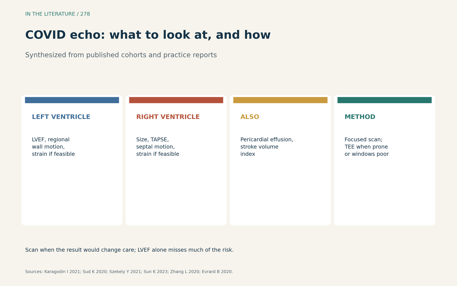
Figure 3. Look at both ventricles, add strain or TAPSE where feasible, and pick the scan for the setting. Synthesized from WASE-COVID, Sud et al., Szekely et al., Sun et al., Zhang et al. and Evrard et al.[1], [2], [3], [5], [8], [9] View larger.

Three takeaways
- Look at the RV as closely as the LV: RV dysfunction was common, and RV dilatation was linked to death in COVID.[1], [4], [5]
- LVEF alone is not enough; strain, TAPSE and stroke volume index carried prognosis more consistently.[1], [3], [6]
- Scan when the result would change care; use focused studies, and TEE when windows are poor in prone patients.[8], [9]
References
- Karagodin I, Carvalho Singulane C, Woodward GM, et al. Echocardiographic correlates of in-hospital death in patients with acute COVID-19 infection: the World Alliance Societies of Echocardiography (WASE-COVID) study. J Am Soc Echocardiogr. 2021;34:819–830. doi:10.1016/j.echo.2021.05.010
- Sud K, Vogel B, Bohra C, et al. Echocardiographic findings in patients with COVID-19 with significant myocardial injury. J Am Soc Echocardiogr. 2020;33:1054–1055. doi:10.1016/j.echo.2020.05.030
- Szekely Y, Lichter Y, Hochstadt A, et al. The predictive role of combined cardiac and lung ultrasound in coronavirus disease 2019. J Am Soc Echocardiogr. 2021;34:642–652. doi:10.1016/j.echo.2021.02.003
- Sprockel Diaz JJ, Rincon JM, Rondon Lopez MA, et al. Echocardiographic 60-day mortality markers in patients hospitalized in intensive care for COVID-19. Heart Lung. 2022;52:123–129. doi:10.1016/j.hrtlng.2021.12.007
- Sun K, Cedarbaum E, Hill CA, et al. Association of right ventricular dilation on echocardiogram with in-hospital mortality among patients hospitalized with COVID-19 compared with bacterial pneumonia. J Am Soc Echocardiogr. 2023;36:558–562. doi:10.1016/j.echo.2022.12.019
- Hegde S, Shnoda M, Alkhadra Y, et al. Prevalence of abnormal left ventricular global longitudinal strain by speckle tracking echocardiography and its prognostic value in patients with COVID-19. Open Heart. 2024;11:e002397. doi:10.1136/openhrt-2023-002397
- Khawandi J, Choaib A, Azzam M, et al. Echocardiogram testing in patients with post-COVID-19 condition: a systematic review and meta-analysis. J Clin Med. 2026;15:4643. doi:10.3390/jcm15124643
- Zhang L, Wang B, Zhou J, et al. Bedside focused cardiac ultrasound in COVID-19 from the Wuhan epicenter: the role of cardiac point-of-care ultrasound, limited transthoracic echocardiography, and critical care echocardiography. J Am Soc Echocardiogr. 2020;33:676–682. doi:10.1016/j.echo.2020.04.004
- Evrard B, Goudelin M, Vignon P. Transesophageal echocardiography remains essential and safe during prone ventilation for hemodynamic monitoring of patients with COVID-19. J Am Soc Echocardiogr. 2020;33:1057–1059. doi:10.1016/j.echo.2020.05.018