ITL-4f1ab6e2f253
Juan Li, MD · Updated November 18, 2026
Key points
- Start from standard cut-offs, used with care. FAC below 35%, TAPSE below 17 mm or S' below 9.5 cm/s define RV dysfunction, but these are not validated in COVID.[1], [2]
- RV findings carry prognosis. RV dysfunction, dilatation and pulmonary hypertension were each linked to higher mortality.[3]
- TAPSE alone misses RV injury. In serial ICU echo, TAPSE-only assessment would have missed many patients.[4]
- Other measures caught what TAPSE missed. FAC, shortening fraction and RV strain separated patients when TAPSE did not.[5], [6]
- Look again, and look for dilation. RV injury appeared within the first two weeks, and dysfunction with dilation carried the risk.[4], [7]
A ventilated patient with COVID ARDS has a TAPSE of 20 mm, but the RV looks large and the septum is flat. Is the RV normal?
Definitions and the bedside approach
RV dysfunction is commonly defined as FAC below 35%, RV ejection fraction below 45%, TAPSE below 17 mm or S' below 9.5 cm/s, and FAC grades RV dysfunction as mild (25%–35%), moderate (18%–25%) or severe (below 18%). RV dysfunction appears particularly common in COVID (20%–39%) (Isgro et al., J Cardiothorac Vasc Anesth 2021).[1]
These quantitative cut-offs have not been validated in COVID, so a pragmatic mix of qualitative and quantitative signs has been proposed; focused intensive care echo often answers qualitative questions, such as whether the RV is dilated (McCall et al., J Cardiothorac Vasc Anesth 2021).[2]
What RV findings mean for prognosis
A meta-analysis of 9 studies (1,450 patients) linked RV dysfunction with higher mortality: 48.5% vs 24.7% (Paternoster et al., J Cardiothorac Vasc Anesth 2021). RV dilatation was linked to mortality of 56.3% vs 30.6% (OR 2.43), and pulmonary hypertension to 52.9% vs 14.8%. The authors note that echo RV assessment may not have been uniform across studies.[3]
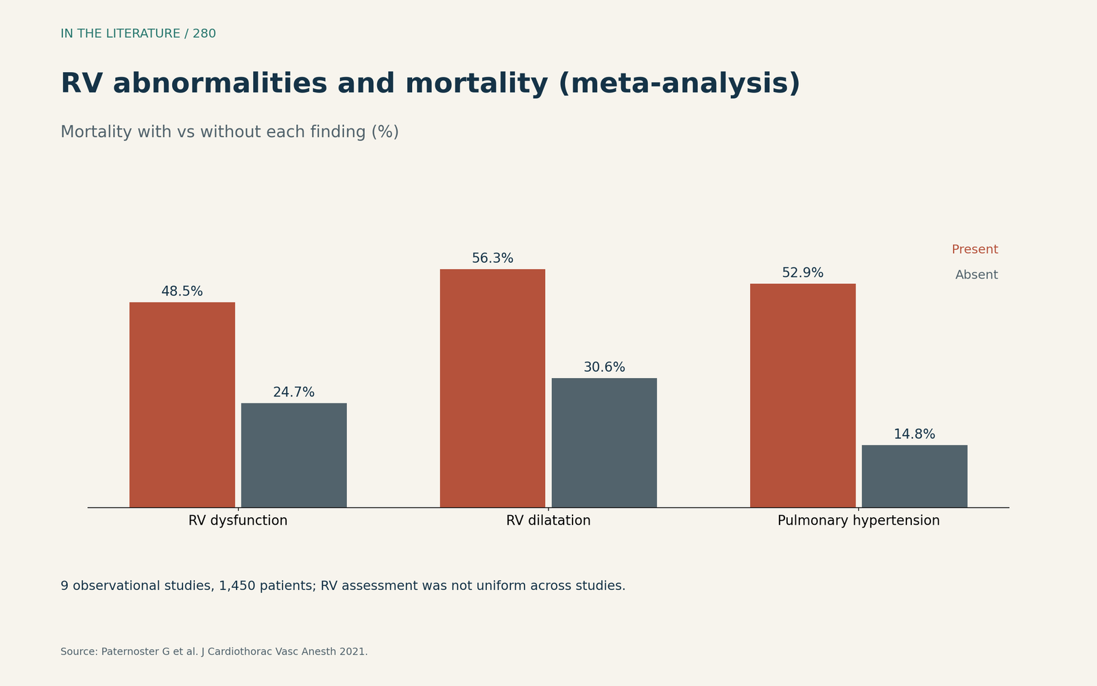
Figure 1. Each RV abnormality was associated with higher mortality in observational studies. Data from Paternoster et al.[3] View larger.

With standardized serial echo in 118 ICU patients (Jozwiak et al., Ann Intensive Care 2024), 62% had at least one RV injury pattern, and Day-28 mortality was 27 vs 7% with vs without RV injury. RV systolic dysfunction alone was probably not associated with mortality; dysfunction with dilation or acute cor pulmonale was.[4]
Why TAPSE alone is not enough
In the same cohort, using TAPSE alone, 63% would have been classed as having no RV injury.[4] In ventilated COVID ARDS patients (Beyls et al., J Cardiothorac Vasc Anesth 2021), TAPSE, S' and FAC did not differ with acute cor pulmonale; RV longitudinal shortening fraction below 20% detected RV dysfunction with 84% sensitivity and 90% specificity, and TAPSE overlapped widely between patients with and without RV dysfunction.[5]
Compared with pre-infection echo, RV dysfunction rose from 12% to 31% during COVID, and abnormal RV strain after COVID carried a hazard ratio of 4.59 for death (Nambiar et al., J Am Soc Echocardiogr 2022).[6]
The course in the ICU
No new RV injury appeared after day 14, but 31% without injury at admission developed it.[4] In 176 ventilated COVID ARDS patients (Tsolaki et al., Ann Intensive Care 2024), RV dysfunction defined by two or more indices was common (69%); RV dilation was present in 86% and acute cor pulmonale in 20%, and right ventriculo-arterial coupling (TAPSE/PASP) was impaired, at 0.8 mm/mmHg. Survival in this intubated cohort was 34.8%.[7]
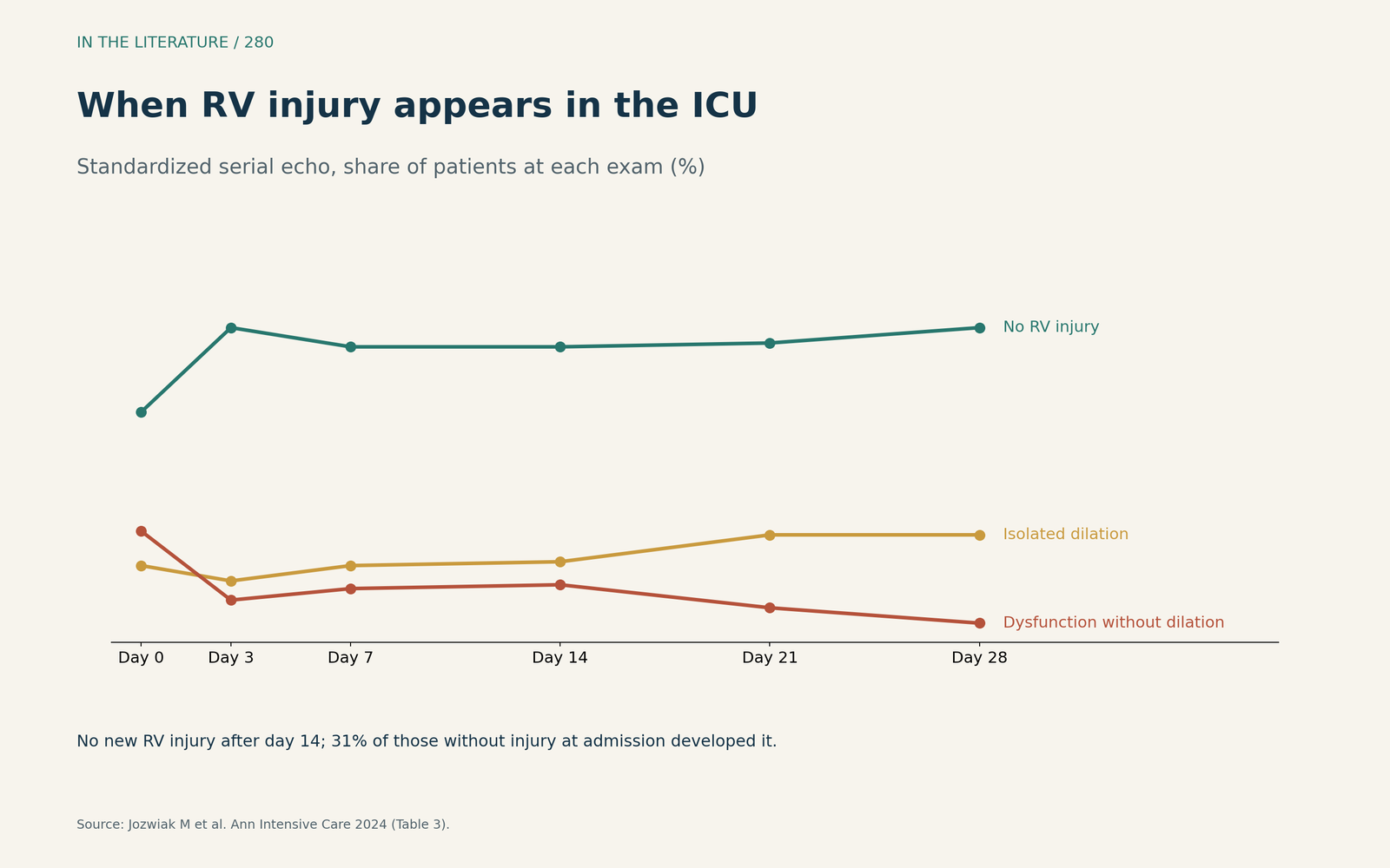
Figure 2. RV injury was most dynamic early; isolated dilation persisted while dysfunction without dilation receded. Data from Jozwiak et al.[4] View larger.

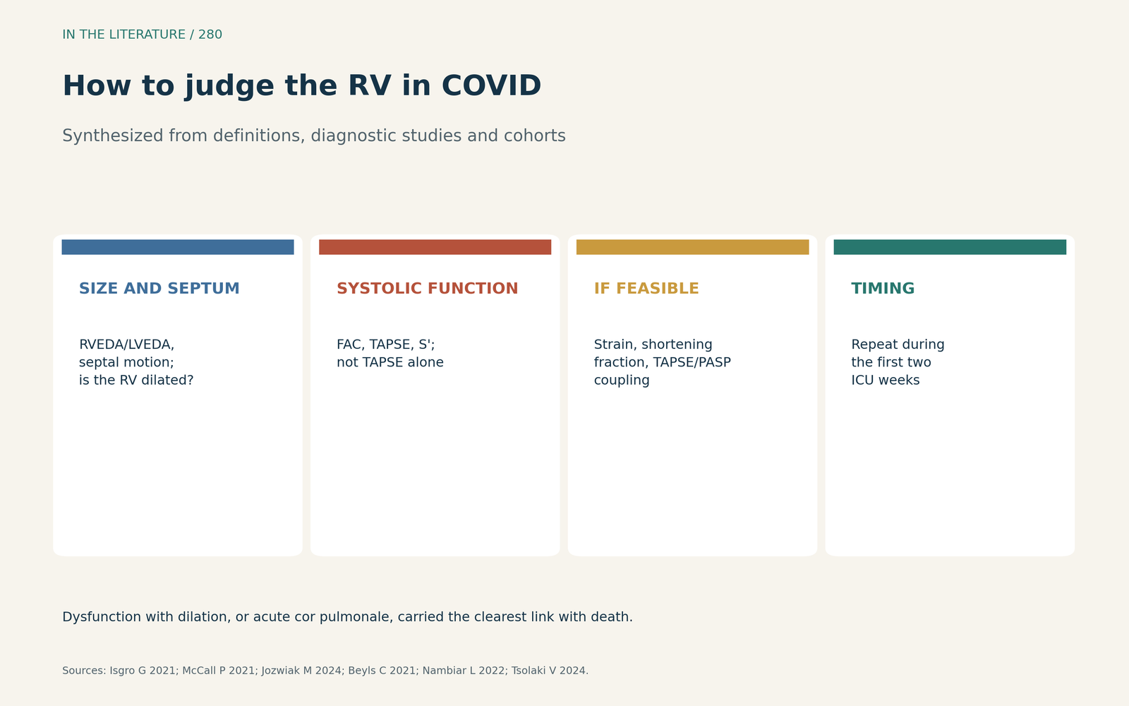
Figure 3. Combine size, septal motion and several function measures, and repeat the study early in the ICU course. Synthesized from Isgro et al., McCall et al., Jozwiak et al., Beyls et al., Nambiar et al. and Tsolaki et al.[1], [2], [4], [5], [6], [7] View larger.

Three takeaways
- Use several measures together (size, septal motion, FAC, TAPSE, S' and strain where feasible); TAPSE alone misses RV injury.[1], [4], [5]
- Dysfunction with dilation, or acute cor pulmonale, carried the clearest link with death.[3], [4]
- Repeat the echo during the first two ICU weeks, when RV injury tends to appear.[4], [7]
References
- Isgro G, Yusuff HO, Zochios V; Protecting the Right Ventricle Network. The right ventricle in COVID-19 lung injury: proposed mechanisms, management, and research gaps. J Cardiothorac Vasc Anesth. 2021;35:1568–1572. doi:10.1053/j.jvca.2021.01.014
- McCall P, Willder JM, McErlane J, Shelley B. A call for pragmatic bedside assessment of right ventricular (RV) function in coronavirus disease 2019 (COVID-19). J Cardiothorac Vasc Anesth. 2021;35:3455–3456. doi:10.1053/j.jvca.2021.03.017
- Paternoster G, Bertini P, Innelli P, et al. Right ventricular dysfunction in patients with COVID-19: a systematic review and meta-analysis. J Cardiothorac Vasc Anesth. 2021;35:3319–3324. doi:10.1053/j.jvca.2021.04.008
- Jozwiak M, Dupuis C, Denormandie P, et al. Right ventricular injury in critically ill patients with COVID-19: a descriptive study with standardized echocardiographic follow-up. Ann Intensive Care. 2024;14:14. doi:10.1186/s13613-024-01248-8
- Beyls C, Bohbot Y, Huette P, et al. Usefulness of right ventricular longitudinal shortening fraction to detect right ventricular dysfunction in acute cor pulmonale related to COVID-19. J Cardiothorac Vasc Anesth. 2021;35:3594–3603. doi:10.1053/j.jvca.2021.01.025
- Nambiar L, Volodarskiy A, Tak KA, et al. Acute COVID-19-associated decrements in left and right ventricular function predict all-cause mortality. J Am Soc Echocardiogr. 2022;35:229–234. doi:10.1016/j.echo.2021.10.002
- Tsolaki V, Zakynthinos GE, Karavidas N, et al. Comprehensive temporal analysis of right ventricular function and pulmonary haemodynamics in mechanically ventilated COVID-19 ARDS patients. Ann Intensive Care. 2024;14:25. doi:10.1186/s13613-024-01241-1