ITL-0f4c4c15ca75
Juan Li, MD · Updated November 17, 2026
Watch the video
Key points
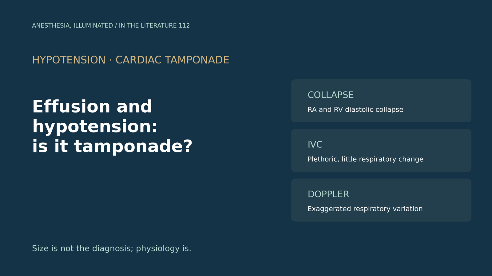
- Tamponade is defined by physiology, not by the size of the effusion. Effusion size correlates poorly with hemodynamics, a rapidly accumulating effusion as small as 250 mL can cause tamponade, and tolerance depends on how fast fluid accumulates.[1], [2]
- The core signs are right atrial collapse lasting more than one third of the cardiac cycle, right ventricular diastolic collapse, which appears when cardiac output has fallen about 20% before blood pressure drops, and a plethoric inferior vena cava. Absence of any chamber collapse has a negative predictive value above 90%.[1]
- In spontaneously breathing patients, an inspiratory fall in mitral E velocity of about 30% supports the diagnosis, but respiratory Doppler should not be used alone and behaves differently during positive-pressure ventilation.[1], [3]
- Atypical tamponade is common in the settings anesthesiologists see: clot after cardiac surgery compressing a single chamber, right ventricular hypertrophy or pulmonary hypertension preventing right-sided collapse, and tamponade during ECMO.[1], [4], [5]
- Physiology can collapse after induction of anesthesia. Maintain preload and vascular resistance, avoid high PEEP and large tidal volumes, induce with the surgeon ready, and avoid vasodilators and diuretics.[3], [2]
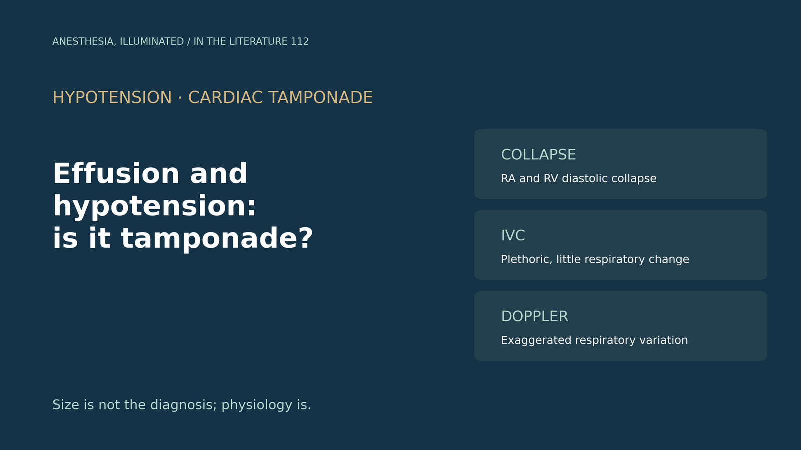
A patient becomes hypotensive and tachycardic in the intensive care unit a few hours after cardiac surgery, and transthoracic images show a moderate effusion. Is this tamponade, which echocardiographic signs matter, and why might the classic signs be missing?[1], [3]
Size is not the diagnosis
The 2013 American Society of Echocardiography (ASE) recommendations grade effusions at end-diastole as small (<10 mm), moderate (10–20 mm) or large (>20 mm), but note that size correlates poorly with hemodynamic impact. Tamponade can occur with volumes as small as 250 mL when fluid accumulates rapidly, and regardless of size it is potentially lethal.[1] The 2015 European Society of Cardiology (ESC) guideline describes tamponade as a "last-drop" phenomenon and notes that tolerance is related more to the rapidity of accumulation than to volume.[2] Hypotension is a late sign, because sympathetic tone maintains blood pressure at first.[1]
The echocardiographic signs
Chamber collapse is the key finding. Right atrial collapse that lasts more than one third of the cardiac cycle is highly suggestive of tamponade. Right ventricular diastolic collapse is seen first during inspiration and appears when cardiac output has fallen by about 20% while systemic pressure is maintained. A dilated inferior vena cava (>2.1 cm) with less than 50% inspiratory collapse was present in 92% of effusions in one series, and the absence of any chamber collapse has a negative predictive value above 90%.[1]
Respiratory Doppler adds support. An inspiratory reduction of about 30% in mitral E velocity is generally considered diagnostic during spontaneous breathing, but these patterns differ under positive-pressure ventilation and respiratory variation should not be used as a stand-alone criterion without chamber collapse.[1] The BJA Education review by Madhivathanan and colleagues adds that Doppler velocities may be inaccurate in mechanically ventilated patients.[3]
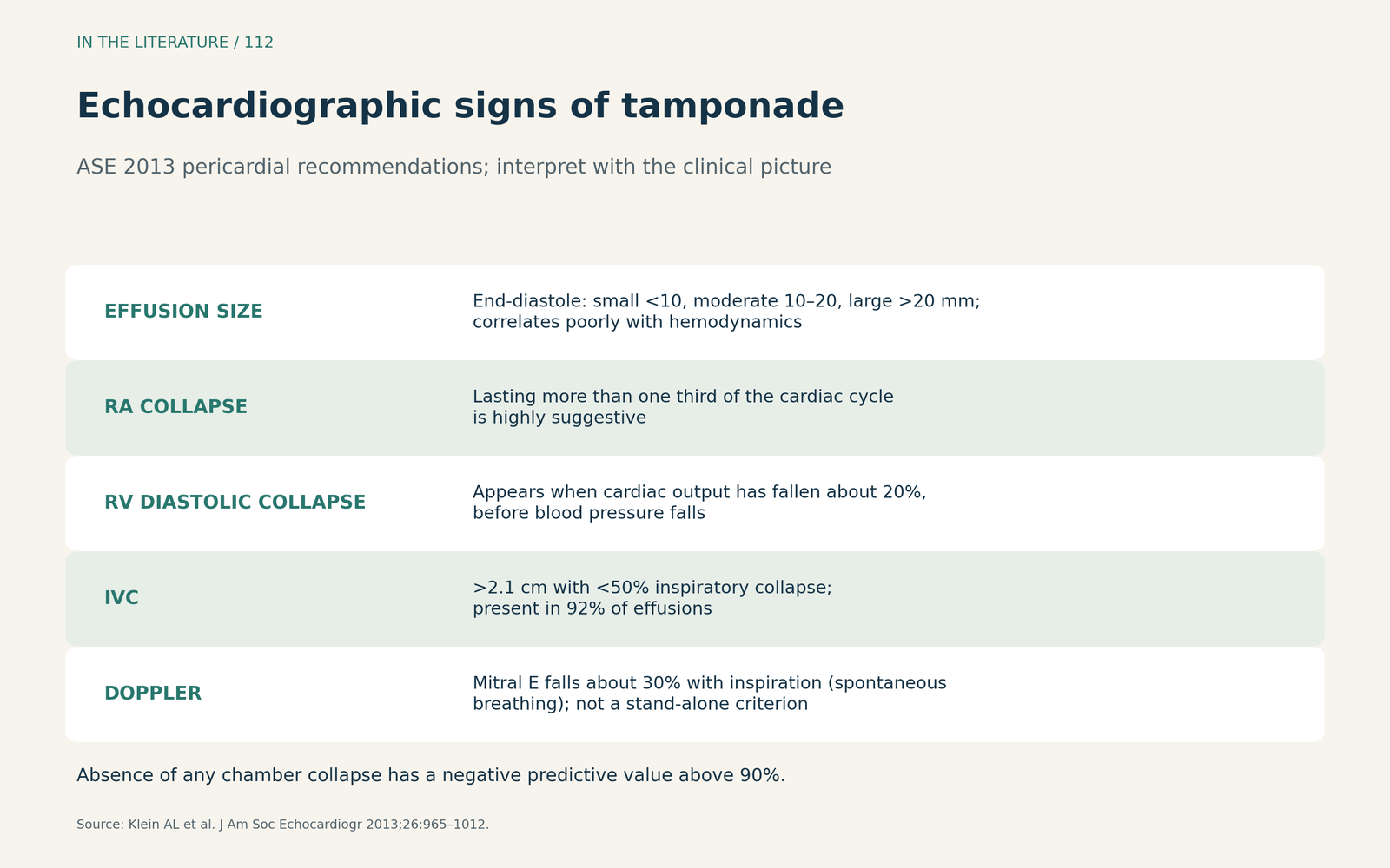
Figure 1. Echocardiographic signs of tamponade. Summarized from the 2013 ASE pericardial recommendations; signs must be interpreted with the clinical picture.[1] View larger.

When the classic signs are missing
After cardiac surgery, clot may form and produce focal compression of individual chambers, and regional tamponade often affects the left-sided chambers; bedside transesophageal echocardiography is often helpful.[3], [1] Right-sided collapse may be absent when the right ventricle is hypertrophied or pulmonary pressure is severely raised.[1] Poorsattar and Maus described a patient with severe pulmonary hypertension and a circumferential effusion in whom there was no right atrial or right ventricular collapse, only left ventricular early diastolic buckling.[4]
ECMO adds another setting. In the Extracorporeal Life Support Organization registry, Basílio and colleagues found tamponade in 2,176 adults (2.6%), with lower hospital survival than in all adults on ECMO (35% vs 49%).[5]
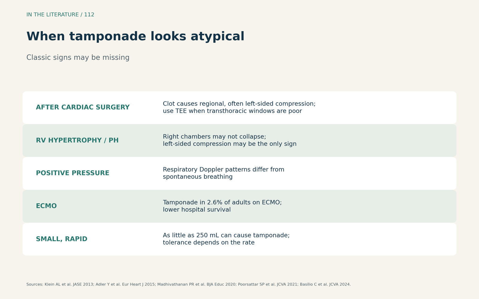
Figure 2. When tamponade looks atypical. Synthesized from the ASE and ESC documents, Madhivathanan et al., Poorsattar and Maus, and Basílio et al.[1], [2], [3], [4], [5] View larger.

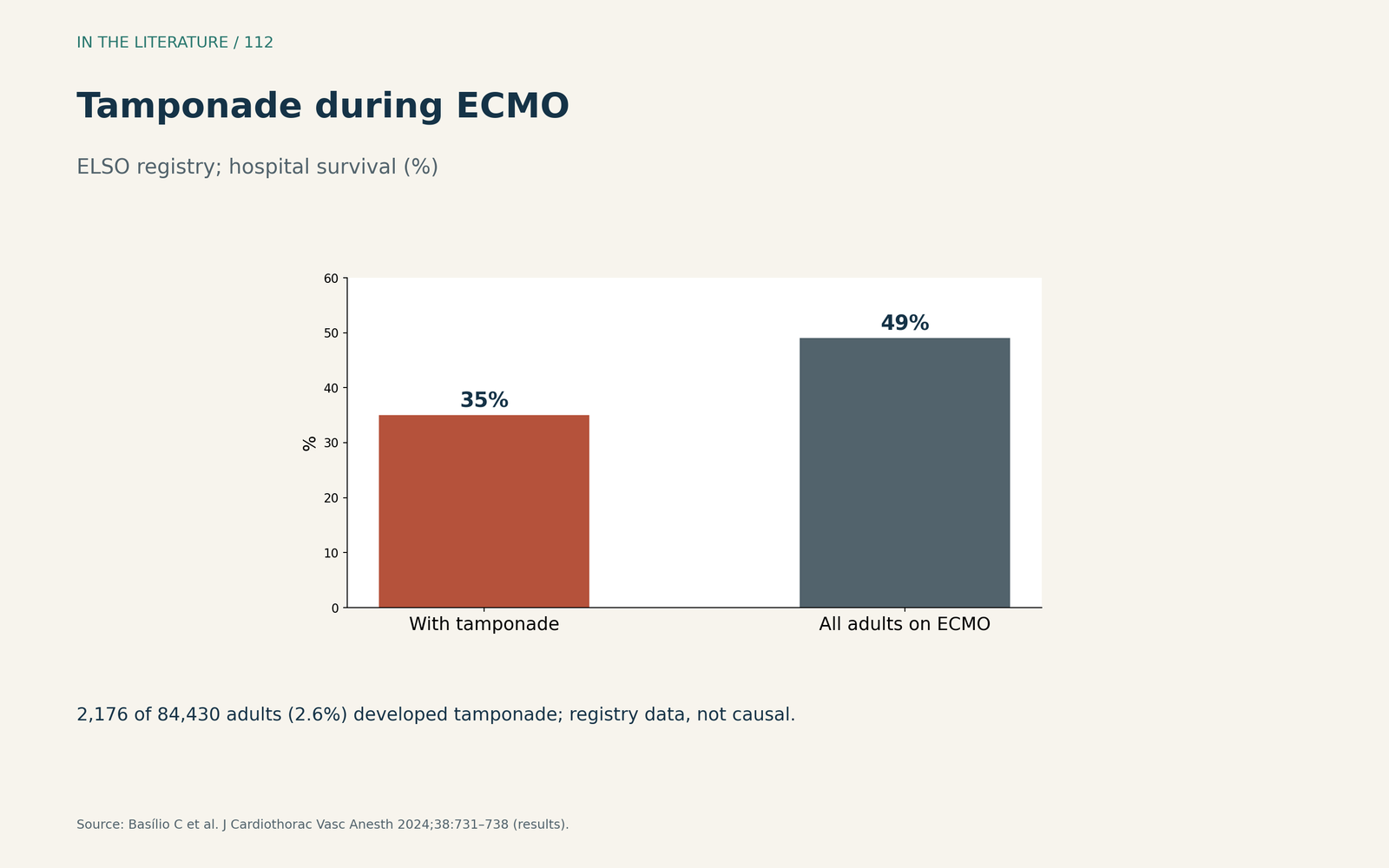
Figure 3. Tamponade during ECMO. Redrawn from the results of Basílio et al. (registry data).[5] View larger.

Anesthetic implications
Clinical signs are unreliable: Madhivathanan and colleagues note that the sensitivity of even one element of Beck's triad was only 50%.[3] The ESC lists tachycardia, hypotension, pulsus paradoxus and raised jugular venous pressure as the classic findings.[2]
The physiology can decompensate precipitously after induction of general anesthesia. The review recommends maintaining preload and systemic vascular resistance, inducing in the operating theatre with the surgeon scrubbed, and avoiding high PEEP and large tidal volumes; removing as little as 50 mL can produce dramatic hemodynamic improvement.[3] The ESC states that vasodilators and diuretics are not recommended in tamponade.[2]
Three takeaways
- Diagnose physiology, not size. Small, rapid effusions can cause tamponade; hypotension is a late sign.[1], [2]
- Look for collapse, the IVC and respiratory Doppler together. Right atrial and right ventricular collapse and a plethoric IVC are the core signs; Doppler variation alone is not enough, especially on the ventilator.[1], [3]
- Expect atypical tamponade. After surgery, with pulmonary hypertension or on ECMO, look for regional compression with TEE, and induce anesthesia only when the surgeon is ready.[3], [4], [5]
References
- Klein AL, Abbara S, Agler DA, et al. American Society of Echocardiography clinical recommendations for multimodality cardiovascular imaging of patients with pericardial disease: endorsed by the Society for Cardiovascular Magnetic Resonance and Society of Cardiovascular Computed Tomography. J Am Soc Echocardiogr. 2013;26(9):965–1012.e15. doi:10.1016/j.echo.2013.06.023
- Adler Y, Charron P, Imazio M, et al. 2015 ESC guidelines for the diagnosis and management of pericardial diseases. Eur Heart J. 2015;36(42):2921–2964. doi:10.1093/eurheartj/ehv318
- Madhivathanan PR, Corredor C, Smith A. Perioperative implications of pericardial effusions and cardiac tamponade. BJA Educ. 2020;20(7):226–234. doi:10.1016/j.bjae.2020.03.006
- Poorsattar SP, Maus TM. Isolated left heart tamponade on echocardiography in severe pulmonary hypertension and right heart failure. J Cardiothorac Vasc Anesth. 2021;35(3):977–978. doi:10.1053/j.jvca.2020.10.029
- Basílio C, Anders M, Rycus P, et al. Cardiac tamponade complicating extracorporeal membrane oxygenation: an Extracorporeal Life Support Organization registry analysis. J Cardiothorac Vasc Anesth. 2024;38(3):731–738. doi:10.1053/j.jvca.2023.12.027