
ITL-2fe8a4a643e7
Juan Li, MD · Updated November 17, 2026
Watch the video

Key points
- Plan a dependable blood-pressure measurement before the vulnerable part of the procedure. ISHLT recommends an arterial catheter when the procedure carries a risk of hemodynamic instability.[1]
- Low displayed flow does not identify the cause of hypotension: venous return, RV performance, vascular tone and pump function must be assessed together.[1], [2]
- Involve the LVAD team before elective surgery and promptly during instability. Unexplained instability can justify rescue TEE or focused cardiac ultrasound when appropriate expertise is available.[2], [3]
- A retrospective study found blood-pressure monitoring gaps differed by device and patient characteristics; it did not establish that routine arterial-line placement improves clinical outcomes.[4]
After induction and the start of positive-pressure ventilation, an LVAD patient's blood pressure falls and the controller shows less flow. Increasing speed may look like a direct way to restore output. But if the LV is receiving too little blood—because venous return has fallen or the RV is failing—more speed can aggravate underfilling and suction. The immediate task is to support perfusion while identifying the reason for the change, rather than treating the flow display as a prescription.[1], [2]
Establish what the blood-pressure reading means
Continuous-flow support can make conventional oscillometric readings difficult, especially with little native pulsatility. A missing or implausible cuff reading requires verification; it is not evidence that the circulation is satisfactory, and it is not itself proof of profound hypotension. Doppler-assisted measurement can be useful for suitable minor procedures, while an arterial catheter provides continuous pressure monitoring when instability is anticipated.[1], [2]
This decision belongs before induction, major positioning or insufflation whenever those transitions are likely to challenge the circulation. Bottiroli and colleagues' 2026 review favors preinduction invasive pressure monitoring for major surgery. The broader ISHLT recommendation is framed around hemodynamic risk, so procedure size, expected fluid shifts, RV function, measurement reliability and the planned anesthetic all matter; “LVAD present” is not the entire assessment.[1], [2]
Vidovich and colleagues studied 115 patients undergoing 187 noncardiac procedures without an arterial line at induction. Their outcome was a blood-pressure monitoring gap of at least 10 minutes. The published abstract reports nearly threefold higher odds of a gap with HeartMate II than with HeartMate 3; morbid obesity and procedures lasting more than 180 minutes were also associated with gaps. Only the formal abstract was available. It supplies no absolute gap rates or confidence interval for that device comparison, and a monitoring gap is not the same outcome as hypotensive injury.[4]
The practical lesson is to anticipate an unreliable measurement and have a workable alternative. The study does not show that an HM3 guarantees reliable cuff measurements, nor does it prove that every minor procedure requires an arterial line.[1], [4]
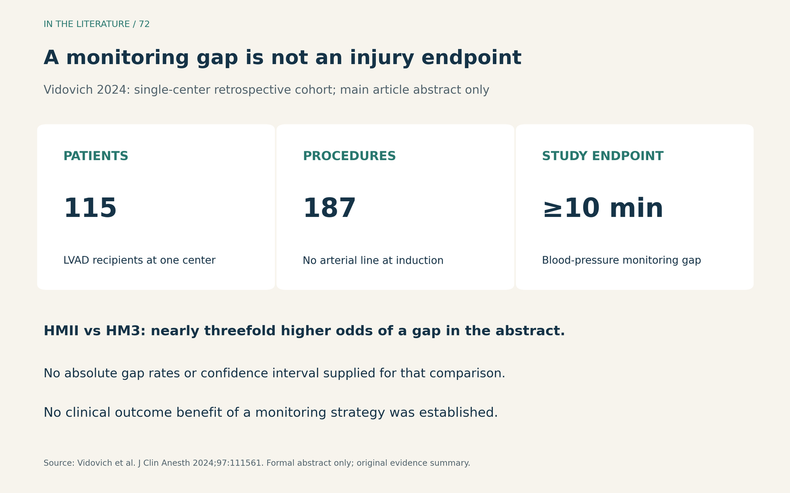
Figure 1. What the monitoring study actually measured. Device and patient associations help frame monitoring plans; they do not establish a clinical benefit from a particular monitoring strategy. Main article: abstract only.[4] View larger.

Match the response to the physiology
At a given speed, pump flow depends on the pressure difference between the LV and aorta. The controller's flow value is an estimate, influenced by device measurements and blood viscosity; it is not a direct measurement of total systemic cardiac output or a stand-alone test of volume status. Interpret changes against the patient's baseline, pressure, rhythm and examination.[2]
Reduced filling is one possibility. Anesthetic-induced venodilation, bleeding, positive-pressure ventilation and changes in position can reduce the blood reaching the LV. Assess the surgical field, volume status and ventricular filling. Fluids may help when insufficient circulating volume is the problem, but low flow alone does not justify repeated boluses.[1], [2]
The RV may be the limiting step. Hypoxia, hypercapnia, acidosis and high intrathoracic pressures can impair RV performance and reduce delivery to the LVAD. A congested, failing RV requires a different response from uncomplicated volume depletion. Review ventilation, oxygenation, rhythm and echo findings; indiscriminate fluid administration can worsen RV overload.[1], [2]
Vascular tone changes pressure and flow differently. Vasodilation after induction may produce hypotension even when displayed pump flow is preserved or increased. Conversely, excessive afterload can reduce pump flow. Support perfusion with appropriately titrated vasoactive treatment when indicated, while identifying concurrent filling or RV problems. Neither a single pressure nor a single flow value distinguishes all of these mechanisms.[1], [2]
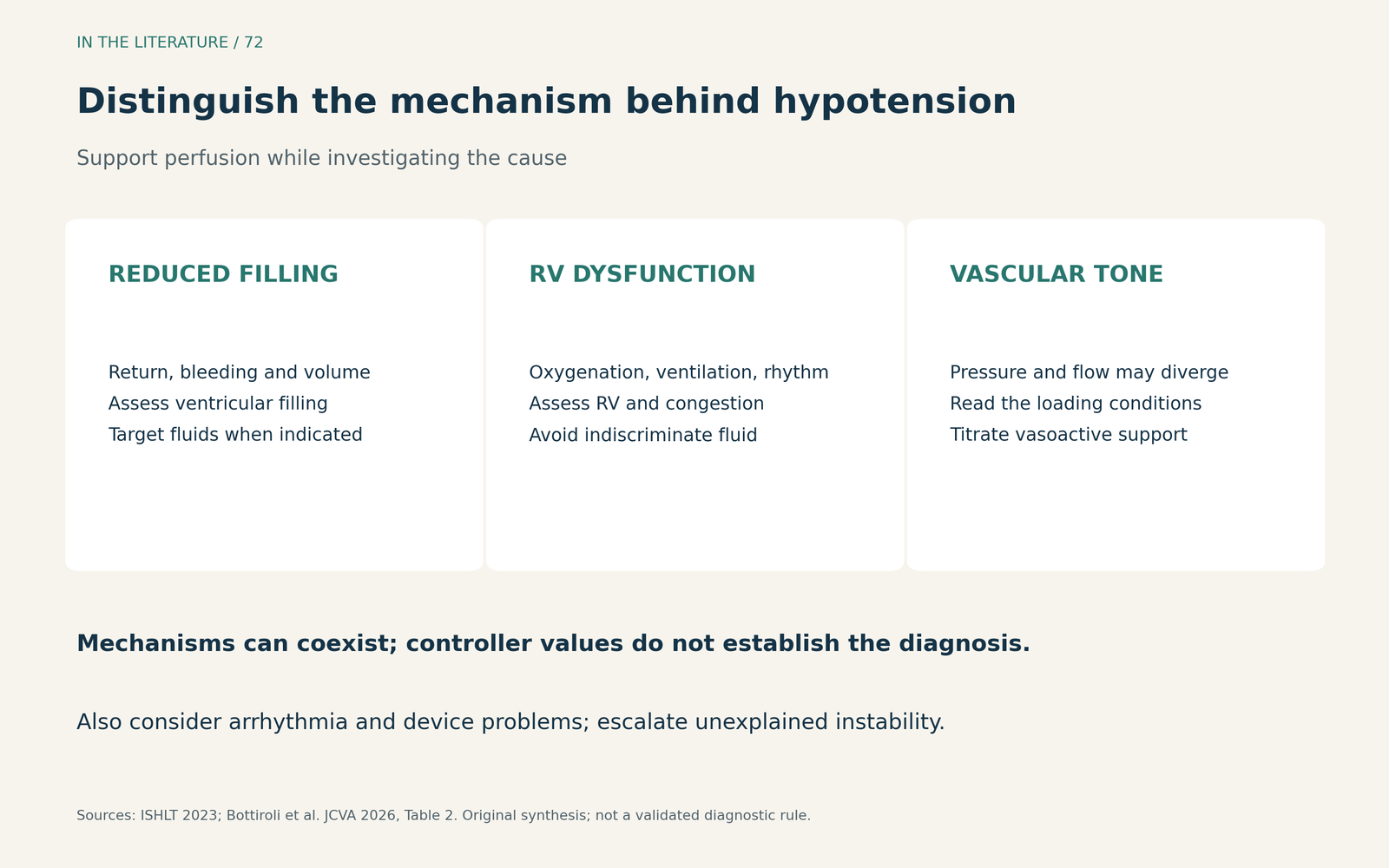
Figure 2. Three mechanisms to distinguish during anesthesia. They can coexist. The diagram is a physiological aid, not a validated diagnostic rule, and does not exclude arrhythmia or device malfunction.[1], [2] View larger.

Make speed changes explainable
Record the usual speed, flow, power and pulsatility index before anesthesia, and arrange continuous device surveillance by appropriately trained personnel. Protect the power supply and driveline through transfers and positioning. These practical details make a change recognizable and permit the LVAD team to interpret it in context.[1], [2]
When suction or excessive unloading is identified, the response may include reducing speed under LVAD-team supervision while correcting the cause of underfilling. Increasing speed reflexively can worsen the mismatch between pump demand and the blood supplied by the right heart. When another mechanism predominates, treatment may instead center on vascular tone, ventilation, rhythm, bleeding or device troubleshooting. The selected speed should have a physiological rationale and an observed response.[1], [2]
For persistent or unexplained instability, TEE or focused cardiac ultrasound can help distinguish ventricular underfilling, RV dysfunction and other cardiac causes when suitable expertise is readily available. ACC/AHA guidance supports such rescue imaging; it does not recommend routine intraoperative TEE screening in patients without relevant patient or procedural risks. The LVAD patient's actual risk and the unresolved clinical question should determine the examination.[3]
The ACC/AHA perioperative recommendations cited here were reaffirmed in September 2026; the societies state that the recommendations and supporting content are unchanged from the 2024 publication. Their LVAD-specific recommendation emphasizes coordination with the specialist team for elective perioperative planning.[3]
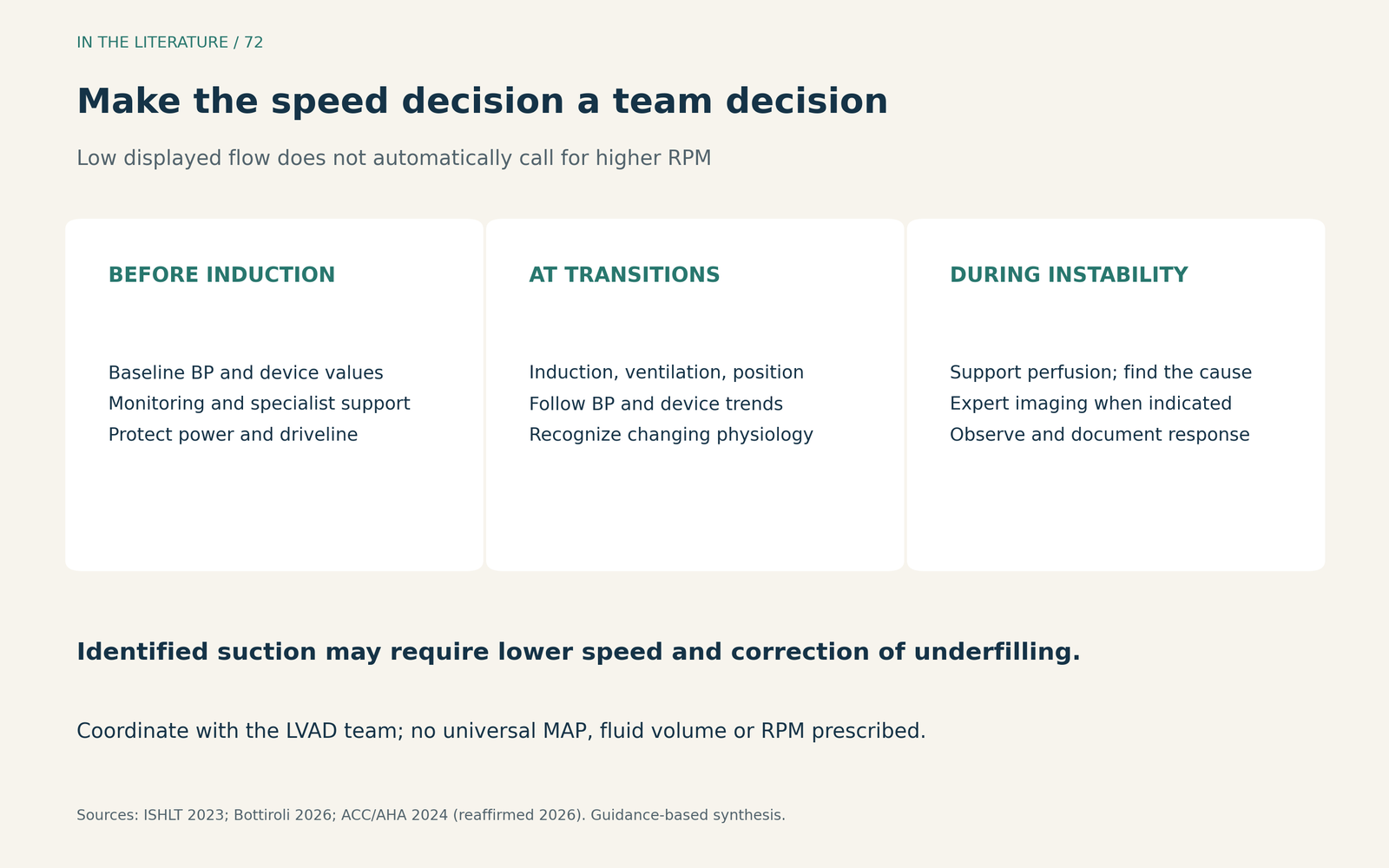
Figure 3. Preparation makes the intraoperative response more coherent. Supporting perfusion and investigating the cause occur together; this sequence does not prescribe a universal MAP, fluid volume or RPM setting.[1], [2], [3] View larger.

Three takeaways
- Make the pressure measurement dependable before you need it most. Plan around the patient's physiology and procedural transitions, with an arterial line when instability risk warrants it.
- Treat low flow as a finding that needs an explanation. Separate reduced filling, RV dysfunction and altered vascular tone; fluids and higher speed are not interchangeable solutions.
- Keep speed adjustment within a coordinated assessment. Support perfusion, use imaging when the cause remains unclear, and document why a speed change helped rather than chasing the controller number.[1], [2], [3]
References
- Saeed D, Feldman D, Banayosy AE, et al. The 2023 International Society for Heart and Lung Transplantation Guidelines for Mechanical Circulatory Support: A 10-Year Update. J Heart Lung Transplant. 2023;42(7):e1-e222. doi:10.1016/j.healun.2022.12.004
- Bottiroli M, Ortoleva J, Pisano D, et al. Perioperative Management of Patients With Durable and Temporary Left Ventricular Assist Devices Undergoing Non-Cardiac Surgery: A Comprehensive Review. J Cardiothorac Vasc Anesth. 2026;40(9):2979–2991. doi:10.1053/j.jvca.2026.05.065
- Thompson A, Fleischmann KE, Smilowitz NR, et al. 2024 AHA/ACC/ACS/ASNC/HRS/SCA/SCCT/SCMR/SVM Guideline for Perioperative Cardiovascular Management for Noncardiac Surgery: A Report of the American College of Cardiology/American Heart Association Joint Committee on Clinical Practice Guidelines. J Am Coll Cardiol. 2024;84(19):1869-1969. doi:10.1016/j.jacc.2024.06.013
- Vidovich C, Laserna A, Bjelic M, et al. Blood pressure monitoring gaps in patients with left ventricular assist devices presenting for non-cardiac surgeries. J Clin Anesth. 2024;97:111561. doi:10.1016/j.jclinane.2024.111561 Abstract only.