ITL-c85cddff6980
Juan Li, MD · Updated November 17, 2026
Watch the video
Key points
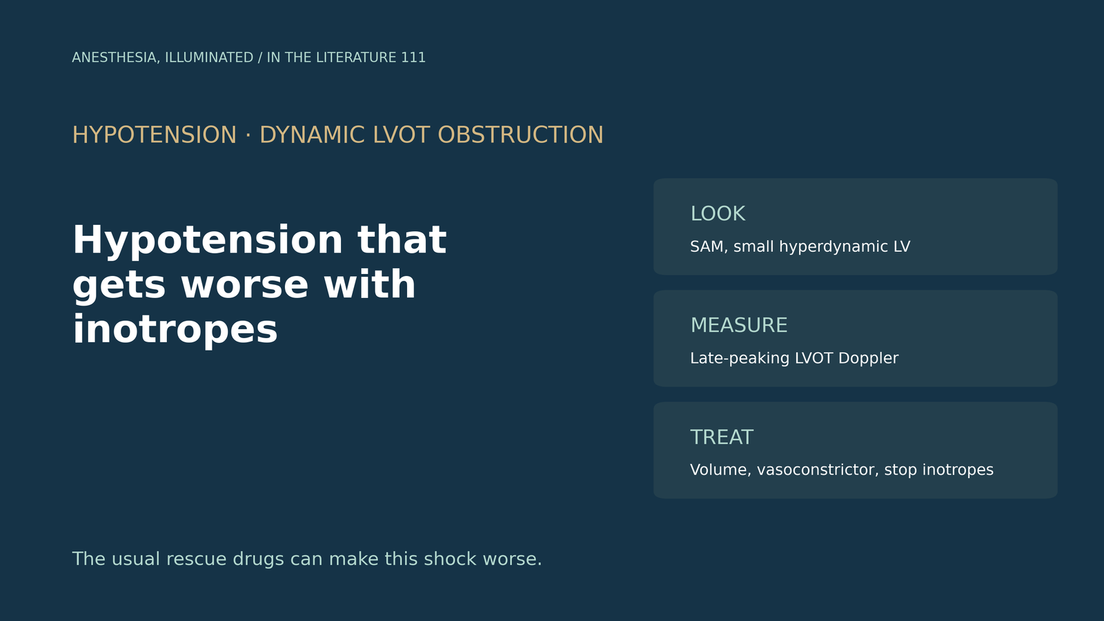
- Suspect dynamic outflow obstruction when hypotension worsens with inotropes or vasodilation. Hypovolemia, low afterload, tachycardia and increased contractility all narrow the outflow tract.[1]
- On echocardiography, look for systolic anterior motion of the mitral valve with leaflet–septal contact, flow acceleration in the outflow tract and a late-peaking continuous-wave Doppler profile. Systolic anterior motion alone is not obstruction, and the mitral regurgitation signal must not be mistaken for the outflow signal.[1], [2]
- It appears outside hypertrophic cardiomyopathy: after anterior myocardial infarction, in takotsubo syndrome (outflow obstruction in 7% to 33%), after mitral repair and during anaphylaxis treated with epinephrine.[3], [4], [5], [2]
- Treatment runs opposite to routine shock care: give volume, use a pure vasoconstrictor such as phenylephrine, reduce or stop inotropes and slow the heart rate. In takotsubo with obstruction, inotropes should not be used and an intra-aortic balloon pump is contraindicated.[1], [4], [5]
- Anesthesia changes what you measure. Gradients under general anesthesia can be much lower than when awake, so reassess with echocardiography after each intervention.[6], [1]
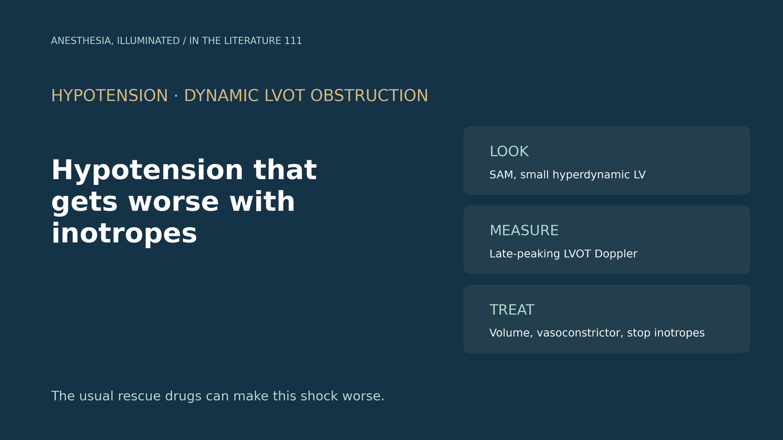
During anesthesia, a patient becomes hypotensive and an epinephrine bolus makes the blood pressure fall further. What should the echocardiogram show if this is dynamic outflow obstruction, which situations predispose to it, and what should be done instead?[1], [2]
Why it happens
The obstruction is dynamic: it depends on loading and contractility. Torre and colleagues explain that hypovolemia, reduced afterload, tachycardia and increased contractility all reduce the ventricular cavity and promote anterior displacement of the mitral leaflet into the outflow tract.[1] Rajendran and colleagues list dehydration, bleeding, diuresis, tachycardia and inotropic agents as precipitating factors.[3]
What the echocardiogram shows
The diagnosis rests on imaging. Obstruction is typically shown by flow acceleration within the outflow tract and a late-peaking continuous-wave Doppler profile, but systolic anterior motion alone should not be considered synonymous with significant obstruction.[1] The mitral regurgitation jet is a pitfall: its signal usually reaches higher velocities, begins earlier and lasts longer than the outflow signal.[1] In the anaphylaxis case reported by Nooli and colleagues, transesophageal echocardiography showed systolic anterior motion with a posteriorly directed mitral regurgitation jet and a late-peaking outflow waveform with a peak gradient of 57 mmHg.[2]
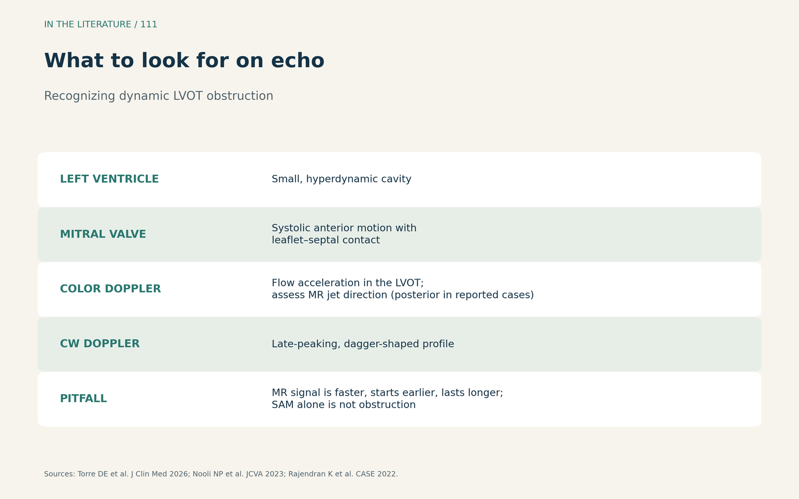
Figure 1. What to look for on echocardiography. Synthesized from Torre et al. and the case reports by Nooli et al. and Rajendran et al.[1], [2], [3] View larger.

Settings where it appears
Dynamic obstruction is not limited to hypertrophic cardiomyopathy. Rajendran and colleagues describe it after anterior infarction, where apical dysfunction is combined with compensatory basal hypercontractility; their patient had a peak gradient of 43 mmHg.[3] In takotsubo syndrome, Kato and colleagues report outflow obstruction in 7% to 33% of patients and cardiogenic shock in 1% to 20%.[4] After mitral valve repair, Crescenzi and colleagues found systolic anterior motion in 9.8% of 608 repairs.[5] During anaphylaxis, epinephrine can provoke obstruction through its chronotropic effects.[2]
Treatment runs the other way
The usual response to hypotension can make this shock worse. Torre and colleagues describe volume as one of the first interventions, phenylephrine as physiologically attractive because it raises afterload, reducing or stopping inotropes, and short-acting beta-blockers such as esmolol.[1] In takotsubo with obstruction, Kato and colleagues recommend fluids under close monitoring as first-line therapy, warn that positive inotropes may worsen obstruction, and state that an intra-aortic balloon pump is contraindicated.[4] In the anaphylaxis case, beta-blockade was not useful while the patient was hypotensive.[2]
After mitral repair, Crescenzi and colleagues used a two-step approach: volume expansion and stopping inotropes, then increasing afterload by manual aortic compression while giving esmolol. It resolved systolic anterior motion in 92% (55 of 60) of patients, and they recommend transesophageal echocardiography before extubation.[5]

Figure 2. What makes it worse, what makes it better. Synthesized from Torre et al., Kato et al., Nooli et al. and Rajendran et al.[1], [2], [3], [4] View larger.

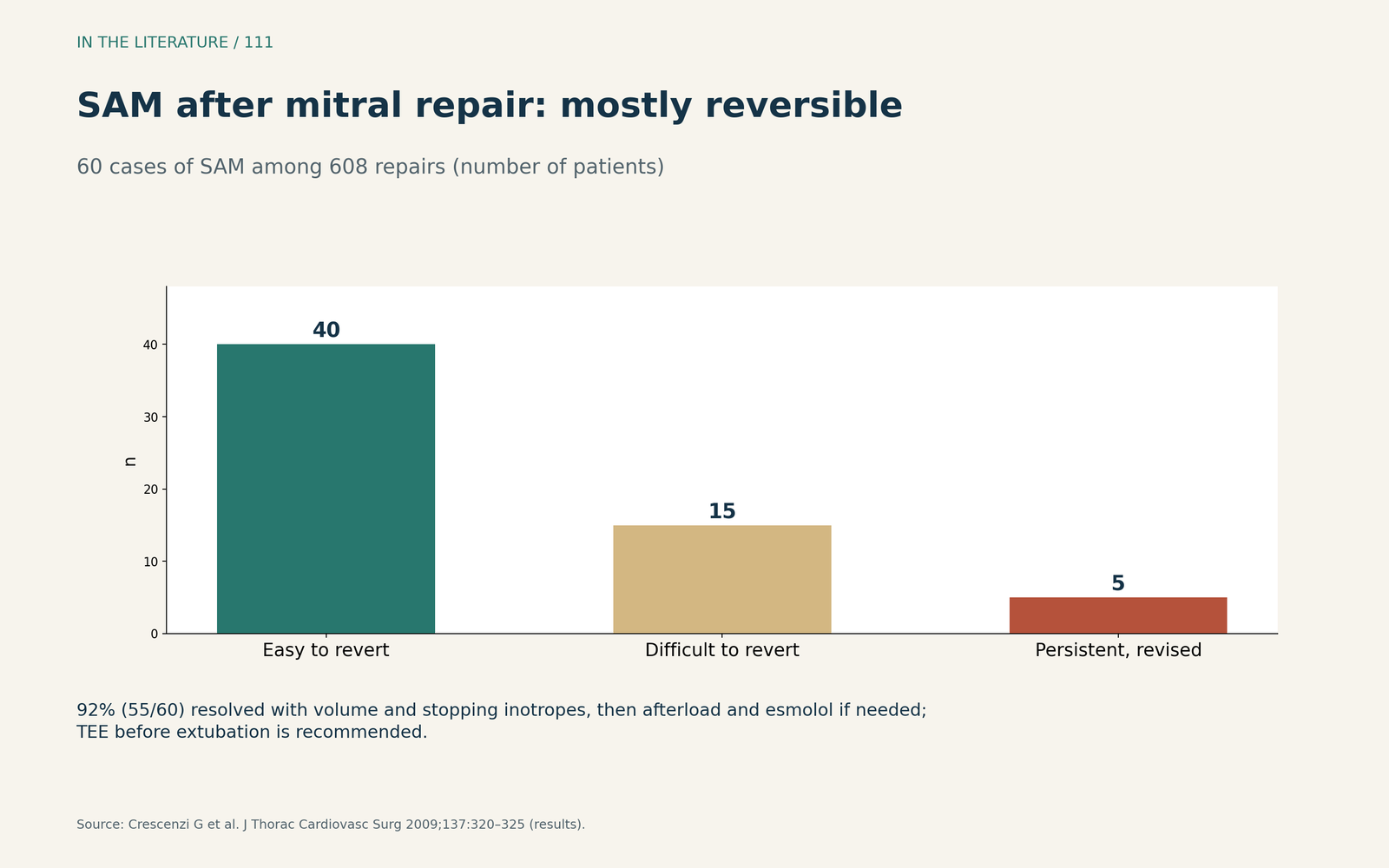
Figure 3. Systolic anterior motion after mitral repair. Redrawn from the results of Crescenzi et al.[5] View larger.

Anesthesia changes the gradient
Gradients are not fixed numbers. In patients with obstructive hypertrophic cardiomyopathy undergoing myectomy, Lentz Carvalho and colleagues found that up to 40% had low intraoperative resting gradients, which they attribute to preoperative fasting, anesthetic agents, volume shifts and positive-pressure ventilation; phenylephrine was required before bypass in 88.4%.[6] Torre and colleagues add that a gradient above 50 mmHg is a marker rather than a validated indication, and recommend repeating echocardiography after every intervention.[1]
Three takeaways
- Think of it when inotropes make hypotension worse. Hypovolemia, vasodilation, tachycardia and inotropes all provoke obstruction.[1], [2]
- Confirm it on echocardiography. Systolic anterior motion with septal contact, a small hyperdynamic ventricle and a late-peaking outflow profile; do not confuse it with the mitral regurgitation signal.[1], [3]
- Treat in the opposite direction. Volume, a pure vasoconstrictor, stopping inotropes and slowing the heart rate, with repeat imaging after each step.[1], [4], [5]
References
- Torre DE, Mangino D, Zoffoli G, et al. Systolic anterior motion after mitral valve repair: echocardiographic prediction, surgical prevention and perioperative management. J Clin Med. 2026;15(18):6958. doi:10.3390/jcm15186958
- Nooli NP, Jensen MA, Lawson P Jr, et al. Role of rescue transesophageal echocardiography during intraoperative anaphylaxis complicated by dynamic left ventricular outflow tract obstruction. J Cardiothorac Vasc Anesth. 2023;37(4):565–569. doi:10.1053/j.jvca.2022.12.014
- Rajendran K, Kunjukrishnanpillai S, Rajan B. Acquired dynamic left ventricular outflow tract obstruction: a rare complication of acute myocardial infarction. CASE (Phila). 2022;6(2):77–82. doi:10.1016/j.case.2021.12.006
- Kato K, Di Vece D, Kitagawa M, et al. Cardiogenic shock in takotsubo syndrome: etiology and treatment. Cardiovasc Interv Ther. 2024;39(4):421–427. doi:10.1007/s12928-024-01031-3
- Crescenzi G, Landoni G, Zangrillo A, et al. Management and decision-making strategy for systolic anterior motion after mitral valve repair. J Thorac Cardiovasc Surg. 2009;137(2):320–325. doi:10.1016/j.jtcvs.2008.08.018
- Lentz Carvalho J, Ashikhmina E, Abel MD, et al. Hypertrophic obstructive cardiomyopathy: discrepancy between hemodynamic measurements in the cardiac laboratory and operating room is to be expected. J Cardiothorac Vasc Anesth. 2022;36(2):422–428. doi:10.1053/j.jvca.2021.05.041