ITL-69a5d12aff35
Juan Li, MD · Updated November 18, 2026
Key points
- Common substrate. A PFO is present in up to 30% of people and raises the risk of hypoxemia and paradoxical embolism on LVAD support.[1]
- The gradient flips. Starting the device decompresses the left heart and can create a right-to-left interatrial gradient, compounded by pulmonary hypertension or RV dysfunction.[2]
- It can grow after surgery. Rising pump speed and inotrope weaning can increase the shunt, and RV failure may unmask or reverse an existing one.[3], [4]
- Easy to miss. Transthoracic bubble study sensitivity has been reported as only 88%, and intraoperative TEE can also fail to detect the shunt.[2], [4]
- Guideline action. ISHLT recommends TEE for interatrial communication before implant and closure of a significant interatrial shunt (Class I, Level C).[5]
Shortly after LVAD start, oxygen saturation falls and does not respond to more oxygen. Why would a shunt that was silent before surgery now cause hypoxemia?[2], [3]
Why the shunt reverses
In advanced heart failure, left atrial pressure is high, so a PFO or ASD stays closed or shunts left to right. Saeed et al. (2023 ISHLT guideline) note that a left-to-right shunt may reverse, with a larger shunt fraction, after LV unloading.[5] Scheiber et al. (J Cardiothorac Vasc Anesth 2024) describe how device start decompresses the left heart, and pulmonary hypertension or RV dysfunction compounds the new right-to-left gradient.[2] Shibao et al. (J Cardiol Cases 2023) state the rule plainly: if right atrial pressure stays above left atrial pressure, as with RV dysfunction, a right-to-left shunt results.[6] Sukhavasi and Merlo (J Cardiothorac Vasc Anesth 2024) add that rising pump speed and inotrope weaning can enlarge the shunt after surgery,[3] and Pillai et al. (Cureus 2024) note that raised right-sided pressures with RV failure may unmask or reverse an existing shunt.[4]
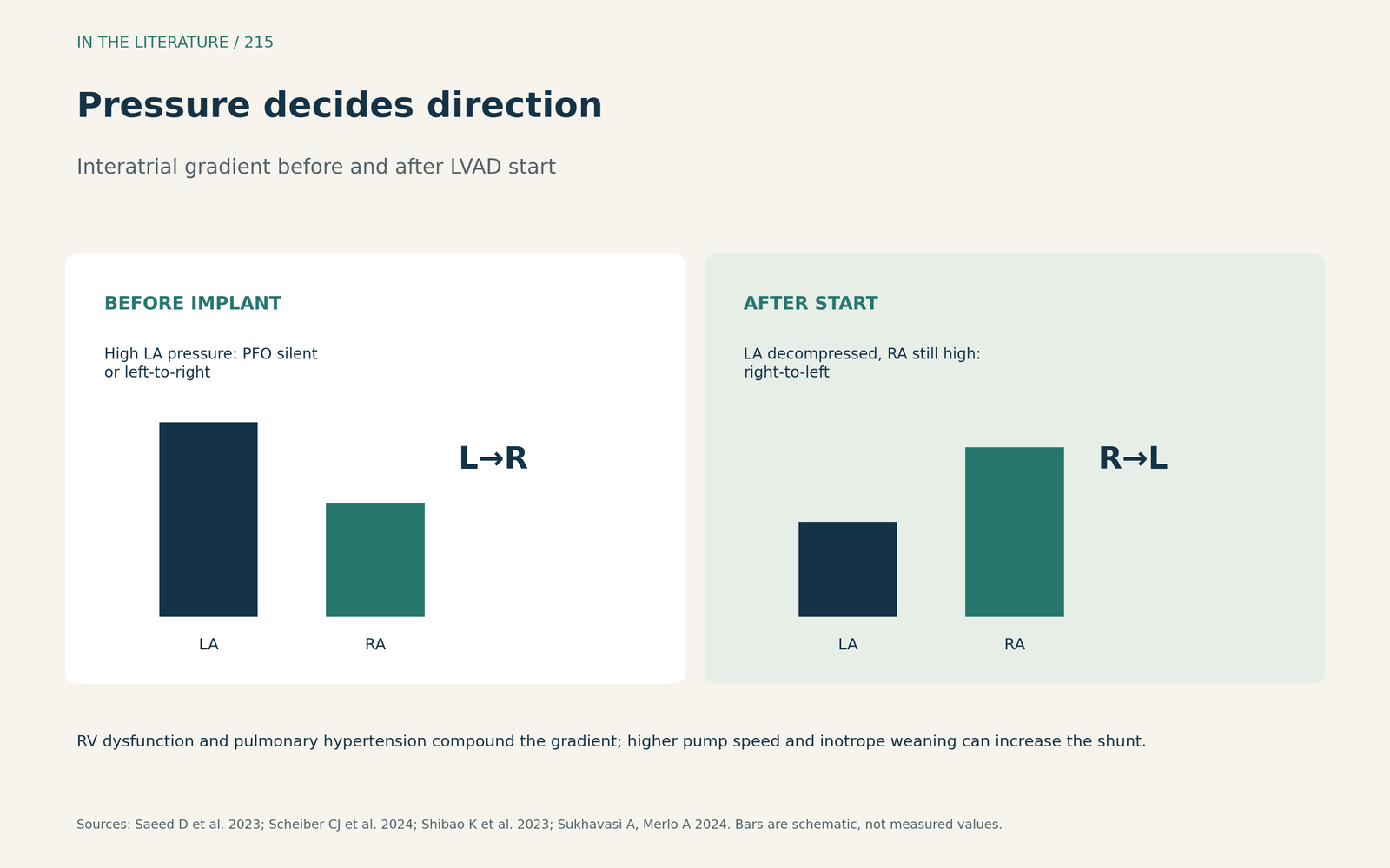
Figure 1. Pressure decides direction. Synthesized from Saeed et al., Scheiber et al., Shibao et al. and Sukhavasi and Merlo.[5], [2], [6], [3] View larger.

Finding the shunt
A PFO occurs in about 1 in 4 people,[2] and iatrogenic ASD is present in 43–82% of patients one month after MitraClip.[7] Stainback et al. (ASE recommendations, 2015) advise finding shunts before surgery and repairing them to avoid sudden desaturation after device activation; when standard testing is inconclusive, agitated saline with a ventilator Valsalva maneuver may help, and post-implant TEE should rapidly assess for an unmasked PFO.[1] Pre-bypass TEE should look for a PFO or any other intracardiac shunt.[5] TEE gives the best spatial resolution of the septum, whereas transthoracic bubble study sensitivity has been reported as only 88%.[2] Transthoracic echo tends to underestimate shunt severity,[6] and a temporary LV pump beforehand may not predict reversal.[4]
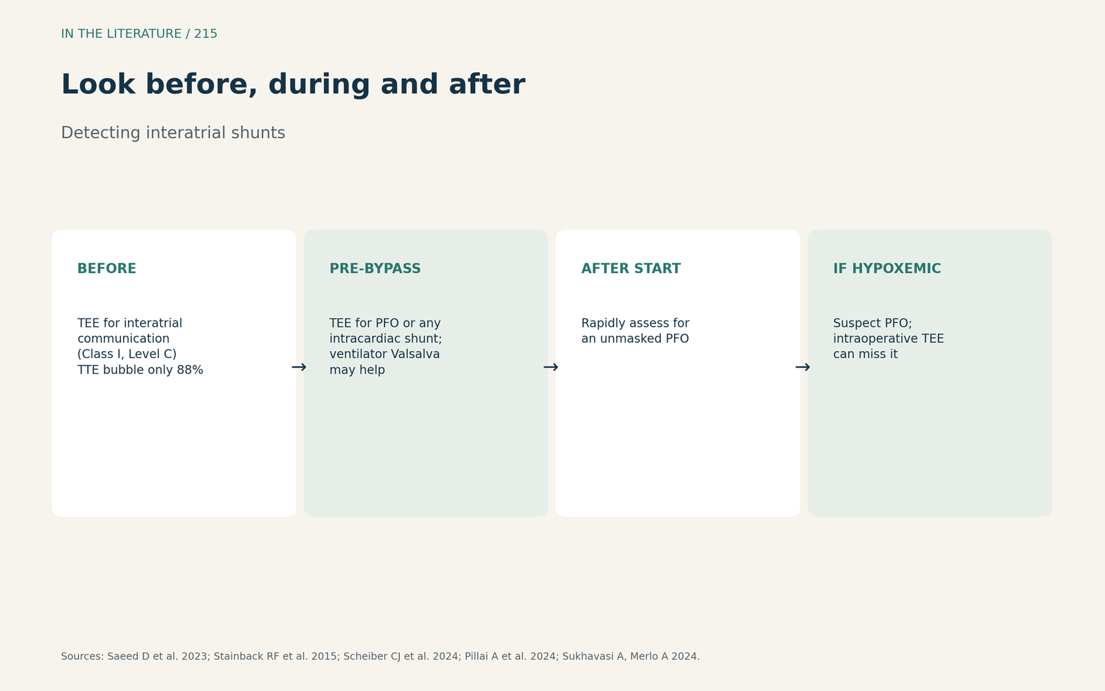
Figure 2. Look before, during and after. Synthesized from Stainback et al., Saeed et al., Scheiber et al., Shibao et al. and Pillai et al.[1], [5], [2], [6], [4] View larger.

To close or not
ISHLT recommends TEE assessment of interatrial communication before implant and closure of a significant interatrial shunt (Class I, Level C).[5] The trade-off is real: any added bypass time can worsen postbypass RV function. Sukhavasi and Merlo advise a high index of suspicion for PFO in hypoxemic LVAD patients and note good outcomes after percutaneous closure in case reports.[3] Because decompression occurs only after implant, Scheiber et al. suggest shunt significance may be judged after bypass.[2] The evidence is expert opinion and case reports.
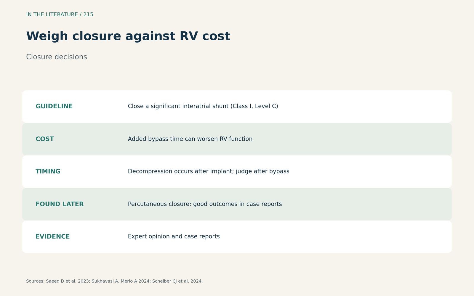
Figure 3. Weigh closure against RV cost. Synthesized from Saeed et al., Sukhavasi and Merlo and Scheiber et al.[5], [3], [2] View larger.

Three takeaways
- Expect a silent PFO or ASD to reverse when LV unloading drops left atrial pressure below right atrial pressure.[5], [2], [6]
- Screen with TEE before implant and look again after device start and with any unexplained hypoxemia.[1], [5], [3]
- Close significant shunts, weighing added bypass time against RV function.[5], [3]
References
- Stainback RF, Estep JD, Agler DA, et al. Echocardiography in the management of patients with left ventricular assist devices: recommendations from the American Society of Echocardiography. J Am Soc Echocardiogr. 2015;28(8):853–909. doi:10.1016/j.echo.2015.05.008
- Scheiber CJ, Teeter EG, Smeltz AM. An intracardiac shunt in a patient undergoing left ventricular assist device implantation. J Cardiothorac Vasc Anesth. 2024;38(5):1260–1264. doi:10.1053/j.jvca.2023.12.030
- Sukhavasi A, Merlo A. Commentary on an intracardiac shunt in a patient undergoing left ventricular assist device implantation. J Cardiothorac Vasc Anesth. 2024;38(5):1265–1267. doi:10.1053/j.jvca.2024.02.021
- Pillai A, Jedeon Z, Hammond J, et al. Intracardiac shunt reversal and early right ventricular failure after left ventricular assist device implantation. Cureus. 2024;16(7):e65320. doi:10.7759/cureus.65320
- Saeed D, Feldman D, El Banayosy A, et al. The 2023 International Society for Heart and Lung Transplantation guidelines for mechanical circulatory support: a 10-year update. J Heart Lung Transplant. 2023;42(7):e1–e222. doi:10.1016/j.healun.2022.12.004
- Shibao K, Murasato Y, Araki M, et al. Acute hypoxemia caused by Impella in a patient with fulminant myocarditis and patent foramen ovale. J Cardiol Cases. 2023;27(1):36–40. doi:10.1016/j.jccase.2022.10.003
- Naveed H, Haj-Ahmad LM, Loyalka P. Emergency bedside transoesophageal echocardiography-guided atrial septal defect closure without fluoroscopy after left ventricular assist device implantation. Interv Cardiol. 2025;20:e29. doi:10.15420/icr.2025.19