
ITL-6ac686c6448c
Juan Li, MD · Updated November 17, 2026
Watch the video

Key points
- Measure from the aortic annulus to the middle of the inlet: approximately 3.5 cm for Impella CP and 5 cm for Impella 5.5. These are model-specific reference distances.[1]
- Appropriate depth and apparently normal console waveforms do not exclude malrotation away from the LV apex.[2]
- A suction alarm requires assessment of both position and ventricular filling; right-heart failure, tamponade and thrombus can also contribute.[3]
- Suspected malposition calls for imaging and experienced-device-team assessment. Adjustment requires continuous imaging; a device that has completely migrated above the valve must not simply be pushed back blindly.[4]
The inlet appears to be at the expected depth, yet unloading remains unsatisfactory or suction alarms recur. Before declaring the position satisfactory, ask a second question: where is the inlet facing, and what surrounds it? A correctly measured distance can coexist with an inlet directed toward the lateral wall or mitral apparatus.[1], [2]
Measure the correct structure, then inspect the whole device
The 2024 ASE recommendations specify the distance from the aortic annulus to the middle inlet point. The distal end of a CP pigtail is not that endpoint. CP has a distal pigtail; Impella 5.5 has a different distal configuration, so one generic depth should not be applied to both.[1]
Device | Approximate annulus-to-middle-inlet distance |
Impella CP | 3.5 cm |
Impella 5.5 | 5 cm |
Depth is one part of the examination. ASE also describes a stable, apex-directed device, a freely positioned inlet away from the mitral subvalvular apparatus and LV walls, the cannula bend at the annulus, and an unobstructed outlet in the ascending aorta well above the valve. Use views that establish these relationships rather than relying on one reassuring measurement.[1]
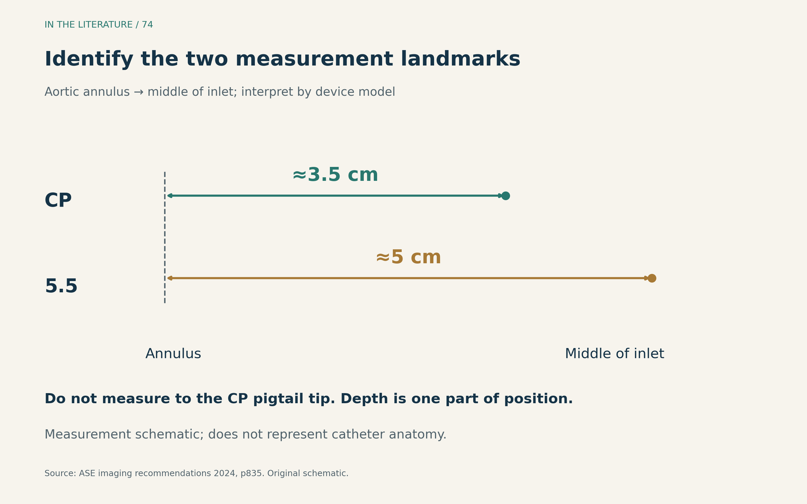
Figure 1. Identify both measurement landmarks before interpreting the number. This is a measurement schematic, not an anatomical drawing or a universal repositioning target. Direction, inlet clearance and outlet location must also be satisfactory.[1] View larger.

Color Doppler can help identify the outlet, but an outlet signal does not establish that the inlet is free of surrounding structures. Examine mitral and aortic valve interaction as well: new regurgitation may reflect tethering, while direct injury is also possible.[1]
Why a normal depth can conceal poor orientation
Baldetti and colleagues studied 100 patients receiving Impella for cardiogenic shock in a single-center retrospective cohort. Malrotation was identified in 36. Their definition deliberately included apparently normal console waveforms and appropriate depth, together with abnormal orientation away from the LV apex and additional anatomical findings. Thus, a “normal” console and distance could be part of the malrotation phenotype.[2]
Among the 85 patients with paired invasive hemodynamic measurements, pulmonary artery wedge pressure at 48 hours was 16.0 ± 8.2 mmHg with malrotation versus 13.0 ± 4.6 mmHg without malrotation (mean ± SD; P=0.033). This association supports looking beyond depth when unloading appears incomplete. It does not prove that repositioning improves survival: the study did not systematically evaluate the effects of repositioning and found no significant differences in major adverse in-hospital outcomes.[2]
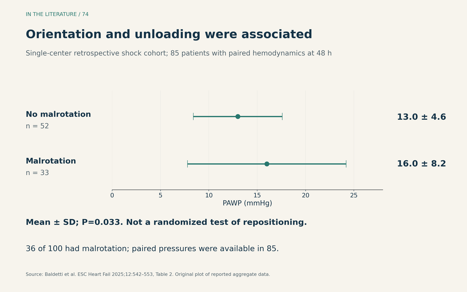
Figure 2. Malrotation was associated with less favorable unloading in an observational cohort. The hemodynamic comparison includes 85 patients, rather than all 100 enrolled patients; bars show mean ± SD. This is not a randomized test of correction. The cohort predominantly used CP and included only three Impella 5.5 recipients.[2] View larger.

Match the image to the patient's physiology
ASE recommends repeat echocardiographic assessment with unexpectedly low flow, persistent suction or position alarms, suspected RV deterioration, hemolysis, significant patient movement or catheter adjustment. Recheck the device and the circulation together: an anatomically acceptable pump can still have inadequate LV filling.[1]
For Impella 5.5, the clinical management review specifically advises integrating arrhythmia, hemolysis and hemodynamic stability into a repositioning decision. A device that is functioning well may not need manipulation merely to make its measured depth look more textbook-perfect. Conversely, recurrent suction should prompt assessment of position, filling, RV function, pericardial constraint and possible thrombus, rather than automatic advancement or withdrawal.[3]
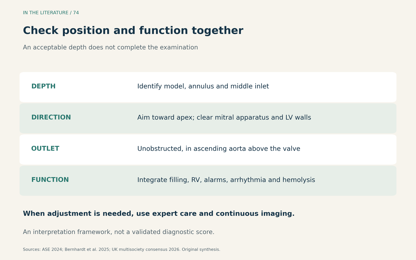
Figure 3. An integrated position check. The framework summarizes imaging recommendations and practical guidance; it is not a validated diagnostic score. Abnormal findings require interpretation in the clinical and device context.[1], [3], [4] View larger.

When repositioning is indicated, the UK multisociety consensus calls for an experienced clinician and continuous echocardiographic guidance, with support reduced to P2 during adjustment while the device still crosses the valve. If echocardiography shows that no part of the device remains below the valve, the consensus recommends returning to the catheter laboratory rather than blind readvancement. These are different situations and should not be managed as the same bedside adjustment.[4]
Three takeaways
- Name the model and the landmarks. A CP pigtail-tip measurement cannot substitute for annulus-to-inlet depth.[1]
- Inspect direction even when depth looks right. Normal console waveforms do not certify unobstructed, apex-directed inflow.[2], [4]
- Correct a clinical problem, not just a number. Integrate filling, device performance and complications; use specialist imaging guidance when adjustment is needed.[1], [3], [4]
References
- Estep JD, Nicoara A, Cavalcante J, et al. Recommendations for Multimodality Imaging of Patients With Left Ventricular Assist Devices and Temporary Mechanical Support: Updated Recommendations from the American Society of Echocardiography. J Am Soc Echocardiogr. 2024;37(9):820–871. doi:10.1016/j.echo.2024.06.005
- Baldetti L, Romagnolo D, Festi M, et al. Impella malrotation affects left ventricle unloading in cardiogenic shock patients. ESC Heart Fail. 2025;12(1):542–553. doi:10.1002/ehf2.15087
- Bernhardt AM, Blumer V, Vandenbriele C, et al. Clinical management of the Impella 5.5 pump. J Heart Lung Transplant. 2025;44(11):1688–1702. doi:10.1016/j.healun.2025.06.008
- Akhtar W, Bowles CT, Costanzo P, et al. UK multisociety consensus statement on the emergency management and resuscitation of patients with left-sided Impella support. Heart. 2026;112(21):1171–1178. doi:10.1136/heartjnl-2025-326896